
Abstract
Bone regeneration using stem cells and growth factors has disadvantages while needing to use supraphysiological growth factors concentrations. Gene therapy has been proposed as alternative, but also has limitation. Messenger RNA (mRNA)-based transcript therapy is a novel approach that may solve plasmid DNA-based gene therapy limitations. Although much more efficient in delivering genes into the cell, mRNA is unfortunately unstable and immunogenic. However, recent reports indicated that chemical modifications of the mRNA molecule can improve stability and toxicity. In this study, we have combined biomaterials and chemically modified mRNA (cmRNA) encoding Metridia luciferase, eGFP, and bone morphogenetic protein (BMP)-2 to develop transcript-activated matrices (TAMs) for gene transfer to stem cells. BMP-2 cmRNA was produced to evaluate its feasibility in stimulating osteogenic differentiation. Fibrin gel and micro-macro biphasic calcium phosphate (MBCP) granules were used as biomaterials. A sustained release of hBMP-2 cmRNA from both biomaterials was observed during 7 days. This occurred significantly faster from the MBCP granules compared to fibrin gels (92% from MBCP and 43% from fibrin after 7 days). Stem cells cultured in hBMP-2 cmRNA/fibrin or on hBMP-2 cmRNA/MBCP were transfected and able to secrete significant amounts of hBMP-2. Furthermore, transfected cells expressed osteogenic markers in vitro. Interestingly, although both TAMs promoted gene expression at the same level, hBMP-2 cmRNA/MBCP granules induced significantly higher collagen I and osteocalcin gene expression. This matrix also induced more mineral deposition. Overall, our results demonstrated the feasibility of developing efficient TAMs for bone regeneration by combining biomaterials and cmRNAs. MBCP synergistically enhances the hBMP-2 cmRNA-induced osteogenic pathway.
Introduction
S
Despite their known therapeutic potential, these molecules present limitations that often jeopardize their clinical applications. One of the most relevant drawbacks is their short half-life. These factors are very unstable when placed in contact with body fluids or tissues [8,9]. Hence, they are often administered at supraphysiological concentrations in the effort of achieving the desired therapeutic outcome. As a consequence, many side effects have been reported [10,11], such as the risk of malignancies being one of the biggest concerns [12]. Based on this, the FDA has released several warnings on complications associated with the clinical use of growth factors (BMP-2 [13] and PDGF [14]).
Much research has been done in many directions to overcome these limitations. The possibility of using plasmid DNA (pDNA) encoding the growth factor of interest has been a very appealing one. These molecules can be delivered inside the cells by different vectors including, for instance, safe and nonviral lipid-based formulations. The target cell would eventually produce the therapeutic protein as a result of the pDNA uptake, thereby facilitating therapeutic pathways to take place. In addition, proteins can be expressed for longer periods and doses of growth factors can be reduced.
While nonviral delivery of pDNA is preferred over viral vectors due to safety concerns, low gene transfer efficiency of nonviral vectors puts their clinical use at risk. Messenger RNA (mRNA) has recently demonstrated to be an interesting alternative to pDNA [15 –17]. Recombinant mRNA targets the cell cytoplasm resulting in an immediate translation of its encoding therapeutic protein. Thus, in contrast to pDNA (targets cell nucleus), mRNA may represent a more efficient gene transfer approach.
Rudolph and colleagues have recently reported a nucleotide-based chemical modification of the mRNA molecule that resulted in a highly stable and nonimmunogenic chemically modified mRNA (cmRNA) [17]. More recently, our group demonstrated the usability of mRNA transcript gene transfer by directly applying cmRNA in vitro [15] and in vivo [17]. Transcript-based gene transfer technology is highly revolutionary for the gene therapy field. It may accelerate the clinical translation of gene therapy by overcoming safety and efficiency issues. However, while technology has demonstrated its benefits, direct application of cmRNA (eg, without a delivery system) may not satisfy all therapeutic needs.
In this study, we aim to adapt this technology to a tissue engineering approach by combining biomaterials with cmRNAs. Importantly, one of the biomaterials has osteoconductive properties. We hypothesize that the combination of clinically used biomaterials and cmRNA encoding BMP-2 may enhance protein translation of this osteogenic growth factor. Thereby, it may enhance functional outcome in stem cells regarding induction of osteogenic pathways and mineralization. Concretely, we have used human bone morphogenetic protein (hBMP)-2 cmRNA loaded onto ceramic biomaterials and into fibrin gel to produce transcript-activated matrices (TAMs). Our aim was to enhance the sustained release of loaded cmRNAs from a three-dimensional (3D) matrix to continuously transfect stem cells. This may enhance the osteogenic potential of the transfected stem cells in clinically used biomaterials for bone regeneration.
Materials and Methods
Biomaterials
Micro-macro biphasic calcium phosphate (MBCP) ceramic granules (Biomatlante, Vigneux de Bretagne, France) and Tissucol (Baxter, Unterschleissheim, Germany) were used as biomaterials in this study. MBCP is a synthetic resorbable biphasic calcium phosphate ceramic composed of 60% hydroxyapatite and 40% tricalcium phosphate. The granules used in our study were 1–2 mm in size and presented 70% porosity with an interconnected network of macro- and micropores. The granules were sterilized by γ-radiation before use. On the other hand, Tissucol is a fibrin material that is composed by fibrinogen (3000 kIU/mL) and thrombin (4 U/mL). Both components were received sterile. Fibrinogen and thrombin were mixed in equal volumes immediately before the experiments to generate the fibrin gels.
Animals
Female Sprague Dawley rats (N = 15, 250–300 g; Charles River Laboratories, Sulzfeld, Germany) were used for bone marrow stem cell (BMSC) isolation. The animals were euthanized by carbon dioxide asphyxia immediately before bone marrow collection. Procedures were approved by the local legislative committee of the upper Bavarian government (Germany). In addition, all the experiments were performed according to the German and European law for animal protection and in accordance with the Guide for the Care and Use of Laboratory Animals as defined by the National Institutes of Health.
Rat-derived BMSCs
BMSCs were isolated from femurs and tibias following a standard protocol [18]. All cell culture materials were obtained from PAA Laboratories GmbH (Pasching, Austria) unless specified otherwise.
In brief, all harvested bones were opened at both epiphyses and incubated in sterile collagenase type II (Gibco™, Invitrogen, CA) solution (2.5 mg/mL in nonsupplemented Dulbecco's modified Eagle's medium [DMEM]) at 37°C and 5% CO2 for 2 h. Next, the bone marrow was flushed out by using DMEM supplemented with 10% fetal bovine serum and 1% penicillin/streptomycin (further referred to as complete DMEM). The resulting cell suspension was centrifuged. The obtained cell pellet was resuspended in complete DMEM and the mononuclear cell fraction was collected by density gradient centrifugation (500 g, 30 min, Ficoll-Paque™; GE Healthcare Ltd., CT). Collected cells were plated at 3000 cells/cm2.
For transfection and differentiation experiments, the cells were used from passage 2 up to passage 6. Characterization of BMSCs isolated following the described protocol can be found elsewhere [18].
Generation of cmRNAs
hBMP-2 cmRNA, MetLuc cmRNA and eGFP cmRNA were produced following our previously reported protocol [15]. In short, plasmid vectors containing open reading frames of Metridia luciferase (MetLuc) and hBMP-2 mRNA were cloned into the backbone of pVAXA120 between BamHI and EcoRI sites by GeneArt (Life Technologies, CA). eGFP was cloned into pVAXA120 between EcoRI and KpnI sites via semi-blunt ligation. Subsequently, pVAXA120-MetLuc, pVAXA120-hBMP-2 and pVAXA120-eGFP were linearized downstream of the poly-A tail by NotI digestion. In vitro transcription was carried out using the RiboMAX™ Large Scale RNA Production System-T7 (Promega, WI) following manufacturer's instructions. For mRNA capping, an antireverse cap analog (m7,3′-OGpppG; Jena Biosciences, Jena, Germany) was used.
To produce the cmRNAs, 25% modified nucleotides (ie, 5-methylcytidine-5′-triphosphate and 2-thiouridine-5′-triphosphate; Jena Biosciences) were added to the reaction. Purification of resulting hBMP-2 cmRNA, MetLuc cmRNA, and eGFP cmRNA was performed by ammonium acetate precipitation and integrity and sizes were confirmed by native agarose gel electrophoresis.
Fluorescent labeling of hBMP-2 cmRNA
hBMP-2 cmRNA was labeled with fluorescein isothiocyanate (FITC) by using Label IT Fluorescein Labeling Kit (Mirus Bio, WI) following the manufacturer's protocol. The resulting FITC-hBMP-2 cmRNA was purified by ammonium acetate precipitation and washed with ethanol 70%. The absorbance was recorded at 260 nm and at 494 nm (λmax FITC) by using NanoDrop 2000c (Thermo Scientific, Waltham, MA). The density of FITC dye per μg of RNA was calculated using the following equations:
where AFITC represents absorbance value measured for FITC and ∈FITC correspond to the extinction coefficient of nucleic acid-bound dye (for FITC 68,000 M−1 cm−1). In addition, the A260 value for RNA is 40 μg/mL. The results of the FITC labeling of hBMP-2 cmRNA are tabulated in Table 1.
cmRNA, chemically modified mRNA; FITC, fluorescein isothiocyanate.
Loading of FITC-hBMP-2 cmRNA into MBCP granules and fibrin gels. Evaluation of in vitro release
FITC-hBMP-2 cmRNA lipoplexes were formed by mixing cmRNA solution (ultrapure water) and DreamFect Gold (DF-Gold, 4 μL lipid per μg mRNA; OzBiosciences, Marseille, France) at room temperature for 20 min [15]. Subsequently, 50 mg sterile MBCP granules were loaded using 50 μL lipoplexes (containing 1 μg FITC-hBMP-2 cmRNA) and snap frozen in liquid nitrogen. Next, the loaded MBCP granules were lyophilized overnight. In the case of fibrin gels, 50 μL fibrinogen was mixed with 10 μL (1 μg) FITC-hBMP-2 cmRNA lipoplexes. Similarly, the resulting suspension was snap frozen in liquid nitrogen and lyophilized overnight. In both biomaterial compositions, 1% trehalose (Sigma-Aldrich, MO) was used as cryoprotectant. Immediately before the in vitro release studies, the fibrinogen containing FITC-hBMP-2 cmRNA was resuspended in 50 μL ultrapure water and subsequently mixed with 50 μL thrombin to form the cmRNA loaded fibrin gel.
Both cmRNA loaded MBCP and cmRNA loaded fibrin gel were incubated in 500 μL sterile Dulbecco's phosphate-buffered saline (DPBS) at 37°C under gentle shaking (50 rpm) for up to 7 days. At determined time points (ie, 2, 4, 8, 12, 24, and 36 h, 2, 3, 5, and 7 days), 100 μL were taken and replaced with the same volume of fresh sterile DPBS. After each time of observation was reached, samples were photographed using a fluorescence microscope (Biorevo BZ9000; Keyence, Osaka, Japan).
To be able to quantify the released amount of FITC-hBMP-2 cmRNA, decomplexation was performed by adding 10 μg heparin sulfate (50,000 units; Sigma-Aldrich) to the samples and incubated for 15 min at room temperature [19,20]. Subsequently, the amount of FITC-hBMP-2 cmRNA in each sample was quantified by measuring the FITC signal at 485 nm (excitation) and 535 nm (emission) using a Wallac Victor 1420 multilabel counter (PerkinElmer, MA). The cumulative release was calculated as percentage considering the loaded cmRNA dose.
Transfection of BMSCs with cmRNA loaded biomaterials
For transfection, 50 mg cmRNA loaded MBCP granules (lyophilized and containing 1 μg cmRNA: hBMP-2 cmRNA, MetLuc cmRNA, or eGFP cmRNA) were transferred into a 48-well plate and 1 × 10⁵ BMSCs were seeded onto the loaded granules. For seeding, the indicated amount of cells was directly added to the granules using a volume of 50 μL. After 1 h of incubation at 37°C and 5% CO2, complete DMEM was added to reach a final volume of 800 μL.
For transfection using fibrin gels, lyophilized fibrinogen (containing 1 μg cmRNA: hBMP-2 cmRNA, MetLuc cmRNA or eGFP cmRNA) was resuspended in 50 μL ultrapure water and transferred into a sterile Eppendorf tube cap used as mold. Subsequently, 50 μL thrombin containing 1 × 10⁵ BMSCs was added and rapidly mixed for a good cell homogenization. The molds containing the fibrin, cmRNA and BMSCs were incubated at 37°C for 30 min to allow gel formation. Thereafter, the fibrin gel compositions were transferred into a 48-well plate and complete DMEM was added.
Medium change was performed every 3 days. For hBMP-2 cmRNA and MetLuc cmRNA, supernatants were stored at −80°C for further determinations. eGFP cmRNA samples were imaged using a fluorescence microscope as described below.
Cytotoxicity of cmRNA loaded biomaterials
To analyze the biocompatibility of the cmRNA-activated matrices an MTS assay was performed following the manufacturer's instructions (CellTiter 96; Promega, WI). hBMP-2 cmRNA was used for toxicity screening. cmRNA loading and subsequent cell transfection was performed as described above. In brief, 5 and 24 h post-transfection, 200 μL of diluted MTS reagent was added per well and subsequently incubated in the dark at 37°C and 5% CO2, for 3 h. Next, 100 μL supernatant were transferred to a 96-well plate and the absorbance was measured at 490 nm. Latex rubber was used as positive control for cell death [18]. In addition, untreated cells were used as 100% viable cells. Cells seeded into fibrin gels and onto MBCP granules (with no previous cmRNA loading) were also used as controls. Thereby, the effect of the material per se on cell viability can be ruled out.
BMP-2 protein production and gene expression
BMP-2 production (ie, secreted and intracellular) as a result of hBMP-2 cmRNA transfection of BMSCs was evaluated by ELISA (Quantikine; R&D Systems, MN). To quantify the amount of secreted BMP-2, cell culture supernatants were used. To evaluate the intracellular production of BMP-2, cell lysates were prepared with Tris-HCl lysis buffer (25 mM Tris-Hcl, 0.1% TritonX-100, pH 7.8). Supernatants and cell lysates were collected at 24, 48, and 72 h post-transfection. These samples were used immediately after collection for protein quantification. ELISAs were performed according to the manufacturer's protocol and the protein content was calculated using a standard curve.
Metridia luciferase activity was measured in MetLuc cmRNA transfected cells up to 120 h post-transfection by using coelenterazine (50 μM) as substrate and following standard protocols [21]. On the other hand, eGFP-positive cells resulting from eGFP cmRNA transfer were imaged at 24 h post-transfection with the Biorevo BZ9000 fluorescence microscope.
Functional analysis
In vitro osteogenesis
To determine whether the hBMP-2 cmRNA loaded biomaterials resulted in osteogenic stimulation of BMSCs, real-time PCR and mineralization assays were performed at predetermined times of observation (3, 7, and 14 days for PCR and 21 days for mineralization). Expression of hBMP-2, RunX2, alkaline phosphatase (ALP), collagen I (Coll I), and osteocalcin (OCN) was evaluated at days 3, 7, and 14 post-transfection.
Cells were collected with TRIzol (Life technology, CA) and total RNA was isolated by phenol/chloroform method. Total RNA was reverse-transcribed by using First Strand cDNA Synthesis Kit (Thermo Scientific) using random primers. SsoFast Eva Green Supermix (Bio-Rad Laboratories, CA) was used and the PCR reactions were carried out in a Bio-Rad CFX96 thermal cycler (Bio-Rad Laboratories). Rat β-tubulin was used as housekeeping gene and results (ie, fold increase in gene expression) were reported relative to hBMP-2 cmRNA unloaded biomaterials seeded with BMSCs (ie, untransfected cells) using the 2−ΔΔCT method.
Mineralization was evaluated by means of alizarin red staining. The staining was performed at 21 days post-transfection. hBMP-2 cmRNA unloaded biomaterials, with and without seeded BMSCs, were used as controls. In brief, ethanol-fixed samples were incubated with alizarin red solution (5 mg/mL) for 30 min at room temperature. After extensive washing with DPBS, fibrin gels were imaged using a Canon camera model EOS 600D with a resolution of 18 megapixels adapted to a Photo Macroscope (Wild Heerbrugg M400, Heerbrugg, Switzerland). In the case of the MBCP granules, samples were photographed with the Biorevo BZ9000 microscope.
Quantification was performed spectrophotometrically at 570 nm after extraction of the dye with cetylpyridinium chloride (100 mM). In all cases, absorbance was corrected by subtracting the absorbance of cmRNA unloaded biomaterials controls. Thereby, the cell-unspecific staining was not considered during quantification.
Statistical analysis
All experiments were performed at least in triplicates and the obtained values are reported as mean ± standard deviation. Reported data were obtained as a result of multiple repetitions of the same experimental setup. The software used for statistical analysis was GraphPad Prism version 6.00 (GraphPad, CA). Normal distribution of the data was analyzed by D'Agostino-Pearson. Nonparametric Kruskal–Wallis test with Dunn's correction for multiple comparisons was performed to analyze comparison of multiple groups. Two-way analysis of variance corrected by Holm-Sidak for multiple comparisons was performed to analyze the cytotoxicity results. In addition, multiple t-test with Holm-Sidak correction for multiple comparisons was used to compare two groups over time. P < 0.05 was considered statistically significant.
Results
Efficient cmRNA delivery by loaded biomaterials
hBMP-2 cmRNA was successfully labeled with FITC resulting in 181 pmol FITC/μg RNA (Table 1 and Fig. 1A). FITC-labeled cmRNA allowed the evaluation of cmRNA release from fibrin gels and MBCP granules by fluorescence analysis. First, we could visualize the remaining cmRNA into or onto the matrix by fluorescence microscopy after each time of incubation. Next, the cumulative release was determined by fluorescence measurements in collected supernatants. FITC-hBMP-2 cmRNA was released from fibrin gels and MBCP granules up to 7 days in vitro (Fig. 1B).
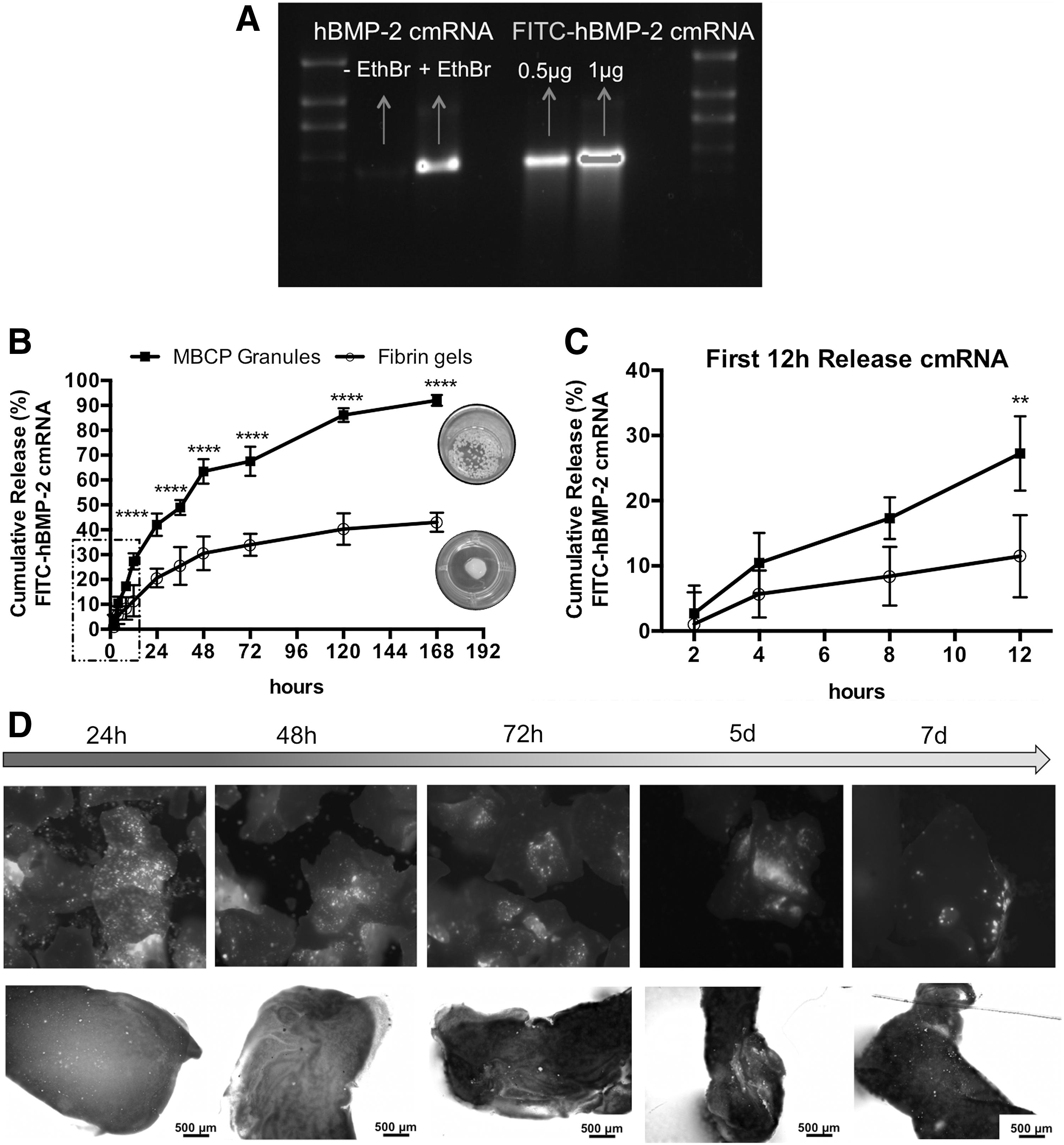
Loading and release of hBMP-2 cmRNA using fibrin gels and MBCP granules.
The release profile shows a sustained release fashion for both matrices, where the FITC-hBMP-2 cmRNA released significantly faster from MBCP granules compared to fibrin gels (Fig. 1B). Twenty-seven percent FITC-hBMP-2 cmRNA was released from the MBCP granules during the first 12 h in comparison with only 12% released from the fibrin gels (P < 0.002, Fig. 1C). At the end of the observation time (ie, 7 days), 92% FITC-hBMP-2 cmRNA has been released from MBCP into the media compared to 43% released from fibrin gels (P < 0.0001, Fig. 1B). This represents 2.14 times more cmRNA released from the ceramic granules in comparison to the gels in vitro.
Figure 1D illustrates the fluorescence images corresponding to both FITC-hBMP-2 cmRNA loaded matrices at different observation times post-release. A sustained release pattern was observed for both matrices. This observation matches the results from the cumulative release calculations. Moreover, remaining FITC-hBMP-2 cmRNA can still be observed in both matrices after 7 days.
Transfection of stem cells by loaded biomaterials
eGFP-positive cells were observed in both eGFP cmRNA/fibrin gels and eGFP cmRNA/MBCP granules at 24 h post-transfection (Fig. 2A, B). On the other hand, BMSCs seeded into MetLuc cmRNA/fibrin gels and onto MetLuc cmRNA/MBCP granules were able to secrete significant amounts of MetLuc over background values (Fig. 2C, P < 0.05 in all comparisons) except at 24 h comparing MetLuc cmRNA/fibrin gels to untransfected BMSCs. In the latter case, although the cells inside the loaded fibrin matrix showed higher gene expression, this increase was not statistically significant (P = 0.1359). Furthermore, 24 h post-transfection, a significantly higher MetLuc expression was obtained for BMSCs transfected in monolayer culture compared to both matrices (Fig. 2C, P < 0.0001).
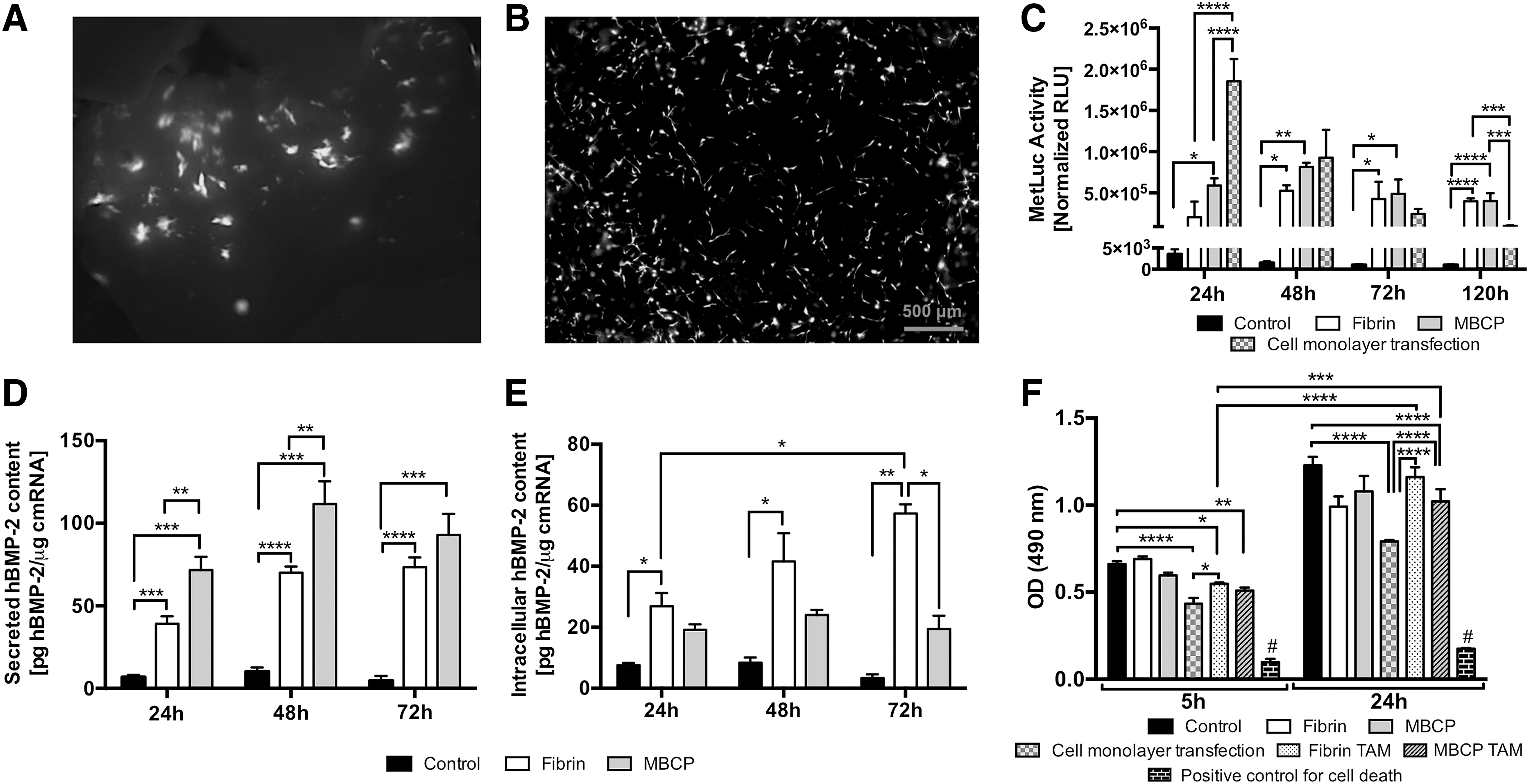
Gene transfer to BMSCs as result of TAMs. eGFP-positive stem cells are observed onto
However, these transfection results were not observed further on. Already at 48 h post-transfection, both matrices resulted in comparable MetLuc expression to the cell monolayer transfection (P > 0.05). At the end of the observation time (ie, 120 h post-transfection), both cmRNA loaded fibrin gels and MBCP granules resulted in significantly higher gene expression when compared to the BMSCs transfected in two-dimensional (2D) monolayers (Fig. 2C, P < 0.001). During the entire observation time, MetLuc expression of BMSCs in fibrin gels remained almost constant in a plateau shape (P > 0.05 over time), whereas for MBCP granules a maximum could be observed at 48 h post-transfection (Fig. 2C, 24 vs. 48 h P < 0.02 and 48 vs. 72 h P < 0.05).
Besides transfection efficiency, another important aspect to consider is the toxicity of the cmRNA loaded biomaterials. At 5 h post-transfection, the viability of the cmRNA-transfected cells decreased significantly when compared to untransfected cells (Fig. 2F, P < 0.021). This early toxicity of the cmRNA was observed for monolayer transfection and for cmRNA loaded biomaterials.
However, hBMP-2 cmRNA/fibrin gels resulted in significantly higher cell viability when compared to cell monolayer transfections (P < 0.05). At 24 h post-transfection, the cell viability increased significantly for both cmRNA loaded biomaterials (comparison of 5 vs. 24 h post-transfection: P < 0.0001 for hBMP-2 cmRNA/fibrin and P < 0.001 for hBMP-2 cmRNA/MBCP). No statistical significance of cell viability was found when comparing untransfected cells and hBMP-2 cmRNA/fibrin gels transfected cells (P = 0.1644). At this time of observation, the cell viability remained significantly lower for the monolayer transfection (P < 0.0001).
hBMP-2 production
BMSCs seeded in both hBMP-2 cmRNA/fibrin gels and on hBMP-2 cmRNA/MBCP granules secreted significantly higher amounts of hBMP-2 when compared to the untransfected cells (Fig. 2D, P < 0.001). At 24 and 48 h post-transfection, BMSCs seeded onto hBMP-2 cmRNA/MBCP granules were more efficient in secreting hBMP-2 than cells seeded into the hBMP-2 cmRNA/fibrin gels (P < 0.005 for 24 h and P < 0.01 for 48 h). However, 72 h post-transfection, BMSCs seeded onto-/into- both matrices resulted in similar hBMP-2 protein production (Fig. 2D, P = 0.07).
Interestingly, considerably higher amounts of intracellular hBMP-2 were found in BMSCs seeded into fibrin gels when compared to the MBCP granules counterpart (Fig. 2E, P < 0.05). Moreover, the intracellular levels of hBMP-2 increased significantly in BMSCs seeded into fibrin gels over time (P < 0.002). In contrast, for the cells seeded onto MBCP granules, the intracellular amounts remained almost constant (P > 0.9). Moreover, when hBMP-2 cmRNA/MBCP granules were used for transfection, intracellular hBMP-2 production was not significant when compared to untransfected BMSCs (Fig. 2E, P > 0.5).
Osteogenesis
As a result of hBMP-2 cmRNA-activated matrix transfections, BMSCs significantly expressed all investigated osteogenic markers (ie, hBMP-2, RunX2, ALP, Coll I, and OCN). The gene expression results are illustrated in Fig. 3A-E. An increase in gene expression was observed independently of the TAM used (ie, hBMP-2 cmRNA loaded fibrin gels or loaded MBCP granules). The levels of gene expression for both TAMs were in the same range.
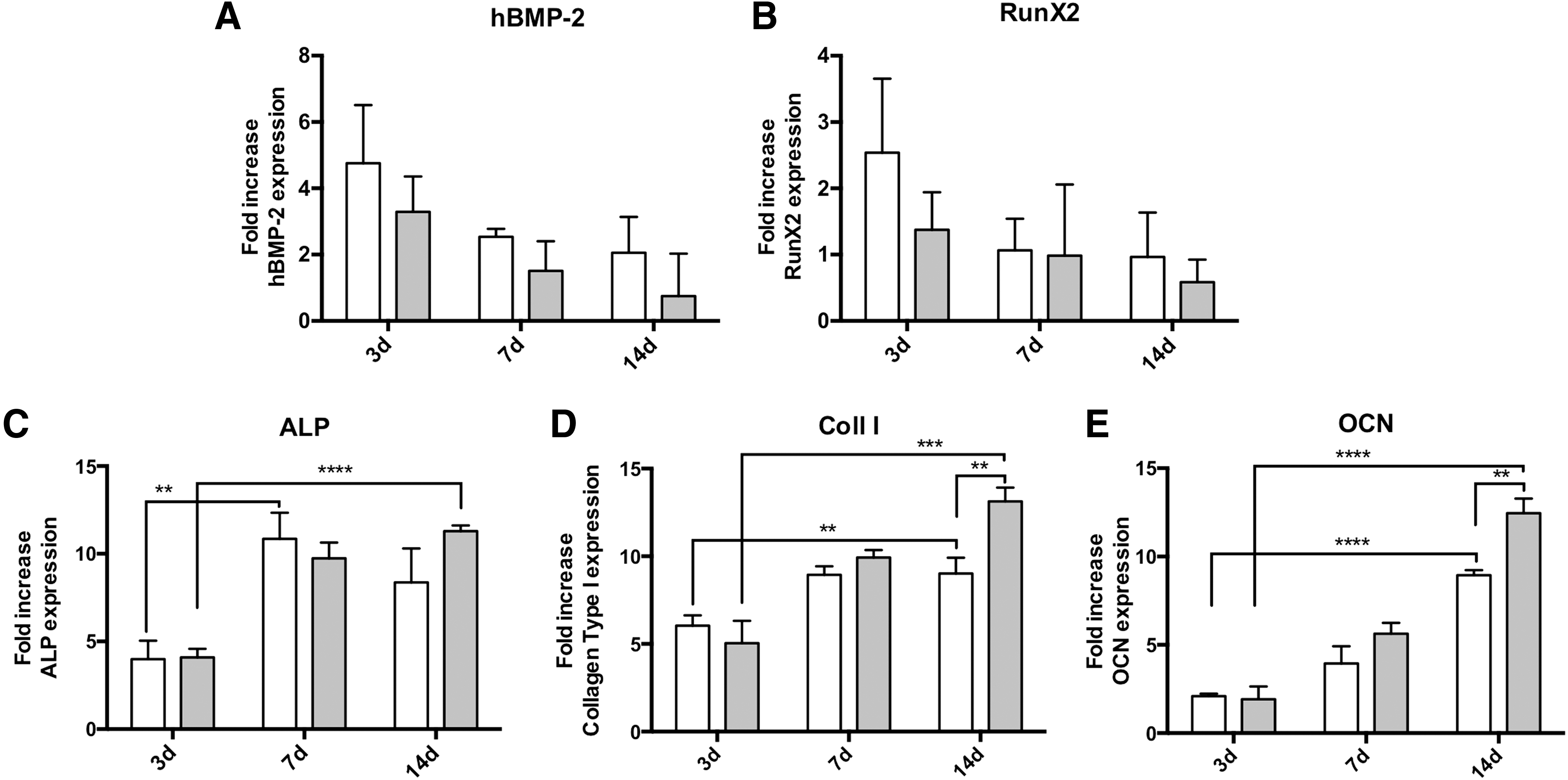
Osteogenic gene expression of in vitro transfected BMSCs by using hBMP-2 cmRNA-activated matrices.
In addition, the expression of ALP, Coll I, and OCN clearly increased over time for cells seeded onto-/into- both matrices (Fig. 3C–E). In the case of ALP, a slight decrease was observed for the cells seeded into hBMP-2 cmRNA loaded fibrin gels from day 7 to day 14 of culture (Fig. 3C, P > 0.5). In the case of Coll I (Fig. 3D) and OCN (Fig. 3E), significantly higher gene expression was induced by hBMP-2 cmRNA/MBCP granules when compared to hBMP-2 cmRNA/fibrin gels at 14 days post-transfection (P ≤ 0.005 for Coll I and P < 0.005 for OCN).
The hBMP-2 cmRNA/MBCP matrix also induced more mineral deposition as indicated by alizarin red staining (Fig. 4C, P < 0.05). Interestingly, BMSCs seeded onto-/into- the hBMP-2 cmRNA-loaded matrices were able to mineralize to a higher extent, 21 days post-transfection, compared to cells transfected in 2D monolayers (Fig. 4C, P < 0.001 for MBCP and P < 0.01 for fibrin).
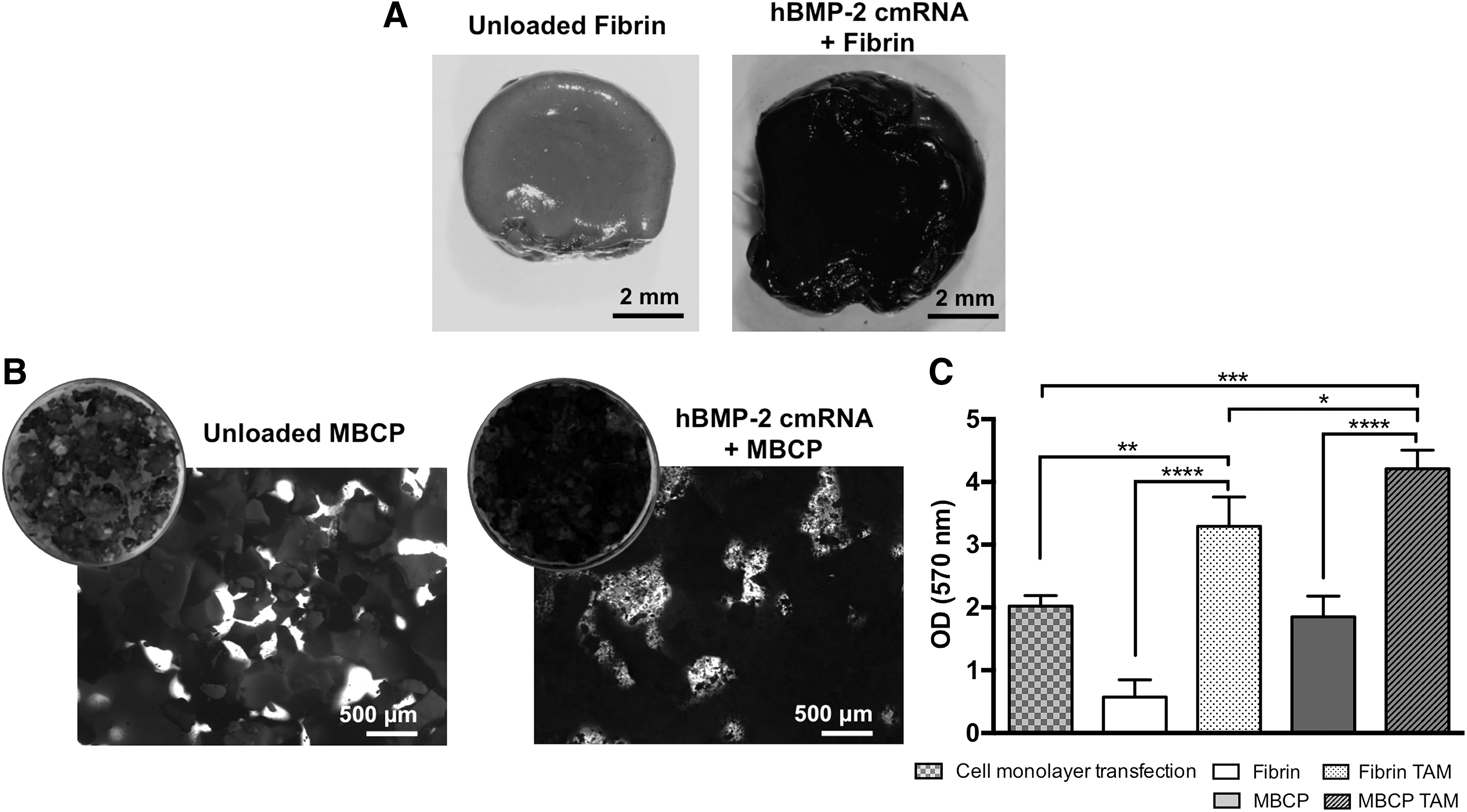
Mineralization of BMSCs seeded into
Discussion
Biomaterials and growth factors are an essential part of the tissue engineering approach aiming at tissue regeneration [22]. Their combination is used to stimulate stem cells with a final therapeutic aim. pDNA and mRNA have been used to overcome growth factor limitations [15,23]. In our study, we have combined two different types of biomaterials with cmRNA to produce TAMs aiming at stem cell differentiation. Moreover, one of the materials is used for clinical bone regeneration. Thus, this approach combines several aspects of the regenerative medicine idea.
Protein-encoding mRNA represents an attractive alternative to pDNA gene transfer. Protein-encoding mRNAs may greatly improve the efficiency of the gene transfer process as it is delivered to the cytoplasm, where it directly exerts its function. However, these molecules have a short half-life and induce strong toxicity. As a consequence, mRNA clinical applications have been narrowed mostly to vaccination. During the last years, several mRNA engineering strategies have been investigated to overcome these limitations. Examples of these strategies are chemical modifications, HPCL-based purifications, synthetic cap analogs, and/or UTRs incorporation, among others [24]. All these approaches aim to increase mRNA stability and avoid its interaction with immune system components. Several successful results have been reported [17,24,25].
Our group has recently optimized a nucleotide-based chemical modification with positive results concerning mRNA immunogenicity and stability [17]. After having demonstrated the benefits of direct in vitro and in vivo cmRNA administration [15,17], we aimed here to optimize the application of this technology by combining cmRNA and clinically used biomaterials. Our idea was that a 3D scaffold-based delivery of cmRNA may synergistically stimulate stem cell growth, differentiation, and extracellular matrix deposition. First, biomaterials can provide support and adequate mechanical properties to the cells. In addition, they also act as a delivery platform for the cmRNA molecules thereby protecting the cmRNA from degradation. Next, hBMP-2 cmRNA stimulates the differentiation of the cells into the desired phenotype for a specific therapeutic outcome, in this case osteogenesis.
MBCP granules and fibrin glue were selected as biomaterials in our study. Both are commercially available and clinically used. MBCP granules are used mainly in orthopedic and maxillofacial surgery, while fibrin glue has been used in most surgical specialties [26]. Both materials are biocompatible, MBCP features additional osteoconductive properties and fibrin is fully resorbable [26]. Additionally, both materials have been used in combination (ie, as a composite) for bone regeneration purposes [27,28].
In our study, we used both MBCP granules and fibrin glue as matrices for the loading and release of cmRNAs. Both matrices allowed the sustained release of the loaded cmRNA molecules up to 7 days in vitro (ie, maximum observation time in our study). However, fibrin glue showed higher affinity for the cmRNA molecules, resulting in slower cmRNA release when compared to the ceramic granules. We speculate that this affinity is the result of strong electrostatic interaction also described for fibrin-pDNA constructs [29]. Furthermore, when preparing a TAM from fibrin glue, the fibrin network is formed in situ, entrapping homogenously suspended lipoplex nanoparticles. When loading MBCP, the lipoplex nanoparticles can diffuse into the ceramic material only to the extent the pore size allows and are afterward not entrapped by a network of fibers. The scaffold composition will ultimately affect cmRNA release profiles.
Lei et al. demonstrated that modifications on the composition of fibrin gels (eg, increasing fibrinogen or aprotinin concentrations) resulted in decreased transfection efficiency of pDNA [30]. In our study, cells entrapped within the gel are expected to gradually degrade the material. Thereby, the cmRNA is released from the matrix and available for cell targeting. In the case of MBCP granules, cmRNA is released mainly by diffusion. These ceramic granules are a composite material constituted of 60% hydroxyapatite and 40% tricalcium phosphate. Also in this case, changes on the hydroxyapatite/tricalcium phosphate ratio and on the type of ceramic material have been reported to impact nucleic acid loading and release [31]. In our study, both cmRNA-activated matrices were developed using clinically employed materials without further modifications in the composition. Thereby, we aimed to be as close as possible to the current clinical use of these materials.
As stated before, mRNA-mediated gene transfer is an attractive approach. When compared to its pDNA counterpart, our previous results showed higher transfection efficiencies, lower toxicity and long-lasting protein expression for the cmRNA [15]. Elangovan et al. also reported the superiority of modified mRNA for in vivo nonviral gene therapy applications [16]. Yet, none or little has been published on cmRNA loading and subsequent release using biomaterials. In our opinion, the use of biomaterials is important to provide a structural support and function as a drug delivery entity. We have previously reported the development of a cmRNA/fibrin-activated matrix [15]. However, the cmRNA release was not evaluated. Elangovan et al. [16] and Badieyan et al. [32] reported cmRNA-activated matrix using collagen scaffolds. In these studies, the loading/release was also not evaluated.
A different scenario is, however, seen for pDNA. The loading and release of diverse pDNAs using fibrin has been extensively evaluated [29,30,33]. Overall, a consensus can be found among authors describing fibrin matrices as providing a slow but sustained release of entrapped pDNAs over days. In some cases, this release may even be over weeks and 100% release may be obtained once the matrix is completely degraded. Our results for cmRNA are in line with these observations for pDNA. One week post-release in vitro, the fibrin matrix released only 43% of entrapped cmRNA in contrast to almost 100% released from the ceramic material. In contrast to fibrin, MBCP has hardly been used as gene-activated matrix and has not been reported at all until today as transcript-activated matrix. One recent study by Plank et al. described the loading and release of pDNA-polyplexes onto and from MBCP granules [34]. Similar to our results for cmRNA, the authors described a rapid pDNA release from the ceramic material.
Transfection of BMSCs using both cmRNA/fibrin- and cmRNA/MBCP-activated matrices resulted in positive gene expression. Interestingly, at early time of observation (ie, 24 h post-transfection), cmRNA transfections of BMSCs monolayers (2D) appeared to be more efficient than both TAMs. This reduced gene expression of cells seeded onto-/into- activated matrices may be due to interactions of cmRNA complexes with the materials (here MBCP and fibrin). This may lead to less initial trafficking of cmRNA-vectors through the cell membrane resulting in less internalization.
Our results are partially in line with those reported by des Rieux et al. for pDNA loaded into fibrin gels [35]. The authors reported significantly higher levels of gene expression in cell monolayer transfection compared to 3D fibrin gels for over 10 days. However, in our study, the superiority of gene expression in 2D monolayer was not observed for periods of observation longer than 24 h. Indeed, 120 h post-transfection, both TAMs resulted in superior gene expression when compared to 2D cell monolayer transfections.
In addition, TAMs may improve gene transfer by decreasing cytotoxicity. Our results showed a significant increase in cell viability when cmRNA-loaded biomaterials were used compared to standard monolayer transfections. This effect was particularly noticeable for cmRNA/fibrin-activated matrices and it is in line with previous reports on pDNA loaded fibrin gels [30]. This increased cell viability may be related to the exposure of target cells to the cmRNA complexes. In the 3D matrices, due to the co-localization of cells and cmRNA, there is a gradual exposure to cmRNA complexes. In cell monolayer transfections, however, the cmRNA complexes are delivered at once into the cells. Thereby, an increased cytotoxicity may be obtained. Overall, our results regarding gene expression and cytotoxicity demonstrate the suitability of both fibrin gels and MBCP granules for extended transgene expression.
Our results are in line with the results reported by other authors on 3D gene-activated matrices [30,36]. Worth to mention is the necessity for a transfection enhancer as part of the cmRNA matrix formulation. In our case, we have used a commercially available lipid. This enhancer is needed to successfully internalize the cmRNA inside the cell due to the limited internalization capacity of naked cmRNA [37]. Although these lipids are associated with some toxicity, great progresses have been achieved in cationic lipids and polymer design that are mild for the cells and being investigated for in vivo use [38,39].
hBMP-2 cmRNA transfected BMSCs were able to secrete high amounts of hBMP-2 in vitro that resulted in subsequent osteogenic gene expression and mineralization. This result was observed to be independent of the matrix used, for hBMP-2, RunX2, and ALP gene expression. However, BMSCs seeded onto hBMP-2 cmRNA/MBCP granules showed higher expression of Coll I and OCN after 14 days. In the same line, those cells also showed more pronounced mineralization in vitro. This may be due to the bone-like structure and composition of MBCP ceramics that are more prone to favor osteogenesis of stem cells than fibrin. Thus, MBCP granules show a synergistic effect.
Overall, our results demonstrate the suitability of fibrin gels and MBCP ceramic as carriers for cmRNA molecules. Both biomaterials allowed hBMP-2 cmRNA sustained release and supported stem cell osteogenic differentiation and mineralization in vitro. Further studies are needed to identify other TAMs for stem cells and to fully understand the mechanism behind cmRNA uptake and internalization by the cells seeded onto-/into- these matrices. Furthermore, in vivo studies are needed to determine the translational value of this technology.
Footnotes
Acknowledgments
Financial support by the German Federal Ministry of Education and Research Go-Bio grant 0315986 and by the Deutsche Forschungsgemeinschaft (DFG) via the Excellence Cluster “Nanosystems Initiative Munich (NIM)” is gratefully acknowledged.
Author Disclosure Statement
C.P. and C.R. are founders and shareholders of Ethris GmbH, a company that develops cmRNA for therapy. J.P.G., C.K., and M.K.A. are employees of Ethris GmbH. E.R.B. and M.v.G. have no conflicts of interest to disclose.