
Abstract
The design of reliable biocompatible and biodegradable scaffolds remains one of the most important challenges for tissue engineering. In fact, properly designed scaffolds must display an adequate and interconnected porosity to facilitate cell spreading and colonization of the inner layers, and must release physical signals concurring to modulate cell function to ultimately drive cell fate. In this study, a combination of optimal mechanical and biochemical properties has been considered to design a one-component three-dimensional (3D) multitextured hydrogel scaffold to favor cell–scaffold interactions. A polyethylene glycol diacrylate woodpile (PEGDa-Wp) structure of the order of 100 μm has been manufactured using a microstereolithography process. Subsequently, the PEGDa-Wp has been embedded in a PEGDa hydrogel to obtain a 3D scaffold-in-scaffold (3D-SS) system. Finally, the 3D-SS capability to address cell fate has been assessed using human Lin− Sca-1+ cardiac progenitor cells (hCPCs). Results have shown that a multitextured 3D scaffold represents a favorable microenvironment to promote hCPC differentiation and orientation. In fact, while cultured on 3D-SS, hCPCs adopt an ordered 3D spatial orientation and activate the expression of structural proteins, such as the α-sarcomeric actinin, a specific marker of the cardiomyocyte phenotype, and connexin 43, the principal gap junction protein of the heart. Although preliminary, this study demonstrates that complex multitextured scaffolds closely mimicking the extracellular matrix structure and function are efficient in driving progenitor cell fate. A leap forward will be determined by the use of advanced 3D printing technologies that will improve multitextured scaffold manufacturing and their biological efficiency.
Introduction
D
In search of the optimal scaffold, innumerable materials and procedures have been proposed for their fabrication. Natural, synthetic, and composite materials have been analyzed for tissue repair and regeneration, and different strategies to functionalize them have been scrutinized [12]. Natural materials, although apparently ideal, are difficult to be extracted and manipulated. They are expensive and potentially carriers of infectious agents. Synthetic materials are easily produced at a low cost, but can release by-products that can modify the microenvironment of the recipient tissue. All biomaterials so far analyzed have been tested using different fabrication procedures like electrospinning, freeze drying, in situ forming gel, and direct writing and printing of photopolymerizable materials [13]. Among others, microstereolithography (μSL) [14,15], a simple technique for 3D modeling and printing, holds promise to allow to easily manufacture suitable scaffolds for tissue engineering. μSL is based on the photopolymerization of a cross-linkable liquid resin by means of a dynamic photomask approach. In this way, it is possible to provide a reconfigurable illumination on the separation surface of a photopolymerizable fluid containing a liquid filter to limit the penetration depth of the light and ensure a real 3D fabrication. μSL offers far shorter manufacturing times, low-cost equipment and materials, and the possibility to easily modify the output structures [16]. The above-mentioned approaches represent a preliminary stage toward the fabrication of biomimetic structures resembling living tissues in their composite bioarchitecture characterized by interlaced cells and materials to maximize the performance of the final assembly. In this context, the intercellular space is filled up with an interstitial matrix in which are embedded fibrillar proteins (mostly collagen) to supply resident cells with structural support. Depending on collagen and elastin concentrations, the extracellular matrix (ECM) can be considered a collation of different microenvironments displaying variable degrees of stiffness and other physical characteristics influencing numerous cell functions. Therefore, biological tissues could be better mimicked using multitextured materials that incorporate single or multiple components to deliver an appropriate array of physical signals to the cells.
In this study, a biocompatible polyethylene glycol diacrylate woodpile (PEGDa-Wp) structure was manufactured by μSL using curcumin as the absorbing dye. Subsequently, PEGDa-Wp structures were embedded in a PEGDa hydrogel to better mimic the ECM microarchitecture, creating an inhomogeneous multitextured structure with improved mechanophysical and biological properties. The Wp architecture represents a reliable structure that allows large deformations with a good specific energy dissipation capacity. Therefore, the optimal combination of this type of in vitro 3D scaffolds and PEGDa hydrogel was expected to favor cell–cell and cell–material interactions, and promote the differentiation of the human Lin− Sca-1+ cardiac progenitor cells (hCPCs). This study allowed to validate the novel concept of multitextured 3D-“scaffold-in-scaffold” (3D-SS), in which 3D structures manufactured on the basis of diverse designs could be combined to obtain scaffolds with unprecedented dynamic characteristics. This study represents the first demonstration of the feasibility of this strategy that can be further enhanced by the application of the emerging 3D bioprinting technologies.
Materials and Methods
PEGDa-Wp fabrication by μSL
The precursor solution used to manufacture the Wp structure was composed of a hydrogel of PEGDa molecular weight (MW) 575 g/mol, curcumin as the absorbing dye (inner optical filter), and 2-hydroxy-4-(2- hydroxyethoxy)-2-methylpropiophenone (also known as Irgacure 819® CIBA) as photoinitiator. All reagents were purchased from Sigma-Aldrich and used without further purification. Curcumin and Irgacure at a concentration of 44 and 0.25 mM, respectively, were dissolved in a PEGDa:ethanol = 3:1 solution. After mixing, the solutions were let stir for 24 h in the darkness. The two-dimensional (2D) models (images) to be printed layer by layer, drawn with a standard software, were projected and focused by means of an overhead projector on the printing stage covered by a thin film of precursor solution. Polymerization occurred only in the lit area during the exposition. After such an exposition, the printed structure was formed on the stage. Then, the platform was lowered down in the liquid solution (by a fixed step of 250 μm) and a new thin layer of unpolymerized solution covered it, ready to be exposed. The inner optical filter (curcumin) ensured that only the top layer received enough light to photopolymerize, while the previously exposed layers remained protected against further photopolymerization due to the absorption of curcumin. The orientation of the polymerized rods was alternated in the subsequent layers and each couple of orthogonally oriented layers could be considered a monolayer of the structure. The final 3D template resulted as the stacking of the sequence of all printed 2D layers. A schematic representation of the experimental 3D printing setup is shown in Fig. 1. At the end of the process, the scaffolds were accurately washed in water and cured under Hg ultraviolet (UV)-lamp for few minutes to stabilize the structure against uncontrolled modification induced by room light. This procedure allowed obtaining structures with a resolution of about 300 μm; they were morphologically characterized by an optical microscope (Nikon L-IM equipped with a digital camera DS-5M), while their stiffness was assessed by a nanoindenter (Nano Test Micro Materials Ltd.), using a diamond Berkovitch tip.
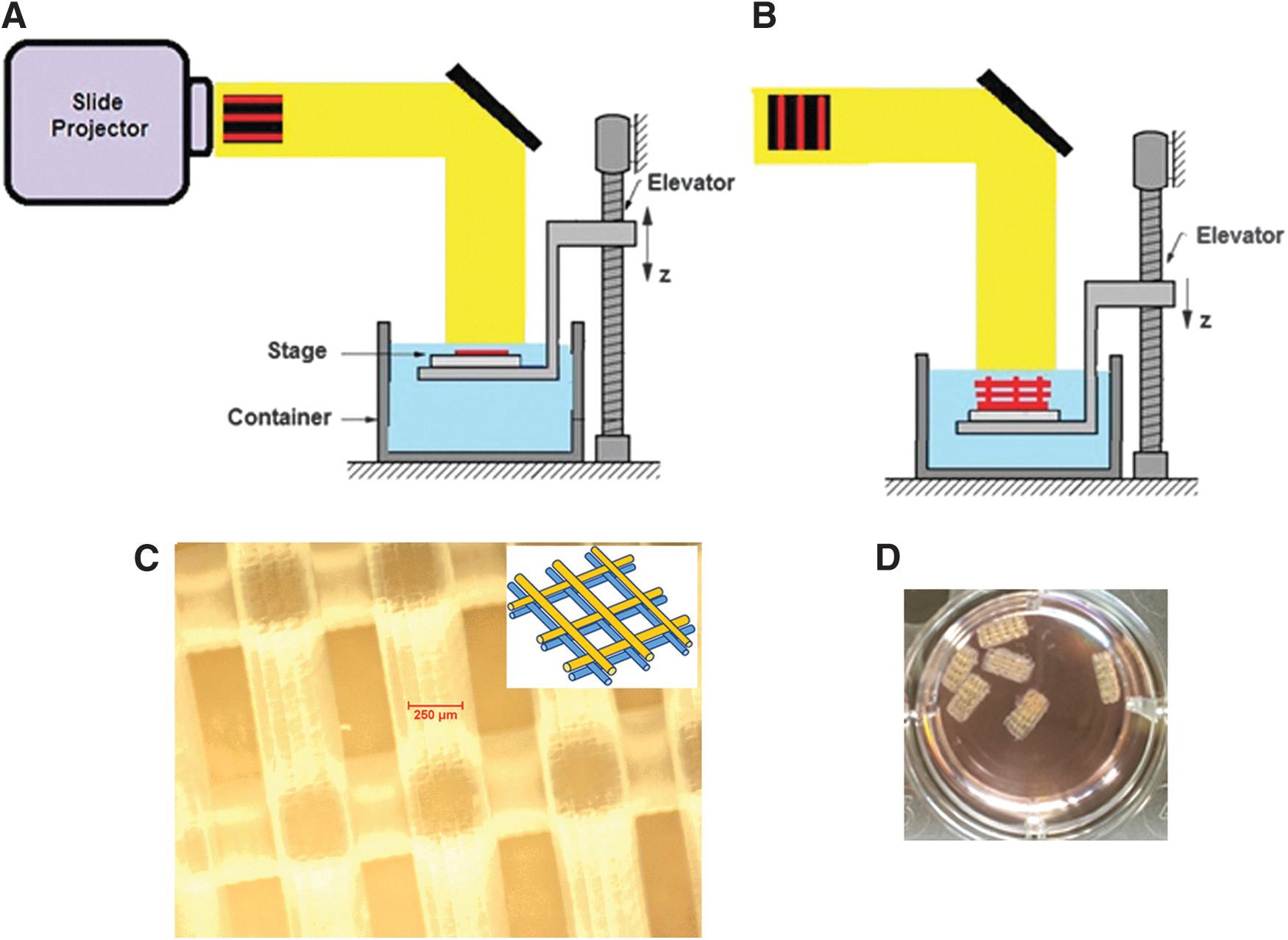
Experimental setup for μSL:
Before further steps, the PEGDa-Wp structures were washed with sterile water for 1 week and exposed overnight to the UV light. They were then conditioned for 1 week in Dulbecco's modified Eagle's medium (DMEM) with high glucose (Gibco) supplemented with 20% v/v fetal bovine serum (FBS; Gibco), 1% penicillin–streptomycin (Sigma-Aldrich), 1%
3D-SS fabrication
The 3D-SS systems were manufactured by embedding the PEGDa-Wp into a PEGDa hydrogel obtained by photopolymerization of a solution of 15% w/v PEGDa 20 kDa MW and 3% w/v PEGDa 6 kDa MW in phosphate-buffered saline (PBS). The hydrogel photopolymerization was achieved using molds with 5 mm diameter and adding 0.1% w/v Irgacure 2959 (Ciba Specialty Chemicals, Basel, Switzerland). Finally, the samples were exposed to UV light (365 nm and 4–5 mW cm−2) for 5 min.
Cell culture and cell proliferation assay
hCPCs were isolated from samples of the right auricles of patients (seven male and six female, 52–83 years) undergoing coronary artery bypass surgery [17] after signing a written consent form according to a joint protocol approved by the Ethic Committee of Ospedale Maggiore della Carità (Novara, Italy). hCPCs (1 × 10⁴ cells in 50 μL of DMEM) were seeded and cultured on PEGDa-Wp in DMEM with high glucose containing 10% FBS, 1% penicillin–streptomycin, and 1%
Recognition of the hCPC phenotype when cultured on 3D-SS
To assess the hCPC phenotype, 3D-SS with 1 × 10⁴ cells Lin− Sca-1pos hCPCs were cultured for 7 days in the complete medium. The scaffolds were then washed in PBS, fixed in 4% paraformaldehyde (PFA) in PBS for 15 min at 4°C, permeabilized with 0.2% v/v Triton X-100 (Sigma-Aldrich) for 10 min, and incubated with phalloidin-Alexa fluorochrome-conjugated (Life Technologies). In a further set of experiments, the hCPC phenotype on 3D-SS systems was assessed after incubation with cardiac alpha sarcomeric actinin antibody (Ab-α-actinin; Sigma-Aldrich) for 9 h and with the secondary antibody [Goat anti-Mouse IgG (H+L), Alexa Fluor® 546; ThermoFisher] for 4 h at room temperature (RT). Nuclei were stained with Hoechst 33342 (Sigma-Aldrich). Confocal microscopy of the cell-seeded constructs was performed using an LSM 700 Confocal Laser Scanning Microscope (Carl Zeiss MicroImaging, Jena, Germany) and acquired by means of ZEN 2010 software.
The differentiating potential of the PEGDA-Wp structure (in the absence of hydrogel) was assessed by seeding hCPCs onto sterile 2D PEGDa-Wp and 3D PEGDa-Wp structures in DMEM supplemented with 10% FBS. One day after seeding, the cells were shifted to DMEM supplemented with 2% FBS. hCPCs were kept in culture for further 11 or 20 days. The medium was changed every 2 days. hCPCs cultured in the same condition in the chamber slides in the absence of structures represented the control. The hCPCs cultured in the absence or presence of PEGDa structures were fixed with cold methanol and the expression of the cardiac alpha sarcomeric actinin (α-actinin) and connexin-43 (Cx43) was assessed after incubation with specific primary antibodies (Sigma-Aldrich) for 1 h at RT and then with the appropriate anti-mouse or anti-rabbit secondary antibodies (Alexa Fluor 546; ThermoFisher) for 1 h at RT. The secondary antibody in the absence of specific primary antibody was used to exclude the occurrence of unspecific signals. Nuclei were stained with 1 μg/mL 4′,6-diamidino-2-phenylindole (DAPI; Sigma-Aldrich). The confocal images were taken using a confocal laser scanning microscopy Olympus Fluoview 1000.
Statistical analysis
The statistical analysis was performed using GraphPad Prism version 5.0 for Windows (GraphPad Software, San Diego, CA). Data from three or five independent experiments were quantified and analyzed for each variable using a one-tailed Student's t-test or analysis of variance one-way test. A P value of <0.05 was considered to be statistically significant. Standard deviations of the mean were calculated and presented for each type of sample.
Results
Scaffold fabrication and characterization
In μSL, the most crucial aspect is the choice of the inner optical filter. It must almost totally absorb the incoming light in a very thin layer. Indeed, growing an object layer-by-layer is possible only if the exposition of each new layer does not affect the already exposed lower structure. A careful choice of dye and its concentration is mandatory to reduce the light to a negligible amount after 250 μm corresponding to the desired growth step. To this end and considering the biological context of the application, the photopolymerization has been carried out using a precursor solution with curcumin, a natural dye extracted from Curcuma longa spice widely used in Middle- and Far-East Asian areas, displaying a total absorption coefficient of 100 cm−1 in the active spectral region (350–470 nm). Curcumin absorption matches well with the absorption spectrum of the Irgacure 819, the photoinitiator used in this study, in the spectral interval of the lamp (Supplementary Fig. S2), while its luminescence does not overlap Irgacure absorption. Moreover, it is endowed with well-known antioxidants, anti-inflammatory, and antitumoral properties [18]. The dye and the photoinitiator were dissolved in the precursor solution with a ratio of about 200:1 to confine the photopolymerization in a thin layer only, just below the exposed surface.
All manufactured PEGDa scaffolds were preliminary characterized by optical microscopy. Figure 1C shows great regularity in accordance with the planned structure sketched in the inset. The average thickness of the segments of the PEGDa-Wp scaffold used in this study is about 300 μm. The mechanical characterization of the PEGDa was accomplished with a nanoindenter. The ratio of the uniaxial stress to the uniaxial strain of a material, the Young modulus, can be experimentally determined from the slope of linear fits to the stress–strain curves in an indentation experiment [19]. Nanoindentation test was performed on a 5 mm diameter disk of 2.5-mm-thick bulk PEGDa samples grown by the same μSL technique, repeating the projection of the disc for ten layers. The measurement of a dried sample yielded a value of the elastic modulus of 5 MPa. A hydrated sample, whose surface was blown in a gentle stream of air before nanoindentation experiments to remove the excess water, was measured yielding a value of 1 MPa. The results are in full accordance with Drira et al. [20]. Noteworthily, depending on the morphology of the 3D cage, the mechanical properties of the entire Wp structures could be greatly different from those related to the bulk material only.
hCPC growth and differentiation on PEGDa-Wp structures
hCPCs (1 × 10⁴ cells/cm2) were seeded on PEGDa-Wp structures (Fig. 2A); after 12 h, the structures were transferred in new wells to analyze the proliferation of cells actually attached. Cell viability and proliferation were assessed by WST assay after 4 days of cell growing (Fig. 2B): about 4 × 10⁴ viable cells were found attached to each scaffold (Fig. 2C). After 8 days of cell growth, the microscope analysis showed that the adhesion to the PEG-Wp was quite good and the cells were mostly clustered around the scaffold struts (Fig. 3). In particular, the confocal microscopy (Fig. 3C, D) showed a spindled and branched morphology of the hCPCs, suggesting a potential effect of the scaffold structure on cell assembly and fate.
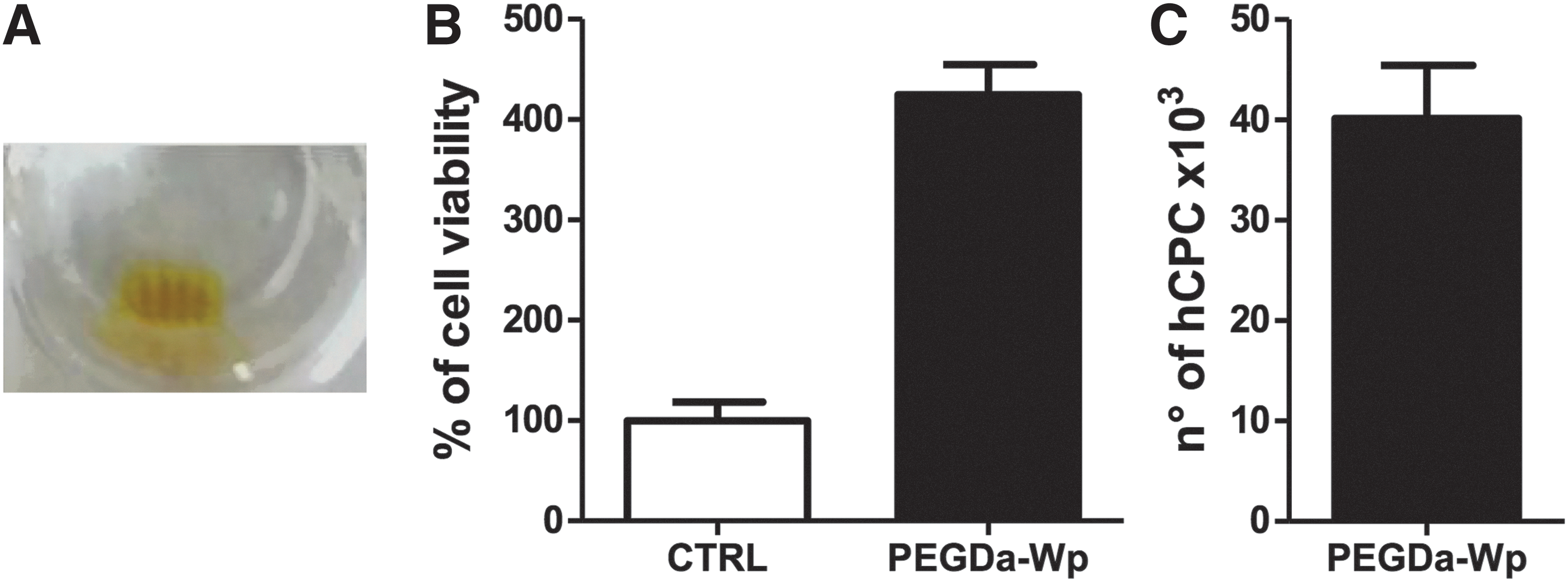
hCPC adhesion and proliferation on PEGDa-Wp structures.
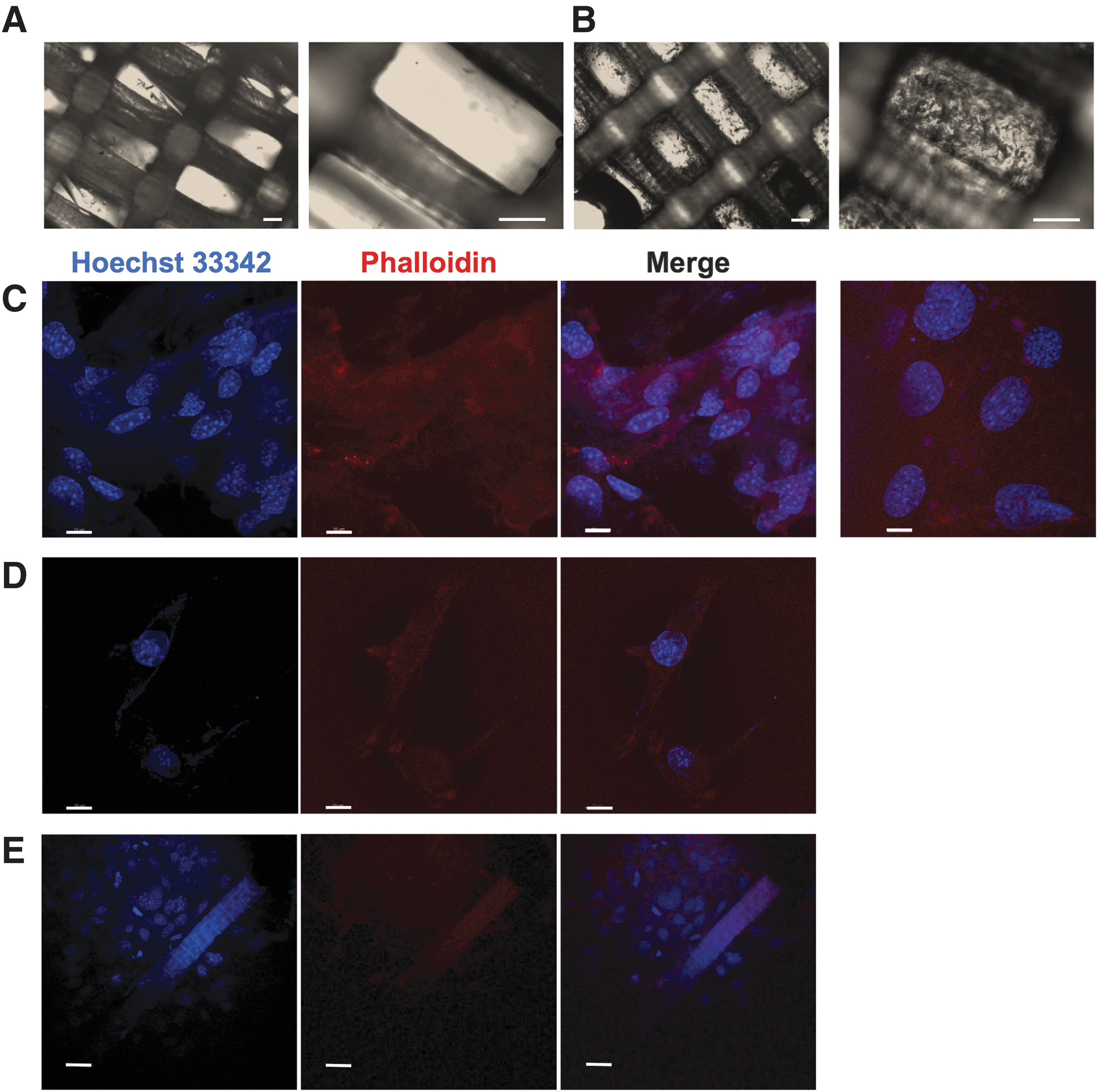
hCPC adhesion, alignment, and elongation are improved by PEGDa-Wp structures. Light micrograph of PEGDa-Wp structures in
The PEGDa-Wp differentiating potential was assessed by seeding hCPCs onto the structures that, after 1 day, were shifted in new wells and cultured with DMEM supplemented with 2% of FBS for 11 days. The evidence of a possible hCPC differentiation toward a cardiomyocytic phenotype was achieved by assessing the expression of specific cardiac differentiation markers, such as α-actinin and Cx43. Figure 4 shows the confocal micrographs of the α-actinin expression in hCPCs seeded on a plate in the absence of the structure (Fig. 4A) and in the presence of 2D PEGDa-Wp (Fig. 5B) or 3D PEGDa-Wp (Fig. 4C). Immunostaining revealed that the α-sarcomeric actinin was widely expressed only in hCPCs cultured in the presence of the microlithographed 3D PEGDa (Fig. 4C), implying that the microstructured scaffold could induce the progenitor cell differentiation toward a cardiomyocytic phenotype. Noteworthily, the microstructure contributed to direct cell spatial-ordered multilayer organization. In addition, Fig. 5B shows that Cx43, the principal gap junction protein of the heart, was highly expressed in the cytoplasm of hCPCs cultured on microlithographed 3D PEGDa (Fig. 5C) rather than in cells cultured on 2D structures (Fig. 5A, B).
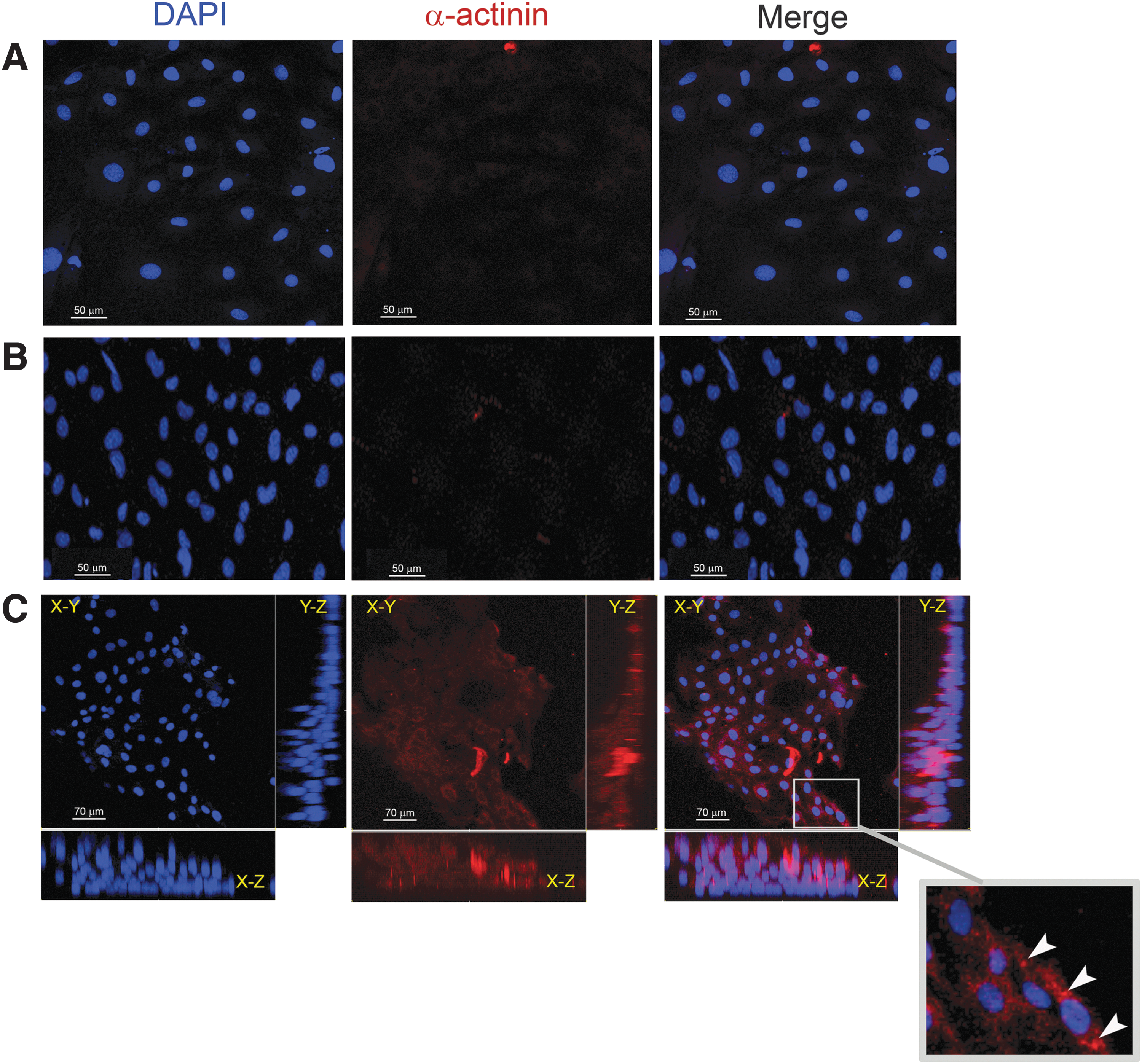
hCPC cardiac commitment on PEGDa-Wp structures. Confocal micrographs of hCPCs cultured for 12 days
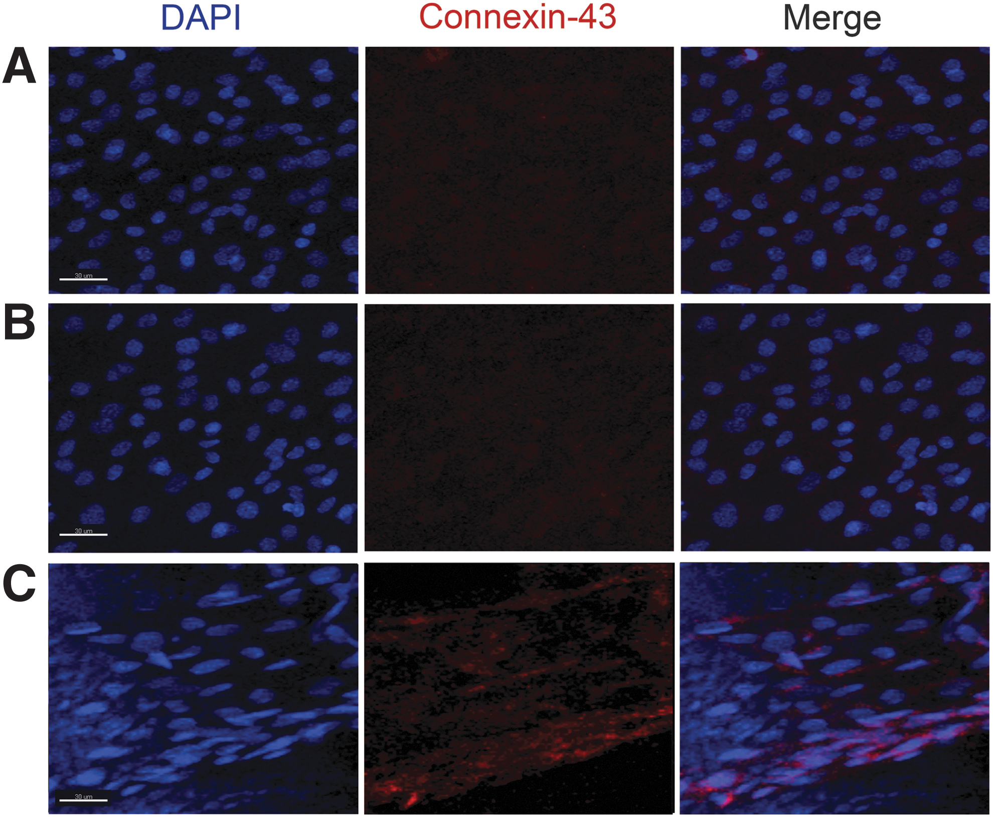
PEGDa-Wp structures enhance connexin 43 expression in hCPCs. Confocal micrographs of connexin 43 expression in hCPCs cultured for 3 weeks in chamber slides in the absence
hCPC growth and differentiation into 3D-SS
In this study, a novel multitextured hydrogel scaffold 3D-SS was manufactured combining a PEGDa-Wp structure with a PEGDa hydrogel mold. 2.5 × 10⁴ hCPCs were seeded into 3D-SS and, after 3 weeks of growth with DMEM supplemented with 10% of FBS, cell viability was checked by WST-1 assay (Fig. 6A), while cell differentiation was analyzed by immunostaining using Ab-anti-α-actinin (Fig. 6B). In Fig. 6B and C, the confocal observation of the hCPCs growing into 3D-SS demonstrated an upregulation of α-actinin, whose expression was undetectable in 2D control cultures (Supplementary Fig. S3); in addition, cells adhering to the construct displayed an elongated morphology. Therefore, the cell alignment on the PEGDa-Wp (Fig. 6B, D) and the expression of α-actinin demonstrated the differentiating potential of the PEGDa-Wp in a more complex system. Thus, 3D-SS was able to stimulate correct stem cell commitment toward cardiac lineage, although a complete sarcomeric rearrangement in these cells was not observed. Figure 7 summarizes the entire 3D-SS/hCPC procedure.
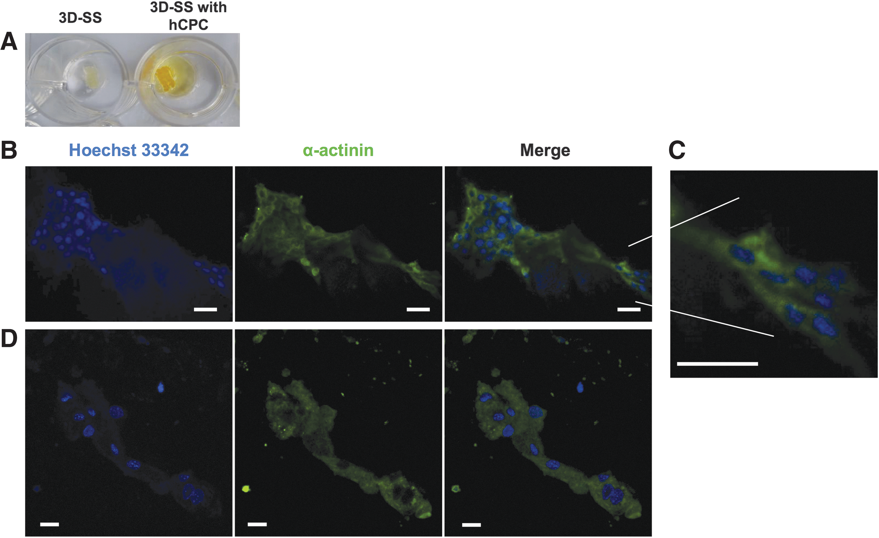
3D-SS composite scaffold showing a differentiation-inducing potential on hCPCs.

3D-SS preparation and hCPC growth into 3D-SS. microstereolithographed PEGDaWp;  hCPCs;
  PEGDa solution;
  hCPCs embedded into 3D-SS.
Discussion
This study demonstrates that biologically efficient multitextured scaffolds mimicking the inhomogeneous microarchitecture of the ECM can be manufactured by combining structures with different physical characteristics. The proof of concept has been achieved by embedding a stiffer Wp structure into a softer hydrogel (SS) to provide cells with resorbable mechanical support and orientation grid for their 3D growth, differentiation, and alignment in a microenvironment rich in oxygen, water, and metabolites flowing through interconnected channel networks, also draining the products of cell catabolism. Indeed, the ECM is made of the matrix in which filaments of different size, structure, and physical characteristics are embedded [21,22]. The validation of this approach has been obtained by growing cardiac progenitor cells in the SS system: cells three dimensionally aligned along the three axes of the scaffold and triggered their differentiation toward the myocyte phenotype. These results pave the way for manufacturing multicomponent composite structures more efficiently, mimicking the 3D organization and function of the ECM. The goal is to manufacture scaffolds in which multiple specifically tuned “stiffer” structures are combined with several “softer” materials characterized by variable textures (pore number, geometry, and interconnection) [23] (Fig. 6). Furthermore, the heterogeneous scaffolds could be enriched with biologically active molecules (cytokines, growth factors, antibodies, etc.) or delivery systems (microbubbles, microbeads, exosomes, etc.) [24] able to convey signals on the basis of a predetermined spatiotemporal schedule.
Many aspects of engineered tissue manufacturing remain to be clarified yet, before a safe and efficient application to the clinical setting could be envisaged. Several materials and procedures to manufacture the scaffolds, on which cells can be grown and differentiated, have been proposed, but optimal solutions are not yet at hand. In general, two major classes of materials have been so far considered to manufacture scaffolds. In the first class are materials extracted from biological tissues (mostly components of the ECM) [25]: they are expensive and not completely safe, since it cannot be prevented that noxious agents could be transferred to the recipient subject. A particular subclass of natural materials is represented by perfusion-decellularized human hearts from which it is possible to obtain acellular cardiac scaffolds with preserved ECM properties, structure, and patent coronary conduits, with an immunologic profile that induces a constructive remodeling response [26]. However, this technologically superb solution does not phase out the need of a donor and the possibility that some viral copies could be passed through the scaffold [27]. To avoid these limits, the only possibility is to learn how to manufacture a suitable scaffold using synthetic or semisynthetic materials. Indeed, in the last two decades, a lot has been learned about biomaterials, but, as a matter of fact, the knowledge is still rudimentary. Synthetic versus natural materials display higher reproducibility and allow an easy control of mechanical properties, degradation rate, shape, composition, and so on. Synthetic materials with different composition have been scrutinized and manipulated using very sophisticated technologies and very complex designs have been conceived to mimic the ECM, but results, even if very encouraging, are far from what is requested for an efficient clinical application of tissue engineering. Two major weaknesses can be envisaged in the extensive research activity so far carried out worldwide. The first deals with the simplistic idea that, using a single homogeneous biocompatible material, it could be possible to mimic the multifaceted characteristics of the ECM. Indeed, living organisms are made of diverse materials and cells assembled to take advantage of the respective characteristics. Moreover, the concentration and spatial arrangement of every component are not homogeneous throughout the same tissue. Renovated efforts must be directed to emulate the complexity of the ECM, assembling also to favor cell attachment and differentiation. Molecular interactions between cells and scaffold are due to different properties of the material, including its chemical composition and concentration, hydrophilicity/hydrophobicity, roughness, porosity, mechanical characteristics, and degradation kinetics that should be analogous to those of the native tissue. Microstructure geometry represents an important aspect for selecting material and fabricating optimized scaffolds able to promote the growth of specific cell types [9]. Three-dimensional microfabricated scaffolds have been manufactured by soft lithography, reproducing cardiac ECM structure using different bioartificial materials [28]. Furthermore, cell adhesion can be increased by honeycomb structures with pores slightly larger than cells [29]. Microstructured scaffolds, in fact, are able to increase the elongation efficiency of human mesenchymal cells as well as polycaprolactone (PCL) scaffolds displaying anisotropic geometry obtained by uniaxial stretch [30]. The second weakness is related to the poor knowledge available on the adaptive mechanisms operated by cells after adhesion to scaffolds with specific characteristics. The present study, however, is intended to suggest a novel solution to improve the scaffold technology to contribute to vanish, at least in part, the first weakness. To this purpose, a novel class of multitextured scaffolds made of a single inhomogeneously concentrated component, as inspired by the ECM structure made of solid filaments embedded in a hydrated matrix, has been hypothesized. A Wp network of solid filaments has been manufactured by 3D μSL in a biocompatible environment characterized by PEGDa and curcumin. To unravel which signals mostly affect cell differentiation, experiments were carried out under low serum condition (FBS 2%). Results confirmed that the PEGDa-Wp structure emanates a strong array of signals to progenitor cells guiding their 3D spatial multilayered organization and differentiation, as detected by α-actinin and Cx43 expression. Noteworthily, the α-sarcomeric actinin as well as Cx43 proteins were widespread throughout the entire cytoplasm and not organized in sarcomeric structures or in gap junctions connecting cells in a head-to-tail manner, respectively. Therefore, a full progenitor cell maturation to cardiomyocyte was not observed. To verify whether a more reliable microenvironment could improve hCPC differentiation, cells were cultured for 3 weeks into the 3D-SS. In this context, the hydrogel could play a role in optimizing cell supply with nutrients and oxygen. In fact, results demonstrated that, in this more complete environment and under standard serum condition (fetal calf serum 10%), cells showed an upregulation of α-actinin and a more elongated morphology, although a complete sarcomeric rearrangement in the cytoplasm was not observed. In recent studies, cells grown on hydrogels of fibrin and Matrigel [31] or PEG [32] have shown differentiation characteristics of native myocardium. Scaffolds have been credited to provide only mere physical support for stem cell adhesion and proliferation, and geometrical guidance in tissue organization, whereas growth factors [33 –35], physical stimuli [36,37], or coculturing with mature cardiomyocytes [22,38] have been considered the major players in driving cell differentiation. Conversely, several studies performed over the years have exhaustively demonstrated the prominent role of scaffold structure in driving cell fate [39 –41]. These data confirm the role of the scaffold structure in inducing cell alignment and differentiation, highlighting the efficiency of a novel, monocomponent multitextured design in creating a suitable microenvironment to cells using a relatively simple and inexpensive method. In fact, the 3D-SS allowed hCPCs to be exposed to the combined stimuli from different geometrical structures and variable stiffness with advantageous effects on cell–scaffold interaction, alignment, and differentiation. This preliminary study suggests that further advantages in manufacturing engineered tissues could be envisaged using multicomponent multitextured scaffolds more closely mimicking the complexity of the ECM microarchitecture and function. Distinct subsets of cardiac adult stem cells can be seeded in different steps on specifically designed multitextured scaffolds, in which the inner structure could drive prevascularization that, in turn, could cooperate with the outer gel to address progenitor differentiation to contractile cells. To this end, novel manufacturing procedures based on advanced 3D printing should be designed and extensively validated before their use in the clinical setting.
Footnotes
Acknowledgments
The authors are grateful to Dr. Elena Romano [Centre for Advanced Microscopy (CAM), Department of Biology, University of Rome “Tor Vergata”] for the scientific and technical support in the microscopy analysis and Dr. Massimiliano Lucci (Department of Physics, University of Rome “Tor Vergata”) for the scientific and technical support in the nanoindentation measurements.
Author Disclosure Statement
No competing financial interests exist.
References
Supplementary Material
Please find the following supplemental material available below.
For Open Access articles published under a Creative Commons License, all supplemental material carries the same license as the article it is associated with.
For non-Open Access articles published, all supplemental material carries a non-exclusive license, and permission requests for re-use of supplemental material or any part of supplemental material shall be sent directly to the copyright owner as specified in the copyright notice associated with the article.