
Abstract
Intervertebral disc degeneration (IVDD) is a major health problem. Although mesenchymal stem cells (MSCs) have been used to promote IVD regeneration, the actual survival time of implanted MSCs in IVDs has never been studied noninvasively and continuously in vivo. To investigate survival of implanted MSCs in vivo, this study used a canine model of degenerated IVD and MSCs transfected with a mutant herpes simplex type-1 virus thymidine kinase and labeled with magnetic iron oxide nanoparticles (MION). One-stage positron emission tomography (PET) and magnetic resonance (MR) imaging were carried out 3 days and 2 weeks, 3 weeks, and 4 weeks after implantation of MSCs into IVDs with surgically induced degeneration. Pfirrmann disc degeneration grade determined from the MR images indicated that the repair progress of degenerated IVD stopped 3 weeks after MSC implantation. Meanwhile, MION signal strength, signal contrast ratio (%), and low signal area (mm2) did not change significantly from that seen 3 days after cell implantation until 4 weeks [751.43 (4 weeks) ±52.67 (3 days) vs. 225.34 ± 35.62; 47.37 ± 5.01 vs. 85.37 ± 10.54; 1.78 ± 0.31 vs. 5.29 ± 1.35; P < 0.01, respectively]. Accumulation of the PET reporter probe, 9-(4-[18F]-fluoro-3-hydroxymethylbutyl)-guanine, was dramatically decreased at 3 weeks after MSC implantation. These results demonstrated that MSCs could survive no more than 3 weeks after implantation into IVDs with surgically induced degeneration, suggesting that MSCs could contribute to IVD repair for the first 3 weeks after implantation. The results also indicate that PET imaging could be used reliably to quantify the survival of implanted MSCs, whereas MION with MR imaging would likely be unsuitable for long-term tracking of MSCs in IVDs.
Introduction
C
Previous studies using MSCs derived from bone marrow, adipose tissue, or synovial tissue in animal models of disease and in humans purported that MSCs can contribute to repair processes to some degree. In IVDD, the mechanisms associated with this repair could involve differentiation of implanted MSCs to acquire functions of nucleus pulposus (NP) cells, stimulation of endogenous IVD cells, or promoting extracellular matrix production in the IVD niche [9 –13].
Despite an increasing number of preclinical and clinical studies, significant obstacles to successful cell therapy for IVDD remain [14 –16]. The specialized microenvironment of the IVD is characterized by low oxygen levels and low pH, along with high osmotic pressure and avascularity associated with poor nutrient supply, which together present a major challenge to insuring the survival and function of implanted cells.
We previously conducted two related studies to clarify the fate and repair capacity of MSCs transplanted into the femoral head of canines with osteonecrosis [17,18]. Other studies have examined the fate of MSCs in IVD [19 –21]. However, the results of these studies were largely limited to histological staining and double-immunofluorescence labeling of specimens. Although these methods can be highly specific and sensitive, the results only provide information about the implanted cells at the time of sacrifice, and thus may not reflect the true functional status or number of live cells in vivo.
Therefore, noninvasive and continuous tracking of implanted MSCs in vivo would provide clinically relevant information about the relationship between MSC survival time and IVDD repair processes, as well as provide a foundation for improving stem cell therapy for IVDD.
Magnetic iron oxide nanoparticles (MION) are often used with magnetic resonance (MR) imaging as a noninvasive method to track the fate of MSCs. This approach provides high-quality multidimensional functional and anatomic information with high soft-tissue contrast [22,23]. However, this method has a limitation in its theoretical inability to distinguish extracellular iron from intracellular iron present in stem cells or tissue macrophages, which could lead to overestimation of stem cell engraftment, as has been suggested in a study by Terrovitis et al. [24]. Amsalem et al. [25] and Ma et al. [26] also found in a rat model of myocardial infarction (MI) that iron-labeled MSCs were retained in macrophages 4 weeks after MI as measured by the macrophage-specific surface marker CD68.
This limitation can be addressed using positron emission tomography (PET) with molecular imaging reporter genes, which trace surviving cells with picomolar sensitivity (10−11 to 10−12 mol/L) [27]. In this study, we used mutant herpes simplex type-1 virus thymidine kinase (HSV1-sr39tk) as the PET reporter gene and 9-(4-[18F]-fluoro-3-hydroxymethylbutyl)-guanine ([18F]FHBG) as the PET reporter probe. HSV1-sr39tk offers quantitative and improved imaging sensitivity over wild-type HSV1-tk and was shown to be effective in cell-based therapies for MI [28]. HSV1-sr39tk integrates into the genome to produce a kinase that phosphorylates [18F]FHBG, causing its intracellular entrapment, which makes this probe an effective standard for the unique identification of surviving implanted cells in vivo. Although PET images generally lack anatomic context compared with MR, integration of these two techniques permits the acquisition of accurate and complementary data [29,30].
We used HSV1-sr39tk-transfected and MION-labeled MSCs together with PET and MR imaging to continuously and noninvasively monitor MSC fate in a canine model of IVDD. To our knowledge, this is the first report describing one-stage imaging of PET and MR, as well as MSCs “colabeled” with HSV1-sr39tk and MION to monitor the potential of stem cell therapy for IVDD.
Materials and Methods
Animal model of IVDD and experiment groups
A total of 27 mature beagles weighing between 15 and 18 kg were used for these studies. All animals were maintained according to the Guidelines for the Care and Use of Laboratory Animals as required by the Shanghai Science and Technology Committee (Shanghai, China). Radiographs (anterior–posterior and lateral) were obtained for every animal before study initiation to confirm the absence of abnormalities. At the end of the study period, all dogs were euthanized by a lethal dose of 120 mg/kg sodium pentobarbital (Hengrui, China).
The IVDD was surgically induced. The dogs were fasted for 48 h before surgery. After anesthesia induction with an intramuscular injection of ketamine hydrochloride (25 mg/kg; Hengrui), sodium pentobarbital (5 mg/kg/h; Hengrui) was administered intramuscularly to maintain general anesthesia. The animals were fixed in the right lateral position with incision sterilization. A lateral left-side retroperitoneal approach was used via a 6- to 7-cm flank incision between the L4 and L5 transverse processes, which can be located by palpation. The external oblique muscle and internal oblique aponeurosis were cut open following the fiber direction. The transverse abdominis muscle was then divided up to the tip of the lumbar transverse processes. The psoas was exposed after a retractor was placed medial to the psoas, and the tips of the transverse processes were exposed by dissection. Using a blunt dissector, the psoas was elevated anteriorly and retracted to move the psoas and peritoneum, and subsequently expose the target IVD. The exposure can be extended in both cranial and caudal directions as needed.
An anterior-lateral annulotomy of L2–L3 (IVD between the second lumbar vertebra and the third lumbar vertebra), L3–L4 and L4–L5 was then made with a number 16 scalpel blade directed transversely through the outer aspect of the annular fibrosus (AF). The NP was partly removed using an IVD rongeur with one bite. The mean removed NP was 13.7 ± 3.5 mg (about 10% of the total volume). The AF was then closed. Each animal recovered in an air-conditioned indoor facility with 24-h observation.
The 27 animals were randomly divided into 3 groups of 9: (1) sr39tk group, in which “HSV1-sr39tk”-transfected MSCs were implanted into the IVDs; (2) MION group, in which MION-labeled MSCs were implanted; and (3) sr39tk+MION group, in which HSV1-sr39tk-transfected and MION-labeled MSCs were implanted. The above three groups were defined as experiment groups, in which MSCs were simultaneously implanted into the IVDs of the same animal of L2–L3 and L3–L4. As such, for each experiment group there were 18 specimens of IVDs for statistical analysis at every time point.
In each animal, the disc of L1–L2 was used as the unoperated control (positive control). The disc of L4–L5 served as the negative control, wherein IVDD was surgically induced but only phosphate-buffered saline (PBS) was injected, and no MSCs were implanted into this IVD.
Isolation and culture of MSCs
Autologous bone marrow, which was harvested by aspiration from the iliac crests of beagle dogs, was carefully added to 20 mL NycoPrep 1.077 (Axis-Shield, Norway) and centrifuged at 600g for 30 min. Bone mononucleated cells were collected from the middle layer using density gradient centrifugation and then washed three times with PBS before culturing.
Analyses of MSCs in vitro
To evaluate the phenotypic characteristics of MSCs in vitro, passage 4 MSCs (before and after transfection/label) were seeded on culture slides and cultured for 4 days. Cells were then detached and incubated in PBS containing 1% bovine serum albumin with the following fluorescent antibodies: mouse anti-dog, BD Biosciences (USA); anti-CD44-FITC, anti-CD105-PE, and anti-CD45-FITC as a panleukocyte marker; anti-CD11b-FITC as a monocyte/macrophage marker; anti-CD90-FITC as a pan-T cell or early bone marrow progenitor cell marker; and anti-CD31-FITC as an endothelial marker (Beckman Coulter). Cells were analyzed on a fluorescence-activated cell sorting (FACSCalibur; BD Biosciences) with 10,000 events stored.
To detect the cell cycle of MSCs, 1 × 10⁶ MSCs (before and after transfection/label) were treated with RNase A for 30 min at 37°C before staining with propidium iodide at a final concentration of 20 μg/mL for 30 min at 4°C. Immediately after staining, cell cycle analysis was performed using flow cytometry (FACSCalibur).
Labeling of MSCs with MION and MSC transfection with HSV1-sr39tk
MION (Molday ION™ Dye Free, Fe3O4 [C6H10O5]n) were purchased from the BioPAL company. Molday ION is a novel superparamagnetic iron oxide (SPIO) nanoparticle.
MSCs were labeled with MION according to the manufacturer's instructions. In brief, MION were added to 20 mL culture media containing MSCs (p4), grown to 70%–80% confluency to a concentration of 15 pg Fe/MSC (labeled at 20 μg Fe/mL) and incubated overnight (∼20 h at 37°C, 5% CO2). After incubation the MION-culture media solution was removed by aspiration. MSCs were then washed twice with PBS to remove extracellular MION and treated with trypsin to remove adherent cells. The MSCs were counted, centrifuged (500g centrifugation for 3 min, 23°C), and resuspended in PBS.
HSV1-sr39tk was provided by the Cellular Pathway Imaging Laboratory (Stanford). Genomic DNA Extraction Kits, Plasmid Extraction Kits, and DNA Gel Extraction Kits were from Qiagen. Lentiviral transfer vector was produced by transfection of 293T cells using routine procedures. Twenty-four hours after plating, 1 × 10⁹ plaque-forming units (pfu)/mL of Ad-CMV-HSV1-sr39tk virus, together with 1 × 10⁹ pfu/mL control virus, were added to separate flasks. Cells were exposed to the lentivirus for 27 h and then replated in 100-mm dishes and grown for an additional 22 h. To assess sr39tk protein expression, lysates from the transfected MSCs were resolved by 10% sodium dodecyl sulfate polyacrylamide gel electrophoresis (SDS-PAGE) and analyzed on western blots (MACBAS, version 4.2; Fuji Photo Film) using antibodies specific for thymidine kinase (TK) (protein). The total protein concentration was 10 μg per lane. MSCs in the sr39tk+MION group were first transfected with sr39tk and then labeled with MION.
Implantation of MSCs
After IVDD was confirmed (6 weeks later after the induction surgery), MSCs (1 × 10⁶) were embedded in atelocollagen gel (Koken, Japan), which was then implanted posterior-laterally into target IVDs in each group percutaneously using a discogram needle (27-gauge insulin microinjector) guided by fluoroscopic imaging while the animal was anesthetized. The correct position of the needle tip was confirmed using a mobile X-ray unit (X-ray energy 60 kV, 2 mAs; Toshiba, Japan). After implantation, the animals were returned to their housing for an additional 4 weeks.
MR measurement
The animals were anesthetized during MR imaging. Signals in T2-weighted images of each disc were evaluated using Pfirrmann Disc Degeneration classification [31], which assigns grades based on changes in intensity and homogeneity of the normally hyperintense white nucleus signal and disc height (1 = normal, 5 = degenerated). The grade was assessed by three independent observers blinded to groups.
MR images were obtained using a PHILIPS INGENIA 3.0T scanner on day 3 after implantation, as well as 2 weeks, 3 weeks, and 4 weeks after implantation in all groups. To detect hypointensities generated by MION-labeled cells, T2-weighted gradient echo sequences were acquired using the following parameters: field-of-view (FOV) 40 × 40 mm, matrix 196 × 196, echo time (TE) 2.83 ms, repetition time (TR) 56.32 ms, resolution (RES) = 560 × 560 × 3,000 μm, slice thickness 1.0 mm, and 20° flip angle in the midsagittal plane for all four time points. Signal contrast ratio (%), low signal area (mm2), and MION intensity were used to identify MSC engraftment in the MION group and sr39tk+MION group.
Regions of interest (ROIs) were determined from the hypointense area and normal IVD (disc of L1–L2) in the slice having maximum hypointensity in T2-weighted images. The signal intensity (SI) and the hypointensive areas were estimated from such ROIs using ImageJ (NIH, USA). The mean SI per ROI was measured and subjected to statistical analysis using the following formula:
PET analysis
Anesthetized animals were placed in a prone position on the gantry before undergoing PET/computed tomography (CT) (GE Discovery STE 16) imaging, which was performed on the same day as the MR examination. After intravenous injection of FHBG (0.2 MBq/kg) 45 min before scanning, a 10-min static PET scan was performed. An ordered subset expectation maximization algorithm provided by the PET scanner manufacturer was used to reconstruct the PET images using an appropriate voxel size. An alignment between PET and CT images was done automatically using scanner parameters and software.
Uptake/background signals were analyzed with an Osirix Viewer (Pixmeo SARL, Switzerland). The uptake values for experimental IVD were obtained using ellipsoid ROIs that were normalized relative to the uptake in an ROI in the first lumbar vertebra (L1) as background.
Statistical analyses
Scheffe's test was used to compare the means ± standard deviations among the three groups at each time point. Statistical tests were performed using SPSS software version 15.0, and P < 0.05 was considered to be statistically significant.
Results
Characteristics of BMSCs in vitro
MSCs appeared as a homogeneous population of fibroblast-shaped cells that maintained a similar morphology after transfection and labeling. Almost all MSCs expressed the bone marrow progenitor cell markers CD44, CD105, and CD90. In addition, most BMSCs did not express CD45, CD11b, or CD31. The HSV1-sr39tk transgenic and nontransgenic MSCs showed similar surface molecule expression patterns for at least 28 days after transfection, as did the MION-labeled MSCs (Fig. 1A). Importantly, cell cycle analysis demonstrated that HSV1-sr39tk transfection and MION labeling did not affect the MSCs cell cycle (Table 1).
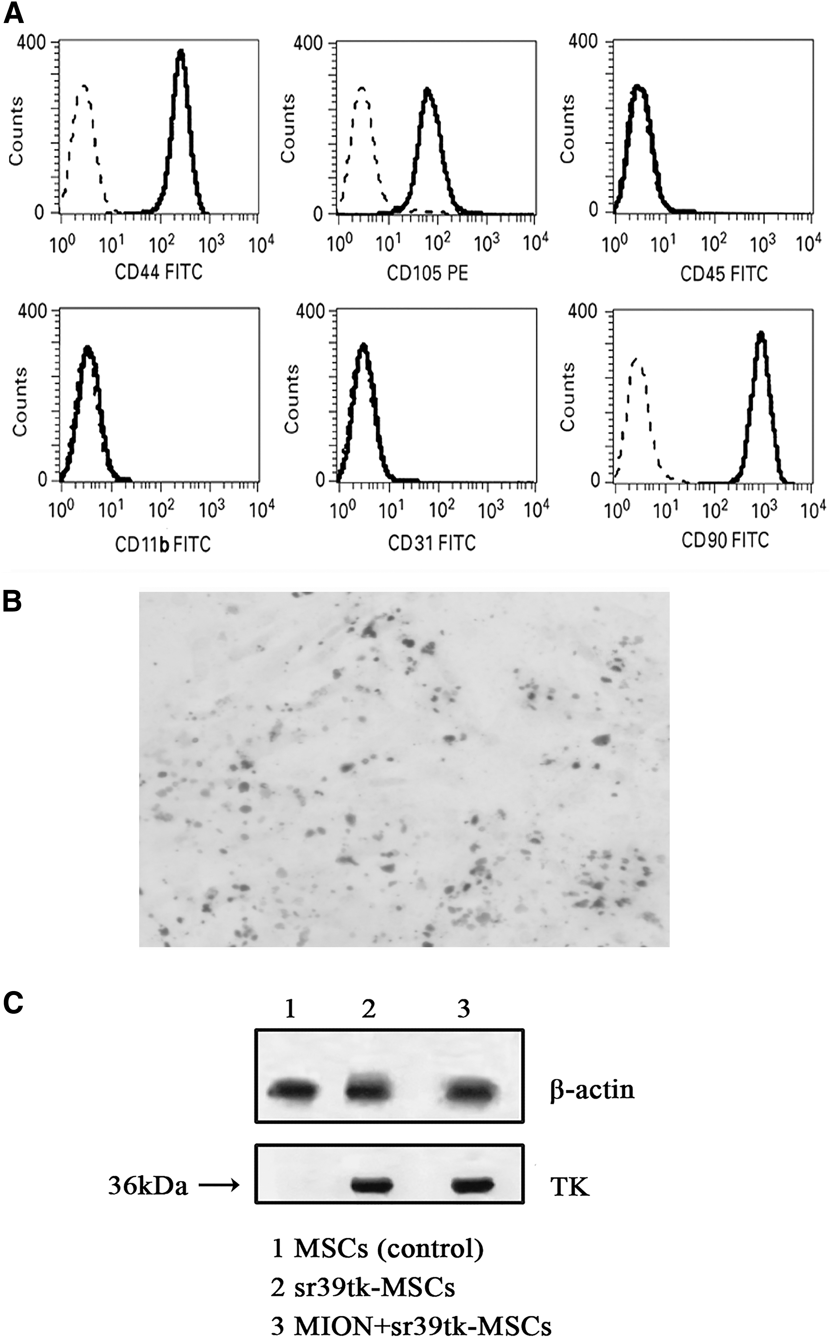
Characteristics of MSCs in vitro.
Means ± standard deviations, n = 3.
MION, magnetic iron oxide nanoparticles.
The labeling efficiency of MSCs with MION at 25 μg Fe/mL reached 75% and clustered blue particles were present in most cells (Fig. 1B). FACS analysis showed that about 37% of the cells were transduced with the lentiviral vector. Western blot analysis of total cellular proteins showed that TK protein expression levels in sr39tk-MSCs and sr39tk+MION-MSCs had similar intensity to the TK band. The predominant band produced by a TK-specific antibody occurred at 36 kDa. Meanwhile, control MSCs showed no significant signal with anti-TK. β-Actin expression was similar for these cell lines (Fig. 1C).
Animal models of IVDD
All animals were healthy throughout the experiment period. The mean blood loss during surgery was 50 ± 5 mL and the average surgery time was 55 ± 10 min.
The L2–L5 IVDs of each animal had an inhomogeneous structure, tissue loss in parts of the NP, and less clear zones at 6 weeks after the induction surgery. The Pfirrmann's classification grade remained between Grades 4 and 5 (4.26 ± 0.24), which revealed a classic phenomenon of disc degeneration (Fig. 2A). Meanwhile, L1–L2 IVD (unoperated discs) showed physiologically and harmoniously bright hyperintense white SI and normal disc height on the T2-weighted gradient echo sequence throughout the experiment period.
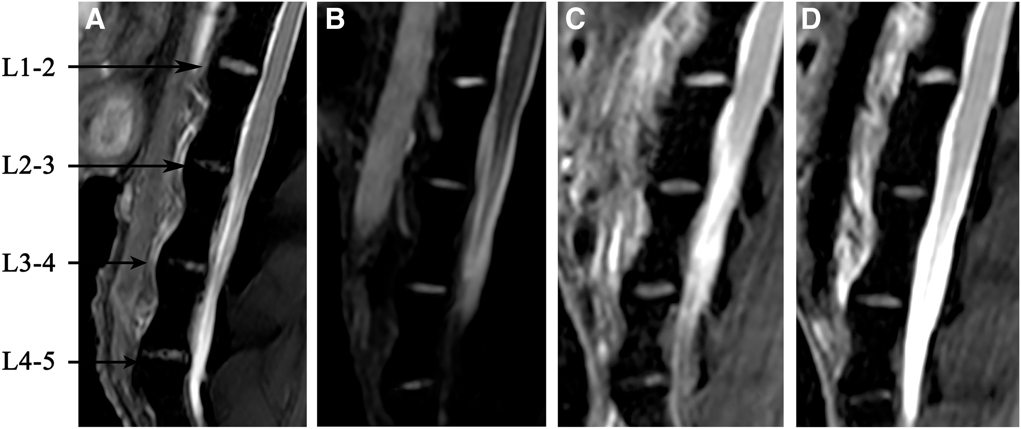
T2-weighted MR images of IVDs.
MR findings
At 3 weeks after MSC implantation, the mean disc degeneration grade in the sr39tk, sr39tk+MION, and MION groups improved from 4.45 to 2.37, 4.36 to 2.33, and 4.20 to 2.09, respectively. All three groups showed a significant improvement in the repair progress of degenerated IVDs, and there were no statistically significant differences among the groups (Fig. 2B–D). By 4 weeks, the disc degeneration grade for the experimental group had not significantly decreased relative to the 3-week time point, but all were significantly lower than that for the positive control group (L1–L2, 1.12 ± 0.11), and all values were similar among the experiment groups.
There were no statistical differences in the signal strength, signal contrast ratio (%), and area of MION (“dark spots,” mm2) among the L2–L4 IVDs relative to the MION groups at 3 days compared with 2 weeks after MSC implantation (225.34 ± 35.62 vs. 251.98 ± 31.43; 85.37 ± 10.54 vs. 78.56 ± 11.31; and 5.29 ± 1.35 vs. 4.76 ± 1.02, respectively, P > 0.05). No statistically significant changes in these three parameters were observed until 3 weeks after cell implantation.
However, at 4 weeks after cell implantation, all three indices changed significantly compared with that seen at 3 weeks (751.43 ± 52.67 vs. 243.76 ± 21.43; 47.37 ± 5.01 vs. 77.79 ± 9.98; and 1.78 ± 0.31 vs. 4.67 ± 1.14, respectively, P < 0.01) (Fig. 3). Similar statistical results were also seen for the sr39tk+MION group at the 4-week time point.

The MR signal of MION signal strength, signal contrast ratio (%), and the low signal area (mm2) in IVDs from the MION group compared with IVDs in the positive control group at four time points after cell implantation. *Indicates P < 0.01; detailed data are described in the Results section.
PET results
PET results demonstrated significantly increased accumulation of [18F]FHBG in L2–L4 IVDs from both the sr39tk group and sr39tk+MION group on day 3 after cell implantation relative to that seen for the background (L1 vertebra). At 2 weeks after cell implantation, retention of [18F]FHBG further increased in L2–L4 IVDs, and there was no apparent difference between the sr39tk and sr39tk+MION groups.
However, the [18F]FHBG signal strength decreased dramatically at the 3-week time point compared with that seen at 2 weeks (P < 0.001), and this difference was seen for both the sr39tk group and sr39tk+MION group. At 4 weeks after cell delivery, the [18F]FHBG signal strength in the sr39tk group and sr39tk+MION group was similar to background levels, indicating the absence of active sr39tk gene expression (Figs. 4 and 5).
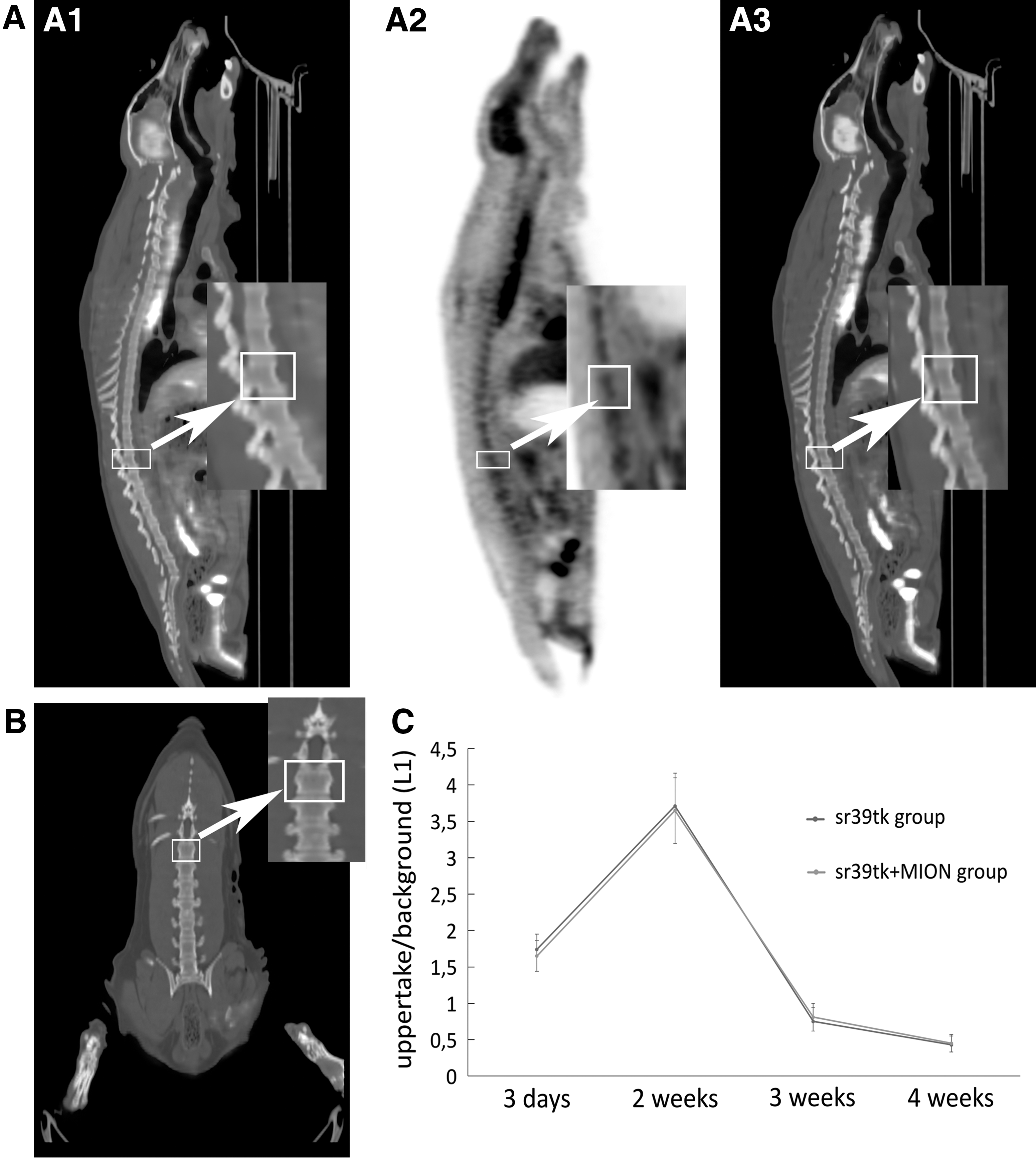
Acquisition of PET/CT (beagles) images and statistical analysis.
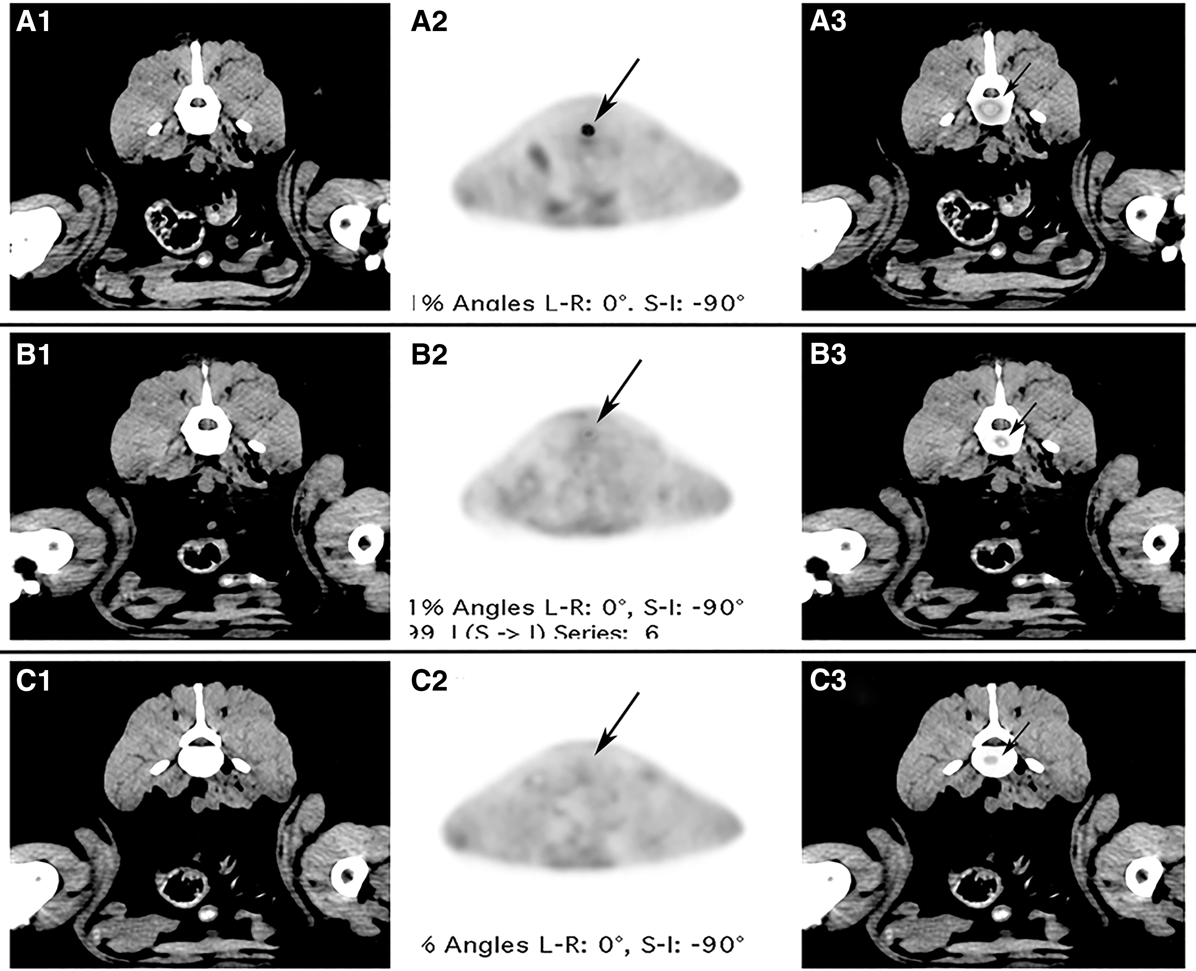
Transverse PET/CT fusion images of IVDs (L2–L3) at 2 weeks, 3 weeks, and 4 weeks after MSC implantation in sr39tk groups.
Discussion
The major findings of this study were that MSCs could survive no more than 3 weeks after implantation in a canine model of surgically induced IVDD, and that MR imaging with MION-labeled MSCs may not be a reliable technique for determining living cell engraftment in IVDs. Nonetheless, MSCs likely did contribute to repair processes in degenerated IVDs during the 3-week period when surviving MSCs were observed.
In contrast to previous studies of MSCs in IVDs, investigations of MSC engraftment in heart are more extensive. In heart, studies using various animal models, transplantation methods, and tracing techniques showed that the MSC survival time was limited to 3–8 weeks [32,33]. These results were consistent with the results of this study.
Some studies reported that transplanted stem cells could survive for more than 6 months in scar tissue in the heart [34,35]. There are also reports that support long-term survival (even more than 6 months) of MSCs in IVDs as measured by double-immunofluorescence labeling of specimens from animals sacrificed at various time points [19 –21]. Although the results of these studies may be valid for the conditions used, we contend that the techniques used in our research may more closely replicate the conditions that occur in vivo.
In addition, recent randomized controlled clinical trials using autologous bone marrow-derived stem cells failed to show significant long-term improvement of left ventricle function in MI patients [36,37]. This outcome may be due to the potential for engrafted cells in infarcted tissue to undergo necrosis or apoptosis over time. Due to the low oxygen levels and nutrient availability, the microenvironment in degenerated IVD would likely be less favorable than that in MI, and as such, MSC necrosis or apoptosis following transplantation into IVDs may be more frequent. The microenvironment for MSCs, or the niche, does indeed appear to be crucial to the fate of MSCs as evidenced by earlier studies showing reduced survival and adaptation of transplanted MSCs [2,38,39].
Dogs commonly experience back pain due to IVDD and surgeries to achieve decompression and spinal fusion are often performed on both chondrodystrophic and nonchondrodystrophic dogs. Given the similar macroscopic and microscopic appearance of IVDD in dogs and humans [40], we used dogs as the animal model for this study. To accurately replicate the typical pathological change seen in human IVDD and accelerate the degeneration process, we surgically induced IVDD by opening the AF anterior-laterally and disfiguring the NP. The IVDD model was successful according to the Pfirrmann Classification grade recorded 6 weeks after the index operation. To avoid leakage of MSCs during injection [41], we removed about 10% (13.7 ± 3.5 mg) of the total NP during the IVDD induction to facilitate later maintenance of the MSCs. Nonetheless, there is still no ideal animal model for IVDD that incorporates the conditions that occur from an erect posture and bipedal walking.
To our knowledge, no study has used HSV1-sr39tk transfection and SPIO nanoparticles labeling to follow MSC engraftment and survival. MSCs can be induced into a chondrogenic/NP phenotype in vitro using several methods, including coculture of MSCs with IVD explants, microencapsulation within alginate beads, or exposure to growth factors [42]. In our experiment, MSC viability, proliferation, and differentiation were not affected as indicated by analyses of phenotypic characteristics and cell cycle before and after the one-stage transfection and labeling procedure. As such, colabeled MSCs could be a useful tool for various studies of MSC therapies.
The repair of IVDD achieved with MSCs was apparent on T2-weighted imaging in all three MSC-implanted groups at the end of the experiment, and there were no significant differences among the groups. Notably, the repair progress was incomplete based on the final results for MR and PET indicating that no MSCs survived beyond the 3-week time point. This result suggests that, in the absence of genetic modifications, MSCs would not be able to promote repair after a certain time point. Moreover, the decisive contributor to the therapeutic effect of MSCs in IVDs would be the MSCs themselves and as well as MSC transdifferentiation, rather than the paracrine stimulation of angiogenesis or activation of endogenous IVD cells. Nevertheless, the full potential of MSCs to treat IVDD remains to be determined. Longer observation periods could yield different outcomes. Moreover, Ho et al. found that there are differences between outcomes for imaging and histology [43], which we did not perform in this study. There are also reports that describe a MION dose-dependent inhibition of chondrogenesis and glycosaminoglycan production [44,45]. Thus, MSCs implanted into IVDs could differentiate into chondrocyte-like cells and in turn affect the apparent repair efficiency of MSCs used in this research.
To date, researchers have become increasingly aware of the potential for overestimation with the MION-MR imaging technique [24 –26,46,47], but most of these studies were performed in hearts, not in IVDs. In this study, we used three evaluation indexes: signal contrast ratio, low signal area, and MION intensity to determine the retention of these iron nanoparticles. Our data showed no significant decrease in the number of nanoparticles up to 3 weeks after the MSC injection in both the MION and sr39tk+MION groups. Meanwhile, the [18F]FHBG signal strength declined dramatically and was accompanied by significant differences between the groups. This kind of deviation indicated that the positive signal strength seen in MR imaging at 3 weeks does not arise from living MSCs since the PET imaging signal is an indicator of cell survival and is a cornerstone of this study.
Integrated sr39tk is minimally regulated by physiological processes in the cell and stable transfection will ensure that the reporter is not diluted upon cell division [48,49]. Meanwhile, both [18F]FHBG and iron oxide nanoparticles are approved by the U.S. Food and Drug Administration as investigational imaging agents. Thus, the “colabeled MSCs” used in this study could have potential clinical applications as novel tools for cell engraftment and viability. Yaghoubi et al. confirmed clinically the accuracy and sensitivity of HSV1-sr39tk and that [18F]FHBG is a high affinity substrate for HSV1-sr39TK that has relatively low affinity for mammalian TK enzymes, which translates to excellent sensitivity for positive detection [50]. In our research, we quantified the ellipsoid ROIs of experimental IVD and found that MSCs could survive no more than 3 weeks in either the sr39tk group or the sr39tk+MION groups.
Due to limited experimental resources, we could not acquire daily PET scans of the animals, which likely would have yielded more detailed results. Instead, for each time point the animals were examined on the same day with separate MR and PET machines. Most recently, new hybrid PET/MR instruments (eg, Biograph mMR, Siemens) have been used in the oncology field to provide a new level of synergistic MR-PET fusion imaging with significantly improved diagnostic accuracy for both research and clinical applications [51]. Future studies should be conducted using these hybrid instruments.
In addition to the limitations of this research, including no ideal animal model for IVDD and lack of daily PET scans of the animals, this method cannot yet be applied in humans due to species-specific differences.
Finally, improvement of the microenvironment (niche) of implanted cells to enhance cell survival as well as use of MSCs that carry relevant gene modifications could be critical for enhancing the success of IVDD therapies that involve MSCs.
Conclusions
In this study, we found that MSCs could survive no more than 3 weeks in a surgically induced canine model of IVDD. PET imaging could be used to reliably determine the survival of implanted MSCs as MION labeling method with MR alone was not suitable for long-term tracking of MSCs. MSCs contributed to the repair process of IVDD during the 3-week period that they survived. Because these results were obtained in a canine model, care must be taken when making clinical applications of these results. Nonetheless, MSCs could be valuable to bridge the gap between symptomatic care and surgical treatment of IVDD.
Footnotes
Acknowledgment
This study was supported by the National Natural Science Foundation of China (grant nos. 81201439 and 81371979).
Author Disclosure Statement
No competing financial interests exist.