
Abstract
Hematological patients who accept chemotherapy always develop secondary tumor or even die of severe infections. As an important central lymphoid organ, the thymus is frequently damaged during chemotherapy. Previous studies showed that the mesenchymal stem cells (MSCs) can promote the proliferation and repair of epithelial cells in thymus. The purpose of our study is to investigate the reparative effects of human adipose-derived mesenchymal stem cells (hADMSCs) in chemotherapy-treated damaged thymus. Eighty mice were randomly divided into four groups: normal group, model control group, hADMSCs untreated group, and hADMSCs treated group. The mice were injected intraperitoneally with dexamethasone sodium phosphate (Dex 20 mg/kg), except the normal group. Then, the chemotherapy models were obtained after 1 week; the treated group was infused intraperitoneally with hADMSCs, whereas the model control group was injected with equal volumes of normal saline. The hADMSC's infusion day was regarded as day 0. The mice were sacrificed at different time points (days 3, 7, 10, and 14). The pathological structure and the function of the thymus, the recovery of T-lymphocyte subpopulation, and the proportion of regulatory T (Treg) cells in spleen and peripheral blood were detected. Additionally, we transfected hADMSCs by lentivirus with green fluorescent protein (GFP) to confirm whether they home to thymus and detected the expressions of cytokines that are associated with the development of thymus in hADMSCs and thymus. The results of the study showed that the hADMSCs treated group had a more rapid recovery in terms of thymic pathological structure and function. The hADMSCs could home to the damaged thymus and secrete cytokines that played important roles in repairing damaged thymus. The results indicated that hADMSCs could repair the damaged thymus caused by chemotherapy and improve the immune microenvironment, which may be a potential treatment for hematological patients.
Introduction
As a member of central lymphoid organ, the thymus plays an important role in the immune system. It is the place which supports the differentiation and proliferation of the lymphoid stem cells [1,2]. The maturity and the perfect function of T cells are supported by the microenvironment in thymus. The development of T cells depends on the interaction with stromal cells in thymus, especially the epithelial cells, which are responsible for positive selection and self-tolerance to generate the naive T cells [3 –5].
Mesenchymal stem cells (MSCs) are nonhematopoietic multipotent progenitor cells, which were first discovered in bone marrow, and then found in nearly all postnatal organs such as liver, lung, brain, spleen, cornea, thymus, umbilical cord, aorta wall, and so on [6 –8]. MSCs have the ability of differentiating into many cells of mesenchymal lineage, such as adipocytes, chondrocytes, osteocytes, fibroblasts, cardiomyocytes, skeletal myocytes, visceral cells, endodermal cells, mesodermal cells, and ectodermal cells [9 –11]. At the same time, they can also secrete cytokines, such as IL-6, IL-7, IL-10, transforming growth factor (TGF), hepatocyte growth factor (HGF), nitric oxide (NO), and express chemokine receptor CCR, CXCR, therefore play an important role in rebuilding the damaged tissue and immune response [12,13]. What is more, MSCs have the potential of self-renewal and repairing ability. They can also home to the damaged tissue [14,15].
Recently, it was found that mesenchyme accounts for the functional development of epithelial cells and the maintenance of epithelial architecture and function [16]. Dysplastic or radiotherapeutic thymus was partly reconstructed and the immune environment was improved through the treatment of MSCs [17]. They could also be used for autoimmune disease, graft versus host disease, and cardiac function improvement [18 –21]. The human adipose-derived mesenchymal stem cells (hADMSCs) which were safe and easy to obtain had also been widely used during clinical therapy [22,23].
However, it is still unclear whether hADMSCs can home to the thymus and repair the damaged thymus caused by chemotherapy. In our study, we infused the hADMSCs to the animal model, which received chemotherapy, to investigate the structural changes of the epithelial cells and thymic cells, to compare the function of thymus, ratio of T-lymphocyte subpopulation, and proportion of regulatory T (Treg) cells in peripheral blood and spleen at different time points, and we traced the hADMSCs to confirm whether they home to the damaged thymus and detected the expressions of cytokines in hADMSCs and thymus. We demonstrated that the functions of thymus and peripheral immune environment were improved, and the immune system recovered greatly. The hADMSCs could home to the thymus and express many cytokines that were considered to be important to the regeneration of thymus. The hADMSCs could repair the damaged thymus. The hADMSCs may be a potential therapeutic choice for the patients who accept chemotherapy.
Materials and Methods
Animal model
The study was approved by the Institutional Review Board of the Second Hospital of Hebei Medical University and monitored by a safety monitoring board. Eighty 6-week-old male BALB/C mice were obtained from the Hebei Medical University and maintained in the animal center of the Second Hospital of Hebei Medical University. The mice were randomly divided into normal group, model control group, hADMSCs treated group, and hADMSCs untreated group. The mice were injected intraperitoneally with dexamethasone sodium phosphate (Dex 20 mg/kg), except the normal group. Then, the chemotherapy models were obtained after 1 week, the treated group was infused with hADMSCs (2 × 10⁵cells/mouse) intraperitoneally, whereas the model control group was injected with equal volumes of normal saline. The hADMSC's infusion day was regarded as day 0. Mice were sacrificed at different time points after the hADMSC's infusion (days 3, 7, 10, 14, respectively, five mice/group and time point).
Isolation and culturing of hADMSCs
The adipose tissue was obtained from hospital with donors' informed consent. Human tissue collection for research was approved by the Institutional Review Board of the Second Hospital of Hebei Medical University. The adipose tissue was cut into 1-mm3 pieces and digested with collagenase I (Sigma) for 30 min, then the digestion was terminated by adding completed medium.
After we removed undigested tissues by filtrating the samples with a nylon mesh (pore size: 100 mm), the filtrate was centrifuged at 1,000 rpm for 3 min. Then the supernatant was discarded and the cells were resuspended with 2 mL 10% fetal bovine serum (FBS; Gibco) and seeded in a 3.5-cm Petri dish. The hADMSCs were incubated in Dulbecco's modified Eagle's medium: nutrient mixture F-12 (DMEM/F12; Gibco) medium containing 10% FBS in a humidified incubator maintained at 37°C in a 5% CO2 humidified atmosphere. When reached 70%–80% confluence, the cells were detached with 0.25% trypsin-EDTA (Gibco). The fourth passage (P4) hADMSCs were used during our study. The phenotype of hADMSC was analyzed by flow cytometry (FCM) using the following monoclonal antibodies: CD34-FITC, CD45-Per-CP, CD44-APC CD105-PE, and CD29-PC-7 (eBioscience). The hADMSCs were cultured in the inducing media for 3 weeks, then stained with Oil Red O solution and Alizarin Red.
Histology and immunofluorescence staining
Fresh thymus tissue was embedded in Tissue-Tek OCT compound (Sakura Finetek, Tokyo, Japan) and was quickly frozen in the freezing microtome. Four micrometers of frozen sections of thymus were obtained and air dried, and then fixed with 4% formaldehyde for 1 min for Hematoxylin and Eosin (HE) staining. The other fixed frozen sections were washed three times with 1 × phosphate-buffered saline for 5 min. One percent Triton X-100 was used as a cell penetration. The sections were blocked with 5% bovine serum albumin for 30 min and incubated with primary antibodies: anti-mouse Keratin 5 (K5; Covance Research Products, Berkeley, CA) and Troma-1 mAb (rat anti-Keratin 8; Rolf kemler, Developmental Studies Hybridoma Bank, University of Iowa, Iowa City, IA) at 37°C for 1 h, then the corresponding second antibodies were incubated for 30 min. Finally, DAPI was used for nuclear staining. Microscopic analysis was performed with Zeiss fluorescence microscope.
T cell receptor excision circle analysis
Total DNA was extracted and purified from 10 mg spleen tissue by using the DNeasy Blood & Tissue Kit (Qiagen). Then, the T cell receptor excision circle (TREC) value was tested with real-time quantitative polymerase chain reaction. The sequences of the primers were as follows: mRec primer (5′-GGACACAGCATGTG-3′), Jα primer (5′-GCAGGTTTG-TAAAGGCTCA-3′), and mRec-Jα fluorescent probe (5′-FAM-CACAACCTGCCCTGTGCA-TAMRA-3′; Invitrogen) [24]. The polymerase chain reaction (PCR) contained 1,000 ng DNA, 0.5 mM of each primer, 0.1 mM fluorescent probe, and TaqMan mix (Roche Applied Science, Penzberg, Germany). Amplifications were performed in triplicate on a Rotor-Gene Q (Qiagen) at 95°C for 10 min followed by 40 cycles of 95°C for 10 s, 60°C for 35 s, 72°C for 1 s, and a final step at 40°C for 30 s.
Flow cytometry
Fresh thymus, spleen, and peripheral blood cells were obtained from each mouse at different time points. The tissues were ground and filtered with a 200-mesh sieve to make a single cell suspension. After staining with antibodies: Per-cp anti-mouse CD45 (eBioscience), FITC anti-mouse CD3 (eBioscience), APC anti-mouse CD4 (eBioscience), and PE anti-mouse CD8a (eBioscience) for 20 min, the thymic cells (1 × 10⁶) were fixed with 3% paraformaldehyde for 10 min. After the red cells were cracked by purified water for 10 min, the thymic cells were washed twice with normal saline at 1,500 rpm for 5 min and analyzed on a flow cytometer (FACS Canto II; BD Biosciences).
The spleen and peripheral blood cells (1 × 10⁶) were stained with antibodies, respectively: Per-cp anti-mouse CD45 (eBioscience), FITC anti-mouse CD3 (eBioscience), APC anti-mouse CD4 (eBioscience), and PE anti-mouse CD8a (eBioscience). The Mouse Regulatory T Cell Staining Kit #2 (eBioscience) were used for the Treg test. Cells were analyzed on a flow cytometer (FACS Canto II; BD Biosciences).
Tracing of the green fluorescent protein-labeled hADMSCs
We transfected hADMSCs by lentivirus with green fluorescent protein (GFP; Genechem) in vitro. When the transfection rate reached 80%, the hADMSCs were injected intraperitoneally into the chemotherapy-treated mice and the normal mice. The mice were sacrificed at 48 h, and the thymus were frozen quickly to make frozen sections. DAPI was used for nuclear staining. Microscopic analysis was performed with Zeiss fluorescence microscope.
Cytokine expression in hADMSCs and thymus
Total RNA was extracted using TRIzol reagents, and complementary DNA (cDNA) was synthesized by PrimeScript™ RT reagent Kit with genomic DNA (gDNA) Eraser (TaKaRa). The sequences of the primers of hADMSCs were as follows: keratinocyte growth factor (KGF) forward primer (5′AAGAACTGCCCTTCCTCA-3′), reverse primer (5′-AAGTCCTCCTCTGCCCTC-3′); Ghrelin forward primer: (5′-CCATGCCCTCCCCAGGGACC-3′), reverse primer (5′-ATCACTTGTCGGCTGGGGCCTC-3′); IL-7 forward primer: (5′-CCCCTTGGGATGGATGAA-3′), reverse primer (5′-ATGATGACCGCAACTGGA-3′); and β-actin forward primer: (5′-ACTTAGTTGCGTTACACCCTT-3′), reverse primer (5′-GTCACCT-TCACCGTTCCA-3′; Invitrogen). The sequences of the primers of thymus were as follows: KGF forward primer: (5′-CAAACGGCTACGAGTGTGAA-3′), reverse primer (5′-ACTGGGTGC-GACAGAACAG-3′); Ghrelin forward primer: (5′-GCTGTCTTCAGGCACCATCT-3′), reverse primer (5′-TTGTCCTCTGTCCTCTGGGT-3′); and β-actin forward primer: (5′-GAGACC-TTCAACACCCGAGC-3′), reverse primer (5′-ATGTCACGCACGATTTCCC-3′; Invitrogen). The cDNA was amplified using PCR in 20 μL reactions (Taq PCR Mastermix; TIANGEN) with the following program: denaturation at 95°C for 2 min followed by 40 cycles of 95°C for 10 s, 55°C for 20 s, and a final step at 72°C for 20 s. The Enzyme-Linked Immunosorbent Assay (ELISA) Kit of KGF (Biorbyt), Ghrelin (Cloud-Clone Corp.) and IL-7 (Cloud-Clone Corp.) were also used to test the expression levels of proteins in thymus.
Statistical analysis
All data were presented as the mean ± standard deviation. Analysis of statistics were carried out with SPSS statistics 21.0 software using one-way analysis of variance and t-test. P < 0.05 was considered statistically significant.
Results
Characterization of the hADMSCs
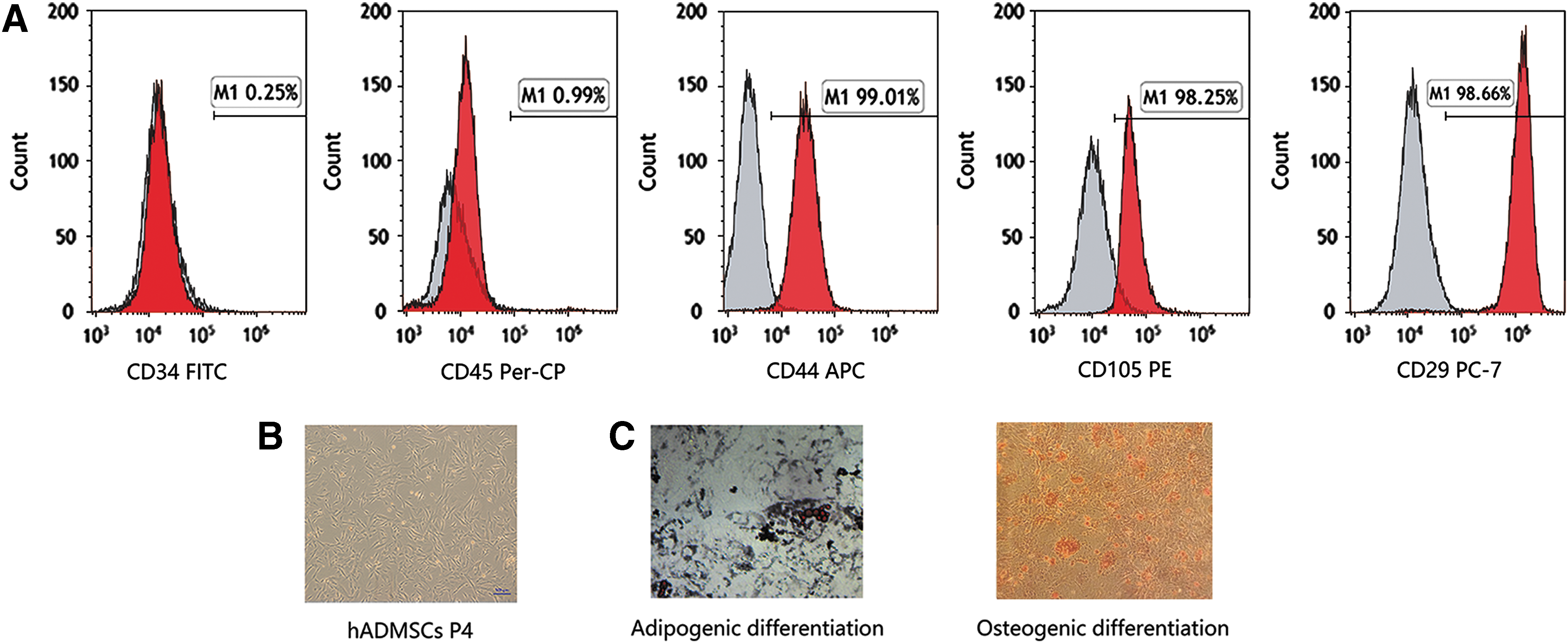
The hADMSCs were isolated from the adipose tissue of donors and cultured in vitro. We used FCM to analyze the surface markers of the P4 hADMSCs (Fig. 1B). The expressions of CD44, CD105, and CD29 were positive in these cells, but negative for CD34 and CD45 (Fig. 1A). Furthermore, we assessed their abilities to differentiate into cells of osteogenic and adipogenic lineages. The lipid was stained with Oil Red O solution, whereas the osteogenic differentiation of hADMSCs were stained with alizarin red (Fig. 1C). All the results of these inspection methods supported the characteristics of stem cells after passage culture in vitro.
The hADMSCs enlarged the chemotherapy-damaged thymus and promoted the recovery of the thymus
The size of the damaged thymus enlarged after the infusion of ADMSCs. We used HE staining and immunofluorescence staining to observe the changes in the chemotherapy-damaged thymus in mice among the four groups. The chemotherapy damage in thymus was diffuse (Fig. 2). The corticomedullary differentiation was not clear under the low-power microscope (40 × ). Under the high-power microscope (400 × ), the number of thymocytes was decreased with irregular cell shapes, the chromatin color became lighter, the nuclei were not clear or disappeared; the number of the thymic epithelial cells (TECs) was decreased with lighter chromatin color and unclear or disappeared nuclear membranes; and the blood vessels were disappeared or incomplete, the endothelial cells appeared swollen, or even showed small necrotic area. However, after the infusion of hADMSCs at days 3 and 7, compared with the untreated group, the corticomedullary differentiation was clear, the number of the thymocytes and the TECs increased, the shapes of the thymocytes and the TECs recovered; and the endothelial cells of the blood vessels appeared integrated, which were almost same with the normal group. But there were little differences at days 10 and 14 between the treated group and untreated group through HE staining, which were all nearly the same with the normal group (Fig. 3). These indicated that the hADMSCs could improve the proliferation and rebuilt the thymic structure, which was damaged by chemotherapy.
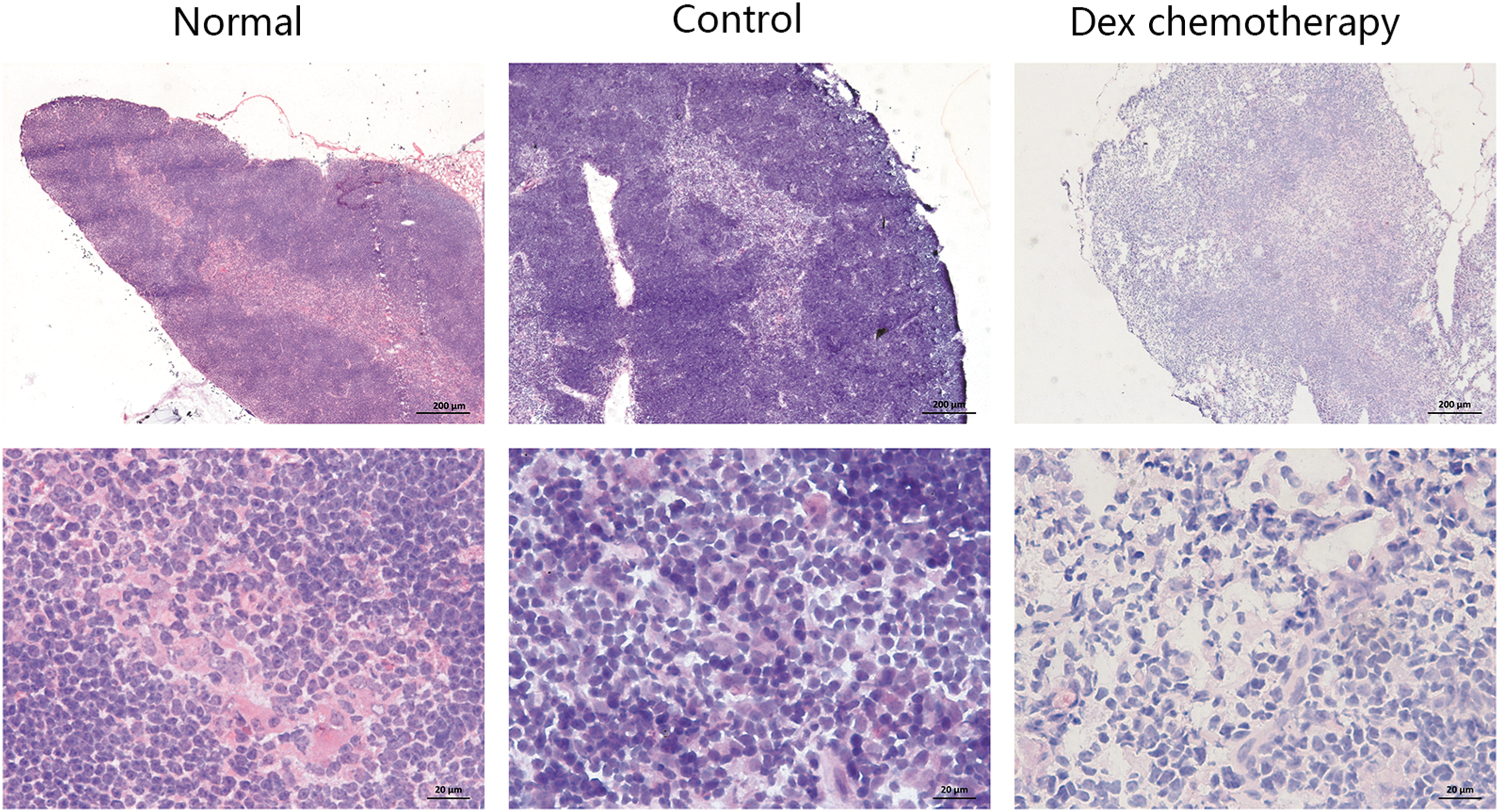
HE staining of thymus in normal mouse, control mouse, and Dex chemotherapy mouse. The top row was under the low-power microscope (40 × ), and the lower row was under the high-power microscope (400 × ). The damage in thymus of the chemotherapy mouse was diffuse, the corticomedullary differentiation was not clear, and the thymocytes decreased with irregular cell shapes. Dex, dexamethasone sodium phosphate; HE, Hematoxylin and Eosin.
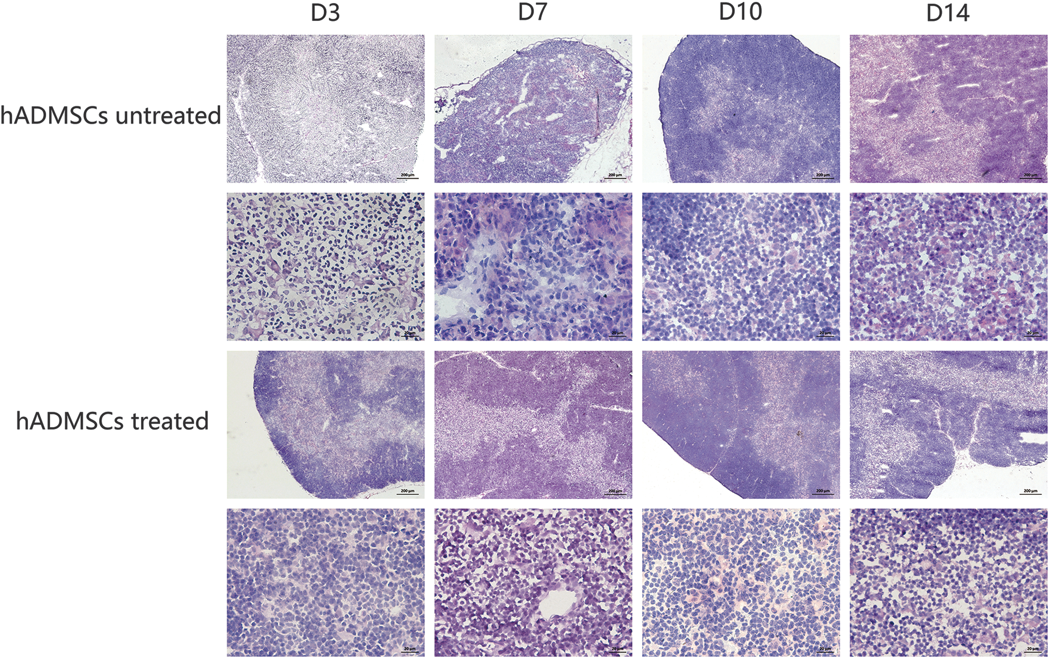
HE staining of thymus in the untreated and treated groups at different time points. The hADMSCs could improve the proliferation and rebuild the thymic structure, which was damaged by chemotherapy. At the first two time points in the treated group, with the treatment of hADMSCs, the corticomedullary differentiation was distinct, the number of the thymocytes and the thymic epithelial cells increased, which was almost the same with the normal thymus. D, day.
The reconstruction of the cortex/medulla structure in damaged thymus
Keratin 8 (K8) and K5 were the markers of the cortical and medullary areas of TECs respectively, so we used them to recognize the right location of TECs. In normal thymus, the K5-positive (K5+) TECs were observed in the center medullary area, whereas the K8-positive (K8+) TECs were observed around the cortical areas. In our study, we analyzed the thymic structure at four time points after hADMSCs infusion (days 3, 7, 10, and 14). At days 3 and7, the untreated groups showed disordered location of fluorescence (D3) or lighter fluorescence (D7), whereas the treated groups showed well-organized and distinct location in the corticomedullary architecture in the thymus (Fig. 4). The quantification of the fluorescence intensity of K8 at day 7 in the untreated group was 3.93 ± 0.8 and the treated group was 22.57 ± 4.35, P < 0.05. The quantification of the fluorescence intensity of K5 at day 7 in the untreated group was 4.82 ± 0.8 and the treated group was 20.47 ± 1.14, P < 0.05. No significant differences were observed among the four groups at days 10 and 14. The findings indicated that the hADMSCs could improve the proliferation of TECs and rebuild the normal structure in the chemotherapy-damaged thymus in mouse.
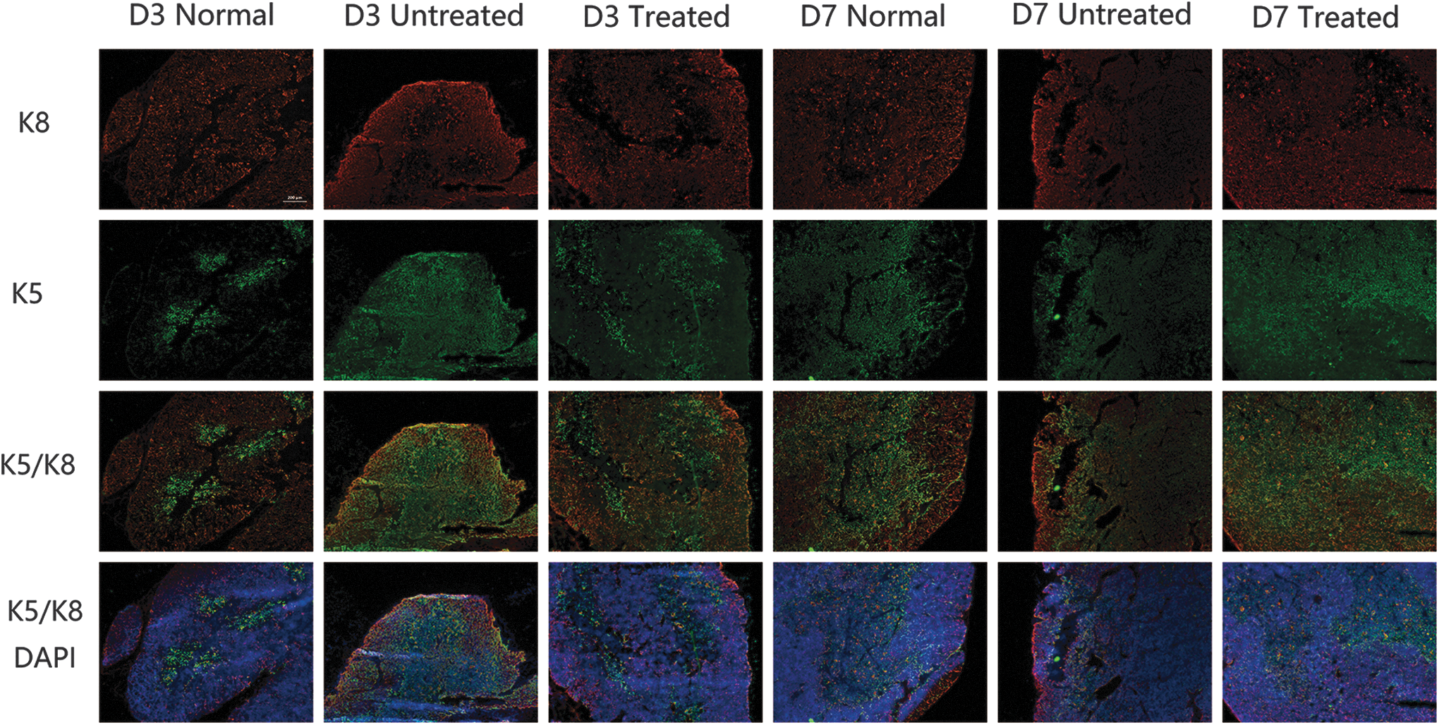
The changes in immune fluorescence in normal group, untreated group, and treated group. The untreated groups showed disordered fluorescence location (D3) or lighter fluorescence (D7) compared with the normal group, whereas the treated groups showed well-organized and distinct location in the corticomedullary structure in the thymus at D3 and D7.
The hADMSCs increased the proportions of mature T cells and improved the export of T cell in the damaged thymus
The CD4+CD8+ T cells are the major cells in thymocytes. The selected CD4+CD8+ immature T cells differentiate to CD4+CD8- and CD8+CD4− T mature cells. The proportions of the CD4+CD8− and CD8+CD4− T mature cells in thymus decreased compared with the normal group at day 3. With the infusion of the hADMSCs, the proportion of the CD4+CD8− and CD8+CD4− mature T cells of the thymus in the treated group was significantly high than the untreated group at day 3 (P < 0.05) (Fig. 5C, D), whereas no significant differences were observed at other time points (P > 0.05). TRECs were formed as the T cell receptor gene recombination, which could be used as an indicator of the output of newly generated naive T cells [25]. We analyzed the TRECs expression in splenocytes through real-time quantitative polymerase chain reaction. At day 3, the expression of TRECs in splenocytes of the treated group were remarkably higher than that of the untreated group (Fig. 5E), whereas no significant differences were observed at other time points (P > 0.05). The results indicated that the infusion of hADMSCs increased the proportions of CD4+CD8− and CD8+CD4− T mature cells and improved the function of the chemotherapy-damaged thymus at day 3.

The flow cytometric analysis diagrams of the thymocytes in the
The hADMSCs recovered the T-lymphocyte subpopulation in peripheral blood of the mice with chemotherapy-damaged thymus
Thymus is the place which supports the mature of T-lymphoid cells. The mature T-lymphoid cells are released to the peripheral blood and immune organs to exert the immune functions. The peripheral T cells include CD3+CD4+CD8− T cells and CD3+CD4−CD8+ T cells, and the proportions of them and the ratio of CD3+CD4+CD8−/CD3+CD4−CD8+ can reflect the normality and stability of the immune system in peripheral. The T-lymphoid cells in peripheral blood and spleen of mice with chemotherapy-damaged thymus were stained with antibodies and analyzed by FCM. The ratio of CD3+CD4+CD8−/CD3+CD4−CD8+ of the treated group in peripheral blood was significantly higher than that of the untreated group at day 3 (Fig. 6) and the proportion of CD3+CD4+CD8− T cells of peripheral blood lymphocytes in the treated group was 34.8% ± 2.1%, which was higher than that of the untreated group 28.3% ± 4.2% (P < 0.05) and was closer to the normal group 40.8% ± 5.5%. However, there were no significant differences in spleen at all time points. The results demonstrated that the hADMSCs could improve the ratio of T-lymphocyte subpopulation and promote the recovery of CD3+CD4+CD8− T-lymphocyte subpopulation in immune environment of peripheral blood in mice with chemotherapy-damaged thymus.
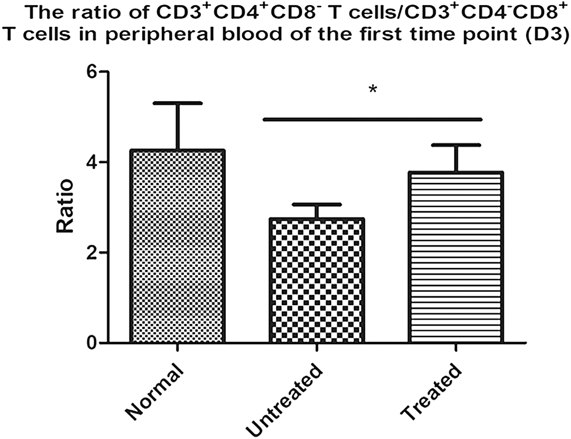
The ratios of CD3+CD4+ CD8−/CD3+CD4− CD8+ in the peripheral blood at day 3. Five mice/group, n = 5; *P < 0.05.
The hADMSCs increased the proportion of Tregs cells in peripheral blood of mice with chemotherapy-damaged thymus
As suppressor cells in the immune system, the Treg cells play an important role in the self-tolerance and immune balance. It derives from the CD4+ T cells. The normal proportion of CD4+CD25+Foxp3+ Treg cells in CD4+ cells of the peripheral and spleen is 5%–10% [26]. We analyzed the proportion of Treg cells both in peripheral blood and spleen in the four groups to estimate the effect of hADMSCs infusion on peripheral immune microenvironment. It showed that the proportion of Treg cells in the treated group was higher than that of the untreated group at day 3 in peripheral blood, and the proportion of the treated group was close to the normal group (Fig. 7). There were little differences among the four groups at other three time points and in spleen.

The flow cytometric analysis diagrams of Treg cells
The hADMSCs homed to the chemotherapy-damaged thymus
We used fluorescence microscope to visualize distribution of the hADMSCs in the chemotherapy-treated mouse to confirm whether the hADMSCs home to the thymus. The hADMSCs labeled with GFP (Fig. 8A) were observed in thymus after hADMSCs infusion at 48 h (Fig. 8B), whereas the hADMSCs could hardly be found in the normal thymus at the same time point (Fig. 8C).
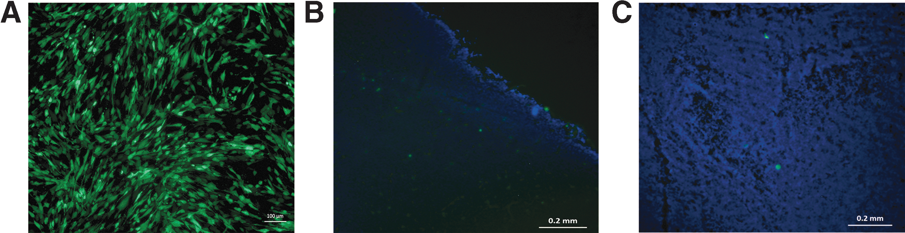
The result of fluorescence in cell tracking.
Expression of cytokines in hADMSCs and thymus
KGF is a 28-kDa member of the fibroblast growth factor family, and is required for postnatal thymic regeneration [27]. Ghrelin is a hormone which can increase the thymic cellularity and differentiation [28,29]. IL-7 is also regarded as an important cytokine for the regeneration of the thymus [30]. Our study showed that the hADMSCs could secrete KGF and Ghrelin, but barely expressed IL-7 (Fig. 9). The levels of KGF and Ghrelin in the treated thymus were also higher than that of the untreated group through reverse transcription polymerase chain reaction and ELISA in day 3 (Fig. 10) and showed statistical significance.
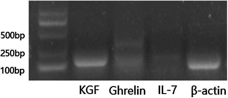
Reverse transcription polymerase chain reaction analysis of the expression of the cytokines in hADMSCs. The hADMSCs could secrete KGF and Ghrelin, especially showed a high level of KGF, but barely expressed IL-7. KGF, keratinocyte growth factor.

The expression levels of KGF and Ghrelin in thymus. The expressions of
Discussion
Our study indicated that the infusion of hADMSCs could repair the thymic corticomedullary structure and epithelial cells in the mice suffering from chemotherapy, improve the function of the chemotherapy-damaged thymus, and promote the ratio of T cell subpopulation and Treg cells in peripheral blood to recover the immune microenvironment. The possible mechanism may be associated with the secretion of various cytokines, especially KGF and Ghrelin that may modulate the thymic microenvironment.
In our study, it showed that the chemotherapy damage in thymus which is caused by chemotherapy was diffuse. The corticomedullary differentiation was not clear, the number of thymocytes decreased with irregular cell shapes, the color of chromatin became lighter, and nucleolus was not clear or disappeared; the number of the TECs decreased with lighter chromatin color and unclear or disappeared nuclear membrane; and the blood vessels disappeared or were incomplete, the endothelial cells became swollen, or even showed small necrotic area. We found that the hADMSCs could home to the thymus as has been previously reported [14]. The thymic structure was rebuilt in the mice with chemotherapy after infusion with hADMSCs at days 3 and 7. We also observed the corticomedullary sector and TECs through the special marked protein K5/K8, and found that the untreated group showed disordered K5/K8 protein expression locations or lighter fluorescence at days 3 and 7. However, after infusion with hADMSCs in the treated group, the expression of the thymic corticomedullary K5/K8 protein was in the corresponding locations which reflected the distribution of the TECs. Moreover, the treated groups were near to the normal groups at the first two time points. At the same time, the proportions of the CD4+CD8− and CD4−CD8+ thymocytes and TRECs expression of the spleen in the treated group were higher when compared with the untreated group at day 3. The ratio of the T-lymphocyte subpopulation, the proportion of Treg cells in peripheral blood of the treated group were also higher than that of the untreated group at day 3. Our data suggested that hADMSCs could home to damaged thymus and secrete cytokines such as KGF and Ghrelin. The hADMSCs had a positive effect of repairing the structure and promoting the function of the thymus, recovered the ratio of T-lymphocyte subpopulation and increased the Treg cells in peripheral blood so as to improve the balance of the peripheral immune microenvironment.
Our findings confirmed that the MSCs can home to and repair the damaged thymus. The hADMSCs could secrete KGF and Ghrelin, especially KGF. The levels of KGF and Ghrelin in thymus in the treated group were much higher than that of the untreated group. The improvement of the structure and function in thymus may have important implications for recovering the immune system of the patients suffering from chemotherapy.
However, on one hand, the reparative effects of hADMSCs to chemotherapy-damaged thymus was significant only at day 3 in thymus. The pathological findings also showed a difference at day 7, although with no statistical difference significant, maybe, the amount of hADMSCs was not enough for this treatment. On the other hand, hADMSCs can increase the proportion of Treg cells, but whether excessive Treg cells in peripheral immune system lead to tumor immune escape will be another problem [31,32]. The mechanism of interaction between hADMSCs and thymus is also unclear, but the cytokines secreted from the hADMSCs really showed their important roles. KGF is produced by cells of mesenchymal origin and binds to FGFR2IIIb that is expressed primarily by epithelial cells. The beneficial effects of KGF are mainly attributed to its protective effects against conditioning-induced epithelial cell injury [33 –35]. Ghrelin is currently known to bind specifically to GHS-R; GHS-Rs are largely expressed in the developing thymocytes [29,36]. Or perhaps, do the hADMSCs have the ability to differentiate into other cells in thymus? Therefore, the treatment amount of hADMSCs and the further repair mechanism between the cytokines and cells will be the research directions in the future study.
It is well known that the patients with malignant hematological disease are doomed to accept chemotherapy, and then inevitably suffer a series of severe infections and organ damage because of chemotherapy drugs, especially in the first 2 weeks after chemotherapy. The ratio of T-lymphocyte subpopulation and Treg cells are important indicators of immune system. Moreover, T cells take an important role in keeping balance of immune microenvironment. In our study, the hADMSCs could rebuild the structure in chemotherapy-damaged thymus and improve the function through increasing the export of mature T cells, so as to improve the ratio of T-lymphocyte subpopulation and proportion of Treg cells in the peripheral immune microenvironment. It can be a potential treatment for preventing patients from severe infections.
In conclusion, our study demonstrated that the infusion of hADMSCs could rebuild the structure and improve the function of the thymus in mice with chemotherapy-damaged thymus, which can recover the immune microenvironment and keep the balance of it in peripheral. The infusion of hADMSCs may provide a valuable method for the patients with severe infections.
Footnotes
Acknowledgments
The work was supported by National Natural Science Foundation of China (grant: GZR81400078). The authors thank Beijing Believe China Biotech Co., Ltd. for the provision of the cells and thank J.Y. and Y.P. for technical assistance.
Author Disclosure Statement
No competing financial interests exist.