
Abstract
Alveolar bone defects can arise as a consequence of trauma, infection, periodontal disease, or congenital alveolar fenestration. Many approaches have been employed in an effort to treat or overcome such defects, but the ability to effectively achieve alveolar regeneration remains elusive. Platelet-derived growth factor-BB (PDGF-BB) has been shown to serve as a key factor capable of orchestrating cell proliferation, angiogenesis, and chemoattraction in the context of osteogenic processes. Exactly how PDGF-BB affects human periodontal ligament stem cells (hPDLSCs), however, requires further exploration. In this report, we utilized a lentiviral construct to achieve PDGF-BB overexpression in hPDLSCs, allowing us to establish that this gene was able to enhance the proliferation of these cells and to mediate osteogenic gene upregulation therein. In addition, we established a rat model of alveolar defects that were implanted using different complexes, and then monitored through histological and micro-CT analyses 4 and 8 weeks postsurgery to assess bone repair outcomes. These analyses revealed that a thermosensitive hydrogel was an effective 3D cell culture scaffold, while PDLSCs overexpressing PDGF-BB enhanced bone growth in the context of alveolar bone defects. Together, these results thus indicate that PDGF-BB represents a potent means of promoting stem cell-based alveolar bone tissue regeneration.
Introduction
Alveolar bone defects can result from trauma, periodontal disease, trauma, [1]. Repairing the alveolar bone remains a substantial challenge, with a range of strategies having been employed to attempt to drive alveolar bone growth including the administration of bisphosphonates, antibiotics, or anti-inflammatory compounds, yet outcomes have been discouraging [2,3]. More recently, however, stem cell therapy has been explored as a potent means of driving the regeneration of alveolar bone through combining stem cells in a scaffold complex to achieve effective bone tissue engineering [4,5]. Multiple different forms of pluripotent mesenchymal stem cells (MSCs) can be employed to achieve periodontal tissue regeneration [6]. Seo et al. demonstrated that human periodontal ligament stem cells (hPDLSCs) are superior in their ability to proliferate and differentiate into osteogenic cells than are bone mesenchymal stromal cells (BMSCs) [7]. As such, hPDLSCs are a potentially ideal means of achieving periodontal bone regeneration. Platelet-derived growth factor (PDGF) is an essential cytokine present in human serum that can mediate the growth of a wide range of cell types, including fibroblasts, smooth muscle cells, and glia. It further acts to drive wound healing both in the bones and in soft tissues [8,9]. There are four PDGF genes that together compose five dimeric isoforms, which are as follows: PDGF-AA, PDGF-BB, PDGF-CC, PDGF-DD, and PDGF-AB [10]. PDGF-BB in particular is known to be released from osteoclasts in the context of bone development, enhancing angiogenic processes and improving bone regeneration such that when PDGF-BB is inhibited this leads to a marked reduction in trabecular and cortical bone mass. [10,11]. PDGF-BB has been shown to directly drive stem cell marker expression on target cells, to promote cell proliferation, and to enhance the ability of BMSCs to mediate bone regeneration through its ability to promote osteogenesis and angiogenesis [12 –15]. Owing to these advantageous properties, there have been many efforts to utilize recombinant PDGF-BB to aid bone healing and defects in a range of contexts including periodontal tissue regeneration [8,16]. Unfortunately, however, recombinant PDGF-BB has a very short half-life of just a few minutes in circulation, making it of limited utility when administered in vivo [13]. It is therefore believed that only by sustainably achieving local PDGF-BB delivery can effective outcomes be achieved in patients. One approach to achieving such delivery involves the use of lentiviral vectors to induce expression of a given gene in target cells, allowing for long-term stable expression of a given gene without the potential for side effects associated with high-dose administration of bioactive compounds [17]. Indeed, lentiviral vectors can readily infect a wide range of cell types and integrate into their respective genomes to achieve long-term and stable expression of a given gene of interest [18]. Such a lentiviral approach to stably overexpressing PDGF-BB may therefore be an optimal means of assessing the ability of this growth factor to facilitate hPDLSC-mediated bone regeneration in vitro and in vivo, thus allowing us to better explore the mechanisms underlying this bone healing process.
The development of hPDLSCs that overexpress PDGF-BB may represent an ideal cell system for mediating periodontal bone regeneration, but the efficacy of such a system depends upon identification of an optimal strategy for delivering stem cells in vivo. A range of biomaterials have been explored for their relative advantages in mediating the 3D encapsulation and delivery of stem cells [19,20]. One popular approach to tissue engineering for gene delivery has focused on the development of scaffold materials that are responsive to their local environment, such as thermosensitive poly (D,L-lactide-co-glycolide)-poly (ethylene glycol)-poly(D,L-lactide-co-glycolide) triblock copolymers (PLGA-PEG-PLGA), which have been shown to exhibit a range of potentially desirable clinical advantages [21]. For example, PLGA-PEG-PLGA exhibits superior biocompatibility while undergoing effective biodegradation, thus ensuring that it can be safely administered in vivo [16]. Importantly, this material is sensitive to temperature such that it undergoes a sol–gel transition at ∼32°C, meaning that PLGA-PEG-PLGA has the potential to be combined with osteogenic drugs to drive bone tissue regeneration and that it can be safely directly injected into sites of tissue defects and can solidify safely within the body without the need to employ any physical or chemical initiators [22]. Alveolar bone defects can take on a wide range of irregular shapes, but when injected as a liquid this thermosensitive hydrogel can fill in the defective region before solidifying at body temperature (37°C). These ideal physiochemical properties have led to the use of PLGA-PEG-PLGA for gene delivery with excellent outcomes.
We therefore sought to use hPDLSCs overexpressing PDGF-BB and encapsulated in a thermosensitive hydrogel to treat alveolar bone defects. This is the first study we are aware of to combine this approach as a means of clinically treating alveolar bone defects.
Materials and Methods
Materials
Reagents used in this study included the following: minimum essential medium alpha (α-MEM; Gibco), fetal bovine serum (FBS; Gibco), CCK-8 kit (Dojindo, Japan), mouse monoclonal antibody against STRO-1 and CD146 (Nouvs), cDNA synthesis kit (TaKaRa, Japan), inverted fluorescence microscope (Leica, Germany), Real-time PCR instrument (Roche), and human MSC Phenotyping Kit (Miltenyi Biotec, Germany).
hPDLSCs culture and identification
We obtained healthy donor periodontal ligament tissue from extracted premolars that were from individuals between the ages of 13 and 18. All aspects of this study pertaining to human sample collection and utilization were approved by the Ethical Review Committee of Shanghai Stomatological Hospital, China (2019-003). Human PDLSCs were grown as in previous reports [23,24]. In brief, periodontal ligament tissues were scraped from the middle of tooth root surfaces and cut into small pieces, followed by addition to 100-mm culture dish. α-MEM culture medium containing 10% FBS and 1% penicillin/streptomycin was used for routine cell culture. Putative stem cells were isolated through the limiting dilution technique. Single-colony-derived cell lines were expanded, with cells from the third passage being used for all experiments. Crystal violet staining was used for assessing cell morphology.
Immunofluorescence staining was used to assess the expression of surface MSC markers, including Stro-1 and CD146 on hPDLSCs. For such staining, hPDLSCs were plated in 24-well plates, fixed for 20 min with 4% paraformaldehyde, permeabilized using 0.25% Triton X-100, and blocked with 5% BSA before addition of primary mouse antibodies specific for human Stro-1 and CD146. In addition, the fluorescence-activated cell sorting analysis was used to confirm the expression of CD105, CD90, CD14, CD20, CD34, and CD45 (Miltenyi Biotec).
Thermosensitive hydrogel preparation and characterization
We prepared a PLGA–PEG–PLGA triblock copolymer through ring-opening polymerization [25]. Specifically, we combined LA, GA, and PEG in an airtight reaction bottle at a 15: 5: 8 ratio, and placed this under a vacuum at 120°C for 30 min. Stannous caprylate (0.2% W/W; Sigma) was then added and heated to 170°C for 8 h, after which vacuum was applied for 30 min for unreacted monomer removal. Samples were then dissolved in water and precipitated at 80°C. This step was then repeated, yielding the final product.
A tube inversion approach was used to assess the temperature at which the hydrogel underwent a transition. In brief, PLGA-PEG-PLGA gel solutions were prepared at 30wt%, 25wt%, 20wt%, and 15wt% copolymer powder concentrations. A range of copolymer solutions in a 500 μL total volume were then added to 10 mm diameter tubes (n = 3), which were then placed in a 25°C water bath. The temperature of the bath was then increased from 25°C to 80°C. The temperature increment was set as 0.5°C/step. Keep the tubes at each temperature for 15 min to further equilibrate. The point of hydrogel transition was identified based on the point at which it ceased to flow upon inversion. We also utilized an advanced rotatory rheological system (ARES-RFS, TA) to measure the polymer storage modulus (G’), loss modulus (G”), and viscosity (η) at different temperatures. For this approach, 20wt% copolymer with a strain amplitude of 1% was used, along with an angular frequency of 1 rad/s and a heating rate of 0.5°C/min.
Lentiviral transduction
The Lentiv-GFP and Lentiv-GFP-PDGFBB vectors were purchased from the Chu Yu company (Shanghai, China). Lentiviral transduction of hPDLSCs was conducted using polybrene based on provided directions. In brief, hPDLSCs from the third passage were plated in 96-well plates overnight and were then treated with the PDGF-BB lentiviral supernatant (hPDLSCs-PDGF-BB group) (MOI = 80) or the negative control GFP lentiviral supernatant (hPDLSCs-GFP group) (MOI = 80). GFP expression in transduced cells was then assessed through fluorescence microscopy, and PDGF-BB expression was assessed through RT-PCR at days 3, 7, 14, and 21 post-transduction. The experiment was repeated three times.
Effect of PDGF-BB on hPDLSCs in vitro
We assessed the proliferation of hPDLSCs expressing PDGF-BB or control lentiviral vectors using a Cell Counting Kit-8 (CCK-8) assay. In brief, cells were plated in 96-well plates (2 × 10⁴ cells/mL) for 24 h, after which CCK-8 solution (1:10) was added for 2 h before assessing absorbance at 450 nm through microplate reader to quantify cell proliferation.
To explore how PDGF-BB affected hPDLSCs gene expression, an RNeasy kit was used to isolate total cell RNA after induction of osteogenesis for 7, 14, and 21 days in the three groups of cells (hPDLSCs, hPDLSCs-GFP cells, and hPDLSCs-PDGF-BB cells). A total of 1 μL RNA was used for cDNA synthesis, and SYBR GREEN Mix was used for RT-PCRs. Primers used in this study are listed in Table 1. The mRNA expression levels of the tested genes were quantified through the 2−ΔΔCt method, and transcript levels were normalized using the GAPDH housekeeping gene.
Primers for Real-Time Polymerase Chain Reaction
For western blotting, hPDLSCs protein was extracted with 2 × SDS loading buffer containing proteinase inhibitors, and was then heated to 96°C for 8 min. A total of 30 μL of this protein from each sample then underwent 10% SDS-PAGE separation, and was transferred to polyvinylidene fluoride (PVDF) membranes, which were blocked using 5% nonfat skim milk probed overnight with primary antibodies specific for OPN (ab8448; Abcam) (1:1,000), OSX (ab22552; Abcam) (1:1,000), COL-1 (ab34710; Abcam) (1:1,000), and GAPDH (1:10,000). Next, membranes were probed with an appropriate secondary antibody (1: 10,000) for 1 h, and Western ECL Substrate was used to visualize protein bands. The protein bands were quantified using an image analysis system (Quantity One). All experiments were repeated three times.
Alizarin Red S and Oil Red staining in vitro
hPDLSCs were plated in six-well plates (1 × 10⁶ cells/well) until 80% confluence, at which time osteogenesis induction medium (10 mM β-glycerophosphate, 50 mg/L L-ascorbic acid, and 0.1 μM dexamethasone) and adipogenic induction medium (10 μM dexamethasone, 200 μM indomethacin, 10 mg/mL insulin, and 0.5 mM 3-isobutyl-1-methyl-xantine) were added to each well for 21 days. Cells from each of the three treatment groups were then fixed for 15 min using 4% paraformaldehyde and stained using 1% Alizarin Red S and Oil Red O solution.
Encapsulation of hPDLSCs and live/dead staining
To achieve 3D cell culture in a thermosensitive hydrogel, we combined suspended hPDLSCs with the prepared hydrogel solution in 24-well plates at 1 × 10⁶cells/mL and agitated the wells for ∼1 s, after which cells were transferred into a cell culture incubator for 5 min. The hPDLSCs and hydrogel suspension changed from a liquid state to a gel state when incubated at 37°C for 5 min. Appropriate amounts of media were then added, with fresh media being exchanged every second day. After a total of 7 days, we washed the cell/hydrogel composite thrice before fixation using 4% paraformaldehyde. Samples were then dehydrated with an ethanol gradient and were subjected to ALTO 1000 scanning electron microscopy (SEM, S-3400N). In this experiment, all added media and PBS were first heated in a 37°C water bath.
A Live/Dead staining kit (Invitrogen) was used to assess hPDLSCs viability within the hydrogel matrix. For this approach, we collected cell/hydrogel composites on day 7 and stained them for 30 min at 37°C with the provided staining reagent, rendering viable cells green and dead cells red when imaged through confocal scanning laser microscopy (A1R, NIKON).
Alveolar bone defect models and surgical procedures
A total of 24 Sprague-Dawley rats (180–200 g) were used for in vitro studies that were consistent with the Fudan University Animal Care and Use Committee guidelines. Animals were housed in a specific pathogen-free environment, with ad libitum food and water access and a 12 h light/dark cycle. For experiments, 2% pentobarbital sodium (0.3 mL/100 g) was intraperitoneally injected into animals to achieve anesthesia, after which critical-sized alveolar bone defects were induced as in previous reports [26 –28]. In brief, animals were initially placed in a supine position, at which time a No. 11 scalpel was used to generate a 1-cm longitudinal incision between the alveolar mucosa and the hard palate. Next, dentoalveolar periosteum was performed to elevate the mucoperiosteum to ensure alveolar bone visualization. A 7 × 4 × 3 mm3 alveolar defect was next generated using a hand-operated low-speed grinder and a side-cutting bit. Vernier calipers were used to ensure that the area of this defect was accurately localized. After generation of these defects, rats were randomized into four treatment groups: a control untreated group, a hydrogel group in which the alveolar bone defect was injected with hydrogel alone, a hydrogel+hPDLSCs-GFP group in which the defect was injected with hydrogel-encapsulated hPDLSCs-GFP (1 × 10⁶ cells/mL), and a hydrogel+hPDLSCs-PDGF-BB group in which the defect was injected with hydrogel combined with hPDLSCs-PDGF-BB (n = 6). After the completion of surgical operations, incisions were closed and sutured using 5-0 vicryl sutures, after which rats were intramuscularly injected with 80,000 units of penicillin. We then collected alveolar specimens at 4 and 8 weeks postsurgery.
Microcomputed tomography and histological analysis
Collected specimens were fixed using 4% paraformaldehyde, dehydrated with a gradient of ethanol concentrations, and a conical beam microcomputed tomography (micro-CT) scanner (Sky-Scan 1076; Bruker-micro CT, Kontich, Belgium) was used to assess bone formation with the following parameters: 0.24 s, 40 kV, and 250 μA. Serial 18-μm-thick coronal sections were obtained from the labial surface of the maxillary incisors to the zygomatic arch. Osteogenic parameters measured included the tissue volume (TV) and the bone volume (BV).
Samples then underwent an 8 week decalcification step through treatment with 0.5 M EDTA (pH 7.2), after which they were paraffin embedded and cut into 5 μm sections. These sections were then analyzed through hematoxylin–eosin (H&E) and Masson's trichrome staining to assess alveolar bone formation, or were used for immunohistochemical (IHC) staining. After deparaffinization, samples were treated with 3% H2O2 to inhibit endogenous peroxidase activity and were then boiled for 10 min in an appropriate antigen retrieval solution. Samples were then washed for 10 min, sealed for 45 min, and probed overnight with appropriate primary antibodies at 4°C. Samples were then probed with secondary horseradish peroxidase (HRP)-conjugated antibodies for 1 h at room temperature, before visualization using DAB, with the ImageJ software used to identify positively stained regions.
Statistical analysis
Data are represented as means ± standard deviation. One-way analyses of variance (ANOVAs) were used to compare groups using PRISM 5. P < 0.05 was the significance threshold.
Results
Characterization of hPDLSCs
Through cell primary culture, we found that hPDLSCs isolated from healthy human donor teeth exhibited a high degree of proliferation and multipotent differentiation, with most cells appearing spindle shaped upon microscopic examination (Fig. 1A, B). When we performed Alizarin Red S staining, many of these cells formed red extracellular mineralized nodules, while a few formed red lipid droplet clusters upon Oil Red O staining after osteogenic and adipogenic induction (Fig. 1C, D). In addition, expression of the MSC markers Stro-1, CD146, CD90, and CD105 was elevated on cultured hPDLSCs, whereas these cells did not express the markers CD14, CD20, CD34, or CD45 as expected (Fig. 1E–G). Cells from passage 3 were harvested and used for the following experiments.
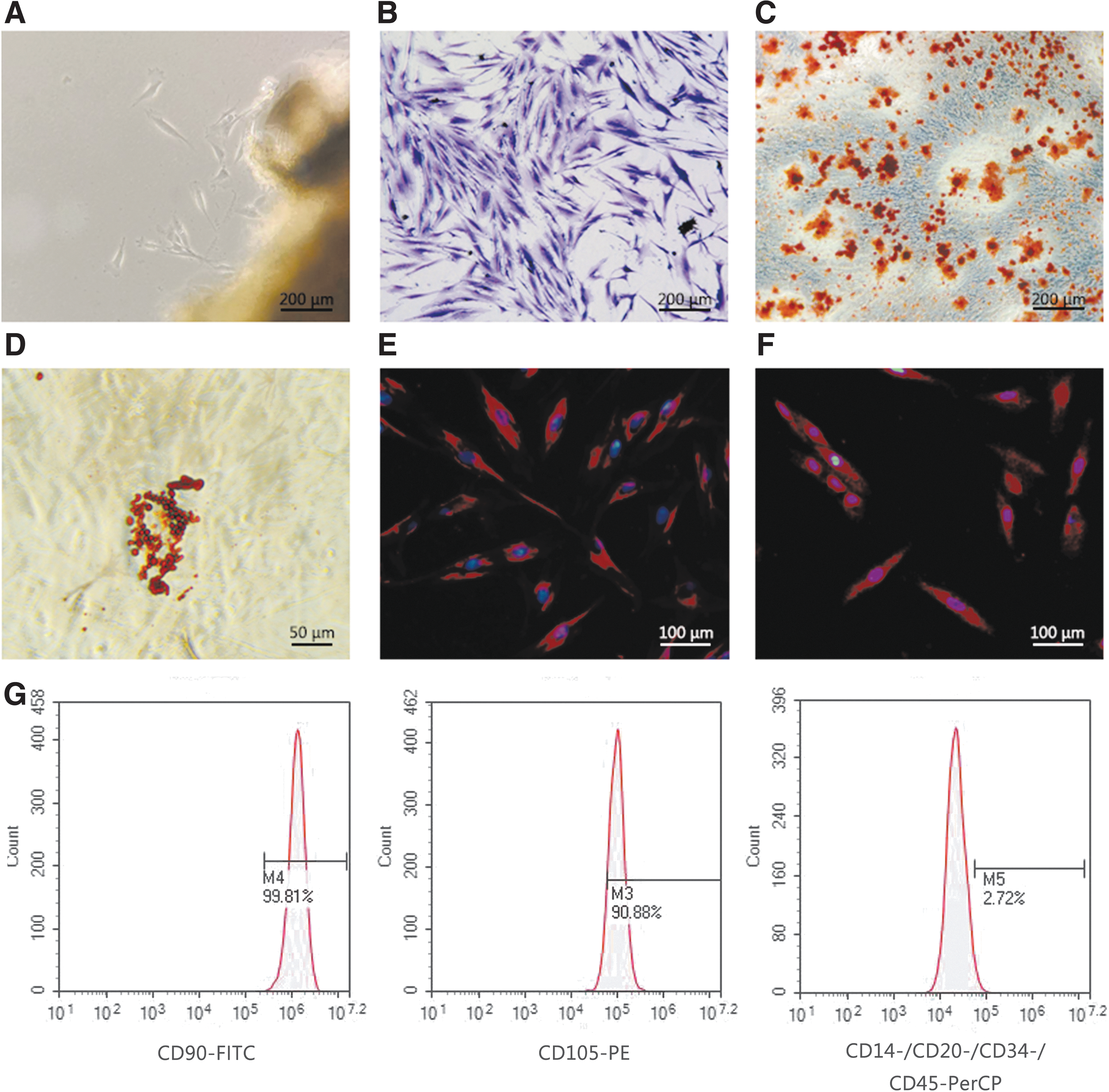
Culture and identification of primary hPDLSCs.
PDGF-BB overexpression in hPDSLCs
Next, we assessed the efficiency of lentiviral transduction in hPDLSCs through assessing these cells for GFP expression 4 days post-transduction, revealing >90% GFP positivity in both negative control and PDGF-BB-overexpressing cells, which was considered to be an acceptable level of transduction efficiency (Fig. 2A, B).
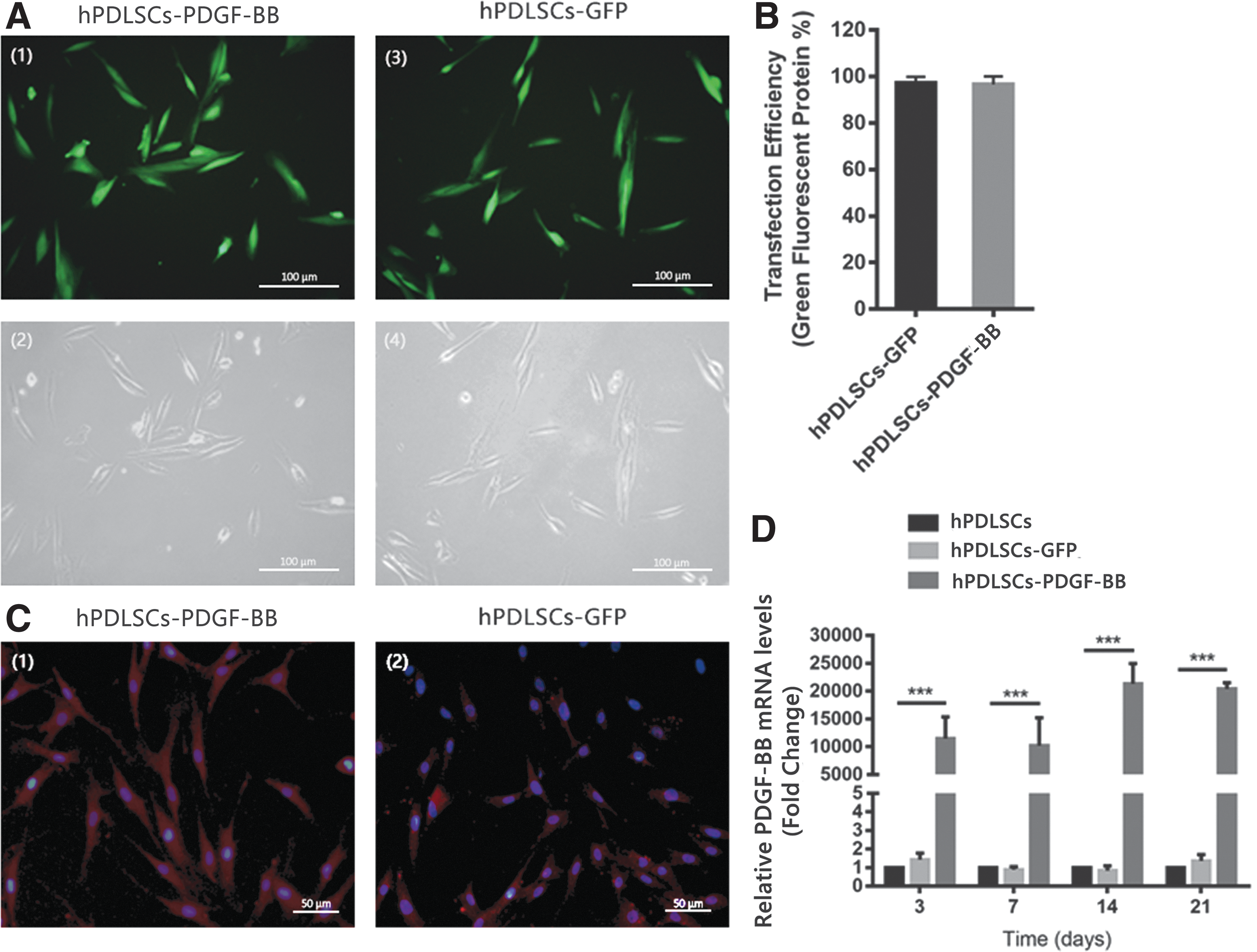
Lentiviral transduction of hPDLSCs.
Immunofluorescent staining further confirmed significant PDGF-BB upregulation in hPDLSCs after transduction (Fig. 2C), with clear evidence of such overexpression at days 3, 7, 14, and 21 after transfection (P < 0.001) (Fig. 2D). Together, these findings thus clearly confirmed that hPDLSCs could be effectively engineered to overexpress PDGF-BB in a stable manner.
PDGF-BB promotes hPDLSC proliferation and osteogenic differentiation
A CCK-8 assay was next used to explore how PDGF-BB affected hPDLSCs proliferation, revealing a significant increase in hPDLSCs proliferation after 3 days for cells in the hPDLSCs-PDGF-BB group (Fig. 3A) relative to the other two groups (P < 0.05), indicating that PDGF-BB enhances hPDLSCs proliferation.

PDGF-BB affected hPDLSCs in vitro.
Subsequently, we used Alizarin Red staining as a means of assessing hPDLSCs osteogenic differentiation after a 21 day culture period. We observed high levels of calcium deposition in all three groups; however, we found using a semiquantitative analysis that the hPDLSCs-PDGF-BB group exhibited the highest number of positive regions (P < 0.05) (Fig. 3B, C).
To explore the influence of PDGF-BB on different markers of osteogenic differentiation of hPDLSCs, we explored the expression of OPN, OSX, COL-1, and BMP2 at the mRNA level. We observed significantly increased expression of OPN and BMP2 in cells of the hPDLSCs-PDGF-BB group on day 7 of osteogenic differentiation (Fig. 4). At later stages of osteogenic differentiation, we also observed significantly increased expression of COL-1 in the hPDLSCs-PDGF-BB group relative to the other two groups (Fig. 4A–D). We further measured VEGF expression, revealing increased expression of this gene in cells overexpressing PDGF-BB at both early and late osteogenic differentiation time points (Fig. 4E). We also assessed levels of osteogenic proteins through western blotting (Fig. 4F–I), revealing OPN and COL-1 protein levels to be consistent with mRNA levels (Fig. 4G–I). With respect to OSX, we found that at the mRNA level its expression in hPDLSCs-PDGF-BB group cells was elevated relative to other groups on day 3, whereas its protein levels were elevated relative to other groups on day 21 (Fig. 4H).

PDGF-BB affects hPDLSC osteogenic gene and protein expression.
Thermosensitive hydrogel properties
In this study, we successfully prepared a thermosensitive hydrogel. The molecular, micro-, and macrostructure of this hydrogel is shown in Fig. 5. The sol–gel transition temperature slightly decreased as polymer concentrations were increased, whereas the gel–sol transition temperature rose substantially as polymer concentrations rose. For the 20wt% copolymer solution, the sol–gel transition temperature was 34.33°C ± 0.57°C, which was nearest to 37°C of tested solutions, and we also found this solution to exhibit good fluidity at room temperature making it ideal for injection purposes (Fig. 5B). The storage modulus (G’), loss modulus (G”), and viscosity (η) of the polymer rose sharply at 32°C, with an intersection at ∼33°C (Fig. 5C, D). These results were consistent with the sol–gel transition temperature for this polymer as measured through the tube inversion method.
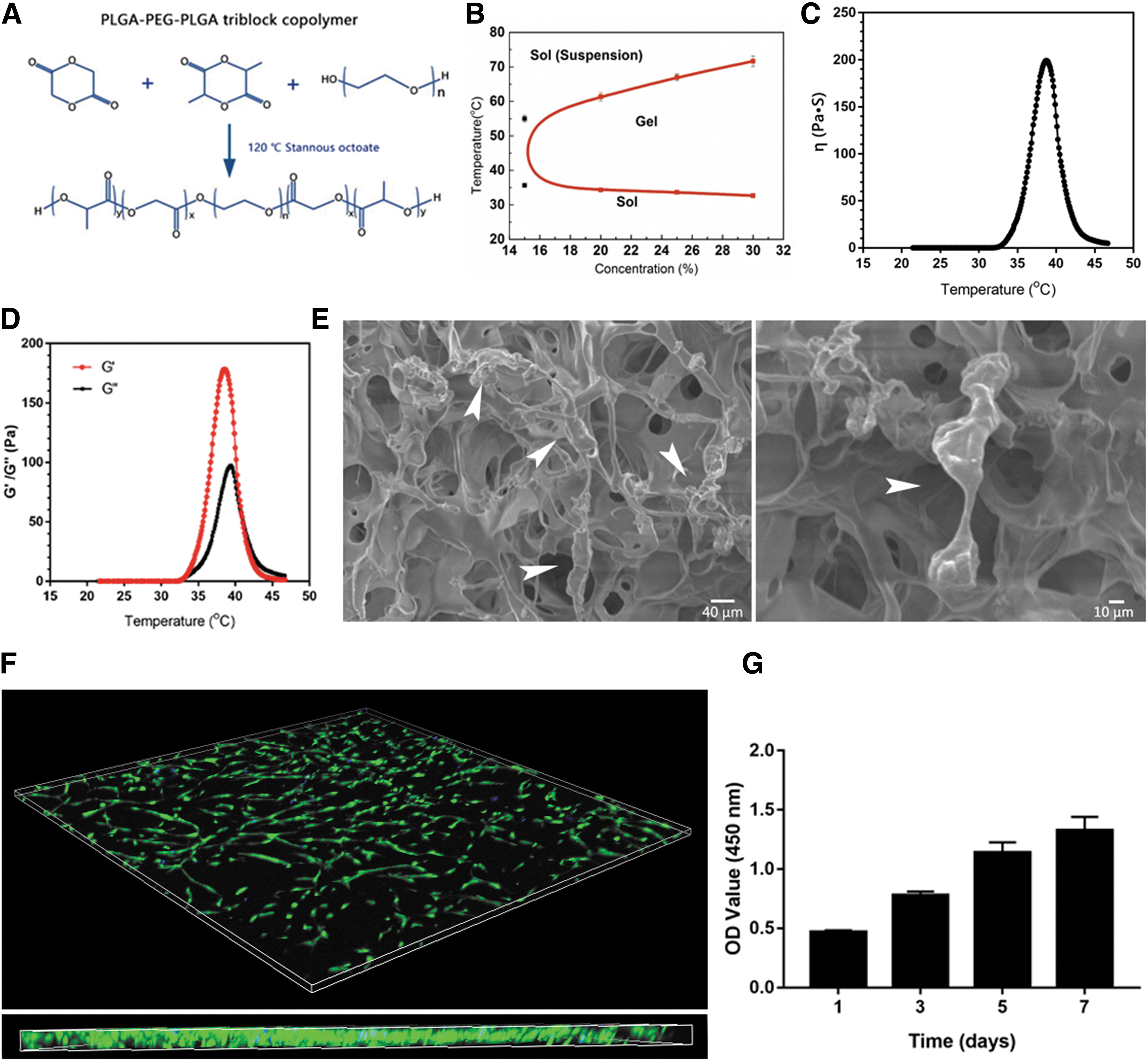
Thermosensitive hydrogel properties and cell 3D culture.
We found that the resultant hydrogel scaffolds exhibited uniform pores that were 100 μm in diameter, and SEM imaging confirmed tight adherence of hPDLSCs to this hydrogel scaffold (Fig. 5E).
Live/dead staining
Next, live/dead staining was conducted to assess cell viability within the hydrogel matrix, revealing the majority of cells to be alive when examined on day 7 of culture through confocal microscopy (Fig. 5F). Through assessment of cell proliferation, we similarly found that this thermosensitive hydrogel was compatible with cell growth (Fig. 5G).
Micro-CT analysis
New bone development in each group was assessed based on micro-CT imaging results. Control untreated animals still exhibited significant alveolar bone defects at 4 and 8 weeks postwounding. Animals treated with hydrogel scaffolds exhibited significantly smaller alveolar bone defects compared with control animals, with animals administered hydrogel containing hPDLSCs-PDGF-BB cells exhibiting the most robust bone regeneration (Fig. 6A). We used the BV and bone ratio (BV/TV) parameters to assess bone formation in these animals (Fig. 6B), revealing that 31.9% ± 5.19%, 37.84% ± 2.19%, 55.99% ± 7.79%, and 62.76% ± 7.59% bone regeneration had occurred in the control, hydrogel, hydrogel+hPDLSCs-GFP, and hydrogel+hPDLSCs-PDGF-BB groups, respectively, at 4 weeks postoperation. At 8 weeks postoperation, the bone regeneration values in these groups were 37.59% ± 3.82%, 49.59% ± 7.56%, 66.73% ± 6.07%, and 78.51% ± 5.46%, respectively. There was thus a clear increase in bone formation in treatment groups relative to control animals.
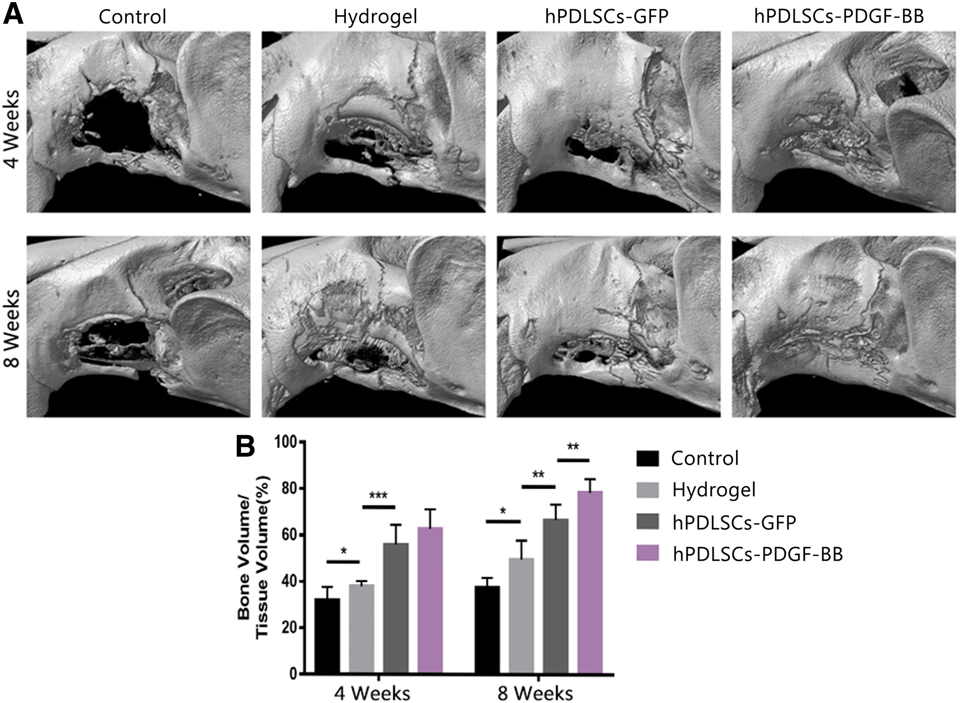
Micro-CT reconstruction of alveolar bone defects.
Histological analysis
After micro-CT analyses, we conducted H&E-based histological examinations of these tissue sections, revealing limited new bone formation in control animals (Fig. 7A). In contrast, hydrogel group animals exhibited increased new bone formation, and the hPDLSCs-PDGF-BB group animals demonstrated the highest levels of new bone formation. We also assessed collagen content and bone maturity in the defect area in treated animals through Masson's trichrome staining. This approach revealed high levels of fibrous tissue but very little bone in control animals, whereas the hydrogel+hPDLSCs-GFP and hydrogel+hPDLSCs-PDGF-BB groups exhibited significantly more new bone formation, thus confirming micro-CT results (Fig. 7A).
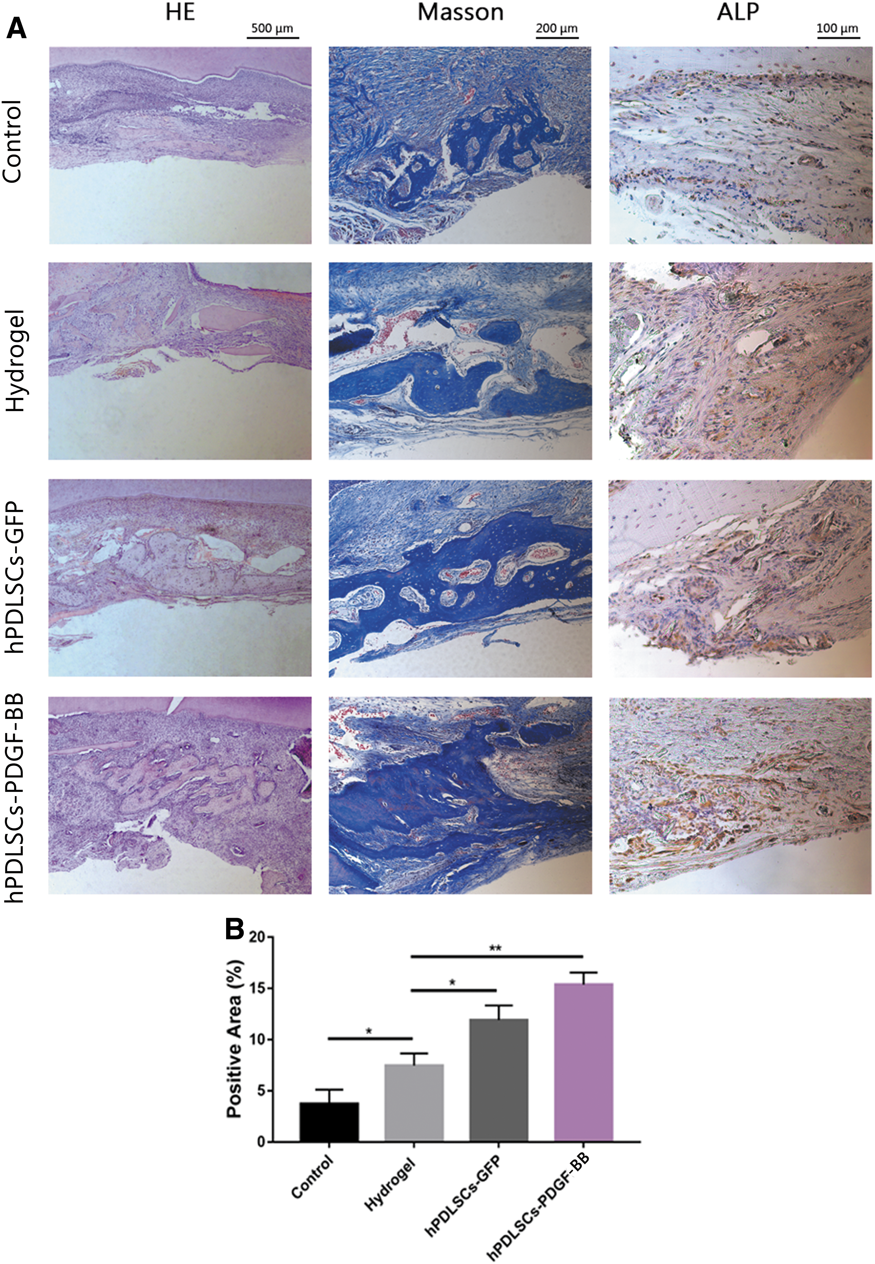
Assessment of alveolar bone repair at 8 weeks.
To further evaluate osteogenesis, we analyzed alkaline phosphatase (ALP) expression through IHC to better explore bone formation and mineralization (Fig. 7A). We observed the lowest frequency of positive (brown) region in control group samples, whereas hPDLSCs-PDGF-BB group samples exhibited the highest rate of positive staining, confirming that these animals exhibited higher rates of osteogenesis than those in other groups, as confirmed by quantitative analyses of these results (Fig. 7B). Together, these results thus suggest that hydrogels offer an ideal physiological microenvironment well suited to stimulating the adhesion and differentiation of hPDLSCs, allowing them to enhance bone repair, particularly when they overexpress PDGF-BB.
Discussion
This study sought to identify an effective and practical approach to achieving in vivo regeneration of alveolar bone defects through the use of a stem-cell-based tissue engineering approach. We opted to use hPDLSCs, as they have been widely studied and found to be ideal for periodontal regeneration approaches [29]. Indeed, hPDLSCs reportedly exhibit superior proliferation and osteogenic activity relative to BMSCs [7], and are capable of differentiating into alveolar bone tissue, making them ideal for use in this study. However, previous reports have shown that on their own PDLSCs are not sufficient to mediate the development of bone tissue in vivo [30]. To overcome such limitations, we therefore assessed the potential for the overexpression of the PDGF-BB growth factor as a means of enhancing alveolar bone regeneration. Indeed, PDGF-BB has been shown to possess potent mitogenic activity [9], and MSCs that are modified to overexpress PDGF-BB have been shown to exhibit enhanced osteogenic differentiation [12 –15]. PDGF-BB is further known to increase the expression of stem cell markers on cells, and to enhance BMSC osteogenic and angiogenic potential to orchestrate bone regeneration [12 –15]. When overexpressed, PDGF-BB can enhance trabecular bone formation and trabecular connectivity, and can drive MSCs toward an osteogenic differentiation pathway [31].The specific impact of PDGF-BB on hPDLSC osteogenic differentiation, however, is not well understood. Previous studies have sought to apply recombinant PDGF-BB as a means of mediating bone or periodontal tissue repair [8,16]. When administered in a recombinant form, however, PDGF-BB is rapidly degraded and cannot be expressed in a sustained manner in vivo [13]. To overcome this issue, we therefore used a lentiviral vector to stably overexpress PDGF-BB, as such vectors are commonly used for similar gene expression approaches [18]. We were able to achieve significant and sustained PDGF-BB overexpression after lentiviral transduction at the RNA and protein level (Fig. 2). We then analyzed the impact of PDGF-BB overexpression on hPDLSC proliferation and osteogenic differentiation, revealing that it both enhanced the proliferative growth of these cells and enhanced their ability to mediate ossification as assessed through Alizarin Red staining, suggesting that these engineered cells may be ideal for bone regeneration approaches (Fig. 3). We further sought to confirm these findings through measuring the expression of the preosteoblast and osteoblast markers Osx and Col1α1, respectively, at the mRNA and protein levels [32 –34]. We found that PDGF-BB overexpression was associated with a significant upregulation of both of these genes, indicating that PDGF-BB facilitates hPDLSC osteoblastic differentiation. Consistent with this, we observed a significant increase in the expression of OPN, which is essential for osteoblast mineralization [33,35], after PDGF-BB overexpression, potentially explaining the observed enhancement of calcium deposition in Alizarin Red staining assays. Together, these results thus confirmed that PDGF-BB was able to promote hPDLSC osteogenic differentiation. Vascular endothelial growth factor (VEGF) is known to be vital both for angiogenesis and for ossification during the process of bone tissue regeneration [36], with both VEGF and PDGF-BB being important regulators of these processes [37]. We found that PDGF-BB overexpression in hPDLSCs was also associated with enhanced VEGF expression in these cells, thus potentially further enhancing the ability of these cells to mediate bone tissue regeneration.
To achieve effective bone regeneration, it is essential that suitable scaffolds be used. Injectable hydrogels are routinely used for such approaches, as they are less invasive than many other procedures. Thermosensitive hydrogels in particular are ideal for repairing alveolar bone defects, as they can be injected as a liquid before safely solidifying at 37°, thereby filling in the defect region [22,38]. As shown in Fig. 5, we were able to produce a PLGA-PEG-PLGA hydrogel that could transition from a gel to solid state as the ambient temperature changed. We further used Live/Dead staining to confirm that hPDLSCs were able to survive with high viability and to proliferate within these hydrogels in vitro, and we also confirmed that they were able to effectively attach to the surface of this scaffolding material through microscopic examination. We then used a rat model of alveolar bone defects [25 –27] to examine the ability of PDGF-BB-hPDLSCs encapsulated in a thermosensitive hydrogel to achieve alveolar bone restoration. We left rats either untreated, administered hydrogel alone, hydrogel+hPDLSCs, or hydrogel+PDGF-BB-hPDLSCs. In subsequent micro-CT analyses, we observed significant differences in bone repair at 4 and 8 weeks postoperation in the four different groups, with bone regeneration in the hydrogel+PDGF-BB-hPDLSCs being superior to that in the other three groups (Fig. 6), particularly in comparison with animals in the hydrogel + hPDLSC group. Consistent with this, BV/TV ratios differed among groups, with those in hydrogel+PDGF-BB-hPDLSC group being higher than those in the other treatment groups (P < 0.05). It is important to note that animals in the hydrogel group exhibited better osteogenic repair than did plank control animals. Hydrogels are capable of filling bone defects in a gel state, thus providing a 3D scaffold that is suitable for cell growth and proliferation, thereby potentially supporting stem cell recruitment and resultant bone tissue repair. It has also been shown that a PLGA-PEG-PLGA hydrogel alone can facilitate bone marrow cavity and fibrocartilage formation within bone defects [39]. PLGA/PEG hydrogels have also been shown to exhibit osteoconductive characteristics that make them ideal for bone tissue engineering [40]. H&E and Masson's trichrome staining further confirmed that the hydrogel + PDGF-BB-hPDLSC group exhibited the highest rates of mature new bone formation in the defect area at 8 weeks postoperation. An IHC analysis of ALP, which is an enzyme essential for bone formation and mineralization and a marker of osteoblast differentiation [41], revealed ALP positivity as highest in the hydrogel+PDGF-BB-hPDLSCs group (Fig. 7). These results thus demonstrate that hPDLSCs are ideal mediators of alveolar bone regeneration, and that overexpression of PDGF-BB can markedly enhance their regenerative potential. Together, our results show that PDGF-BB not only promotes hPDLSC proliferation but also facilitates hPDLSC osteogenic differentiation. PDGF-BB is thus a powerful growth factor capable of enhancing hPDLSC-mediated alveolar bone regeneration.
Conclusion
In conclusion, our results demonstrate that hPDLSCs, which have been modified to overexpress PDGF-BB, can continuously secrete this growth factor, and this overexpression can in turn enhance the proliferation, angiogenesis, and osteogenic differentiation of these hPDLSCs. We further found that a thermosensitive PLGA-PEG-PLGA hydrogel represents an optimal carrier for these hPDLSCs, and we determined that these cells are capable of mediating enhanced alveolar bone regeneration in vivo in a manner directly linked to their overexpression of PDGF-BB. These findings thus offer a source of reference for future studies exploring the efficacy of gene expression-based therapeutic approaches to alveolar bone engineering, and provide a basis for efforts to improve seeded cell survival in recipient tissues.
Footnotes
Author Disclosure Statement
No competing financial interest exists.
Funding Information
This work was supported by the National Natural Science Foundation of China (81470768), Shanghai Science and Technology Innovation Fund (19ZR1445500), Project of Shanghai Municipal Health Commission (201840148), and Shanghai Sailing program (17YF1416500).