




Abstract
Both bone marrow-derived mesenchymal stem cells (BMDMSCs) and extracorporeal shockwave (ESW) have shown promise for enhancing fracture repair. If exposure of BMDMSCs to ESW enhances osteogenic differentiation, these therapies may be combined in vivo or used as a method for preconditioning BMDMSCs. The objective of this study was to determine the effect of ESW on the osteogenic ability of equine BMDMSCs. We hypothesized that ESW would promote osteogenesis evidenced by increased gene expression, alkaline phosphatase (ALPL) expression, slide morphologic score, and protein expression. BMDMSCs were evaluated from six horses. BMDMSCs were culture expanded to passage 3, dissociated, then placed in conical tubes. Treatment cells (“shocked”) were exposed to 500 pulses at 0.16 mJ/mm2 energy. Cells were then reseeded and grown in either growth medium or osteogenic medium. Cellular proliferation and trilineage potential were determined. Cellular morphology was scored and cells were harvested at 1, 3, 7, 14, and 21 days for rtPCR gene expression of osteogenic markers [osteonectin (ONT), osteocalcin (OCN), ALPL, collagen type 3 (COL3), and runt-related transcription factor 2 (RUNX2)]. Media supernatants were evaluated for secretion of BMP-2, VEGF, TGFβ, and PGE2 and cellular lysates were evaluated for ALPL production. There was no difference between the proliferative ability of shocked cells versus unshocked cells in either growth medium or osteogenic medium. ALPL production was greater in shocked cells maintained in osteogenic medium versus unshocked cells in osteogenic medium at day 3 (P < 0.005). Independent of media type, ESW caused a decrease in VEGF and TGFβ production at day 3. No significant increases in gene expression were identified by rtPCR. Exposure of BMDMSCs to ESW does not result in negative effects. An initial significant increase in ALPL was detected but no persistent osteogenic effect was observed with cell expansion.
Introduction
Extracorporeal shockwave (ESW) has shown promise as a noninvasive adjunct therapy to human fracture repair [1,2]. The widespread availability and usage of ESW in horses makes the equine athlete a particularly useful resource for investigation of ESW therapy. In horses, ESW therapy has been investigated and utilized in tendonitis [3,4], desmitis [5], and as a pain-modifying treatment in arthritis [6 –8]. Although ESW appears to be capable of effecting matrix structure in tendon explants and transiently increasing protein and GAG synthesis of equine tenocytes [3,4,9], ESW has not been investigated as a mechanism for bone healing in the horse. Due to the promising osteogenic effects reported regarding ESW in the rat, rabbit, dog, and humans, applicability in the horse should be investigated [10 –13].
Like ESW, bone marrow-derived mesenchymal stem cells (BMDMSCs) have been successful as a treatment of delayed union in people, both children and adults [14,15]. BMDMSCs have been used successfully for musculoskeletal disease in the horse and have been investigated as an adjunct therapy to fracture repair [16,17]. Studies suggest that ESW may enhance osteogenic differentiation in human BMDMSCs by membrane hyperpolarization and Ras activation leading to increased alkaline phosphatase (ALPL) production and osteogenic gene expression [18 –20]. Equine fracture repair remains challenging and techniques to accelerate fracture healing are needed. Long bone fractures challenge implant strength, surgeons are unable to restrict weight-bearing, and minimal soft tissue in the distal limb creates an environment of decreased vascular supply. In equine fracture repair, life-threatening complications are common and include osteomyelitis, surgical site infection, and support limb laminitis [21 –24]. Combination therapies in equine fracture repair such as BMDMSCs and ESW may be used to increase fracture stability and accelerate healing. In addition, investigation of combination therapies may provide the basis for future treatment in humans.
The objective of this study was to determine if ESW promotes osteogenic differentiation of equine BMDMSCs in vitro. We hypothesized that exposure of BMDMSCs to ESW would result in increased osteogenesis as measured by ALPL protein expression, rtPCR of osteogenesis-associated genes, growth factor excretion, and cellular morphology and staining.
Materials and Methods
The experimental protocol described complied with the policies of the Institutional Animal Care and Use Committee at Colorado State University (Protocol 12–3483).
Cell harvest and growth
Bone marrow was harvested from six horses as previously described [25]. All horses were of mixed breed and 2–5 years old. Briefly, horses were sedated with 0.01 mg/kg of detomidine and 0.01 mg/kg of butorphanol, the sternum was clipped and aseptically prepared. An 11-gauge Jamshidi was inserted into the sternum and 10 mL of bone marrow was obtained in 5-mL syringes containing heparin. Red blood cells were removed using centrifugation. The nucleated cells were transferred to tissue culture flasks in low-glucose DMEM, 10% fetal bovine serum (FBS), 0.1% 10,000 U/mL of penicillin–streptomycin–amphotericin B (PSA) and 1 mM HEPES. The medium was changed at 24 h and then every 4 days until MSC formed nearly confluent colonies (colonies were visible within 7–10 days). These colonies were dissociated with Accumax™ and reseeded into tissue culture flasks at 1,200–1,500 cells/cm2 in an expansion medium. Expansion medium contained αMEM supplemented with 10% FBS, 10,000 U/mL PSA, 10 mM HEPES, and 2 ng/mL of fibroblast growth factor-2 (FGF). The cells were passaged a total of three times before cryopreservation in 95% FBS and 5% DMSO. Before exposure to ESWT, cells were allowed to recover from cryopreservation in monolayer culture in expansion medium. Cells were grown to 70–80% confluence before dissociation for ESW.
Exposure to ESW
For exposure to ESW, BMDMSCs were dissociated from monolayer culture using Accumax, placed in 5-mL polystyrene tubes, and exposed to 500 pulses at 0.16 mJ/mm2 energy flux density. Control cells were also placed in tubes but not exposed to ESW. Cells were then reseeded at 30,000 cells/cm2 confluency for 24 h. After 24 h, cells were grown in media according to their treatment group. The four treatment groups were: unshocked cells in growth medium, unshocked cells in osteogenic medium, shocked cells in growth medium, and shocked cells in osteogenic medium. Growth medium contained DMEM supplemented with 10% FBS, 10,000 U/mL PSA, 10 mM HEPES, and 2 ng/mL of FGF. Osteogenic medium consisted of DMEM containing 10% FBS, 10,000 U/mL PSA, 10 mM HEPES, 1 nM dexamethasone, 5 mM beta-glycerol phosphate (Sigma-Aldrich, St. Louis, MO), and 170 μM ascorbic acid (Sigma-Aldrich). Cells were then cultured for 21 days in growth or osteogenic medium with medium changes every fourth day.
Assessment of proliferation
The IncuCyte Live Cell Analysis system (Essen Bioscience) was used to evaluate cell proliferation. Each treatment group (unshocked cells in growth medium, unshocked cells in osteogenic medium, shocked cells in growth medium, and shocked cells in osteogenic medium) were seeded into 24-well plates at a concentration of 30,000 cells/cm2 in triplicate, incubated for 24 h, and then transferred to the IncuCyte Live Cell for 96 h. Percent confluence was measured every 3 h for each well. To normalize small variations in initial confluence, increase in percent confluence was calculated compared with baseline (for each well) and expressed as change in percent confluence.
Morphologic assessment
Wells from each treatment group were photographed at 10x using an inverted microscope (Olympus IX70) on days 1, 3, 7, and 14. Three representative fields were blindly graded for cellular morphology by two individuals (A.C., J.P.) with experience in BMDMSCs culture. Cell morphology grading was adapted from Zachos et al. [26], the grading system used is provided in Table 1. Osteogenic cells appeared smaller than undifferentiated cells and developed a triangular shape with the surrounding extracellular matrix deposition. The highest grades indicate the largest number of osteogenic cells. Grades were averaged between the two individuals.
Cell Morphology Grades
Cell morphology was graded by two independent observers using a scale modified from Zachos et al.[26].
Gene expression
RNA was extracted from samples using an RNeasy Kit (Qiagen, Valencia, CA). RNA was amplified using a one-step SYBR system as per the manufacturer's instructions (Bio-Rad, Hercules, CA). Samples were evaluated at days 1, 3, 7, and 14 for gene expression of Collagen type 3 (Col3A1), ALPL, Osteocalcin (OCN), Osteonectin (OTN), and Run-related transcription factor 2 (RUNX2) (Table 2). All samples were normalized to an 18S housekeeping gene and expressed as relative to the average of the unshocked cells in growth medium for each time point.
rtPCR Primer Sequences for Selected Osteogenic Genes
Protein expression
Supernatants were harvested to determine the secretion of transforming growth factor beta (TGFβ) (R&D Systems, Minneapolis, MN), prostaglandin-E2 (PGE2) (Enzo Life Sciences, Farmingdale, NY), vascular endothelial growth factor (VEGF) (Kingfisher, St. Paul, MN), and bone morphogenic factor-2 (BMP2) (R&D Systems) by commercially available ELISA Kits as per the manufacturer's guidelines. ALPL detection was analyzed from cell lysates using a commercially available kit (Anaspec, Fremont, CA).
Trilineage differentiation
Trilineage differentiation was assessed for both unshocked and shocked cells using exposure to osteogenic, chondrogenic, and adipogenic media. Osteogenic and chondrogenic media were made as previously described [27]. Adipogenic medium was purchased and used according to the manufacturer's recommendations (StemPro™; Thermo Fischer, Waltham, MA). Trilineage differentiation was qualitatively assessed and compared with unstained controls by two observers (A.C., J.P.) both of whom have experience with stem cell culture. Samples were noted to have the presence or absence of staining for each condition. At 21 days, adipogenic cells were identified by dark red lipid staining by Oil Red O demonstrating the presence of fatty aggregates. Osteogenic cells were identified by Alizarin Red stain demonstrating the presence of calcium as well as a change to a cuboidal shape. Chondrogenic cells were identified by uptake of Toluidine Blue demonstrating the presence of proteoglycan-rich extracellular matrix.
Statistical analysis
For assessment of proliferation, percent confluence was evaluated using the IncuCyte Live Cell Analysis system and morphology grading scores were analyzed using a two-way ANOVA with Tukey's multiple comparisons. GraphPad Prism v 7.03 (La Jolla, CA) was used to perform the statistical analysis. Protein expression and PCR results were analyzed using a mixed model ANOVA using animal as a random effect. The mixed model ANOVA allowed for analysis of groups independent of treatment (shocked vs. unshocked) and media type (osteogenic medium vs. growth medium) and the assessment of an interaction term. When a significant interaction term was found, treatment groups were analyzed independently. Pairwise comparisons were analyzed using least squares means with Tukey–Kramer adjustment. Significance was set at P < 0.05. Statistical tests were performed using SAS 9.3 software.
Results
Proliferative ability
There was no difference between the increase in percent confluence between shocked and unshocked cells within the same media type. Both shocked and unshocked cells in osteogenic medium grew at a slower rate than the same cells in growth medium (Fig. 1).
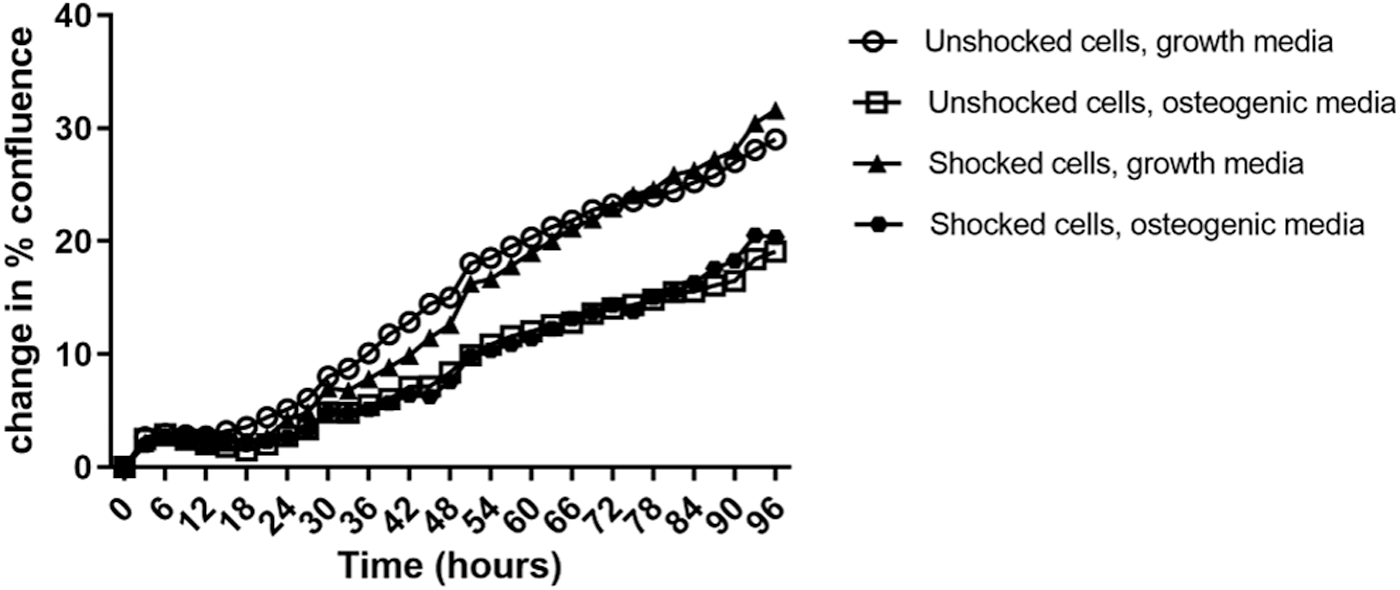
BMDMSCs proliferation. There was no difference in the rate of proliferation of cells exposed to ESW versus unshocked cells. However, both shocked and unshocked cells in osteogenic medium proliferated at a slower rate than the same cells in growth medium. BMDMSC, bone marrow-derived mesenchymal stem cell; ESW, extracorporeal shockwave.
Morphologic assessment
Analysis of morphologic grading scores showed a significant effect of treatment and time (P < 0.0001). The interaction between treatment groups was significant (P < 0.0001). Therefore, treatment groups were evaluated individually using a Tukey multiple comparisons test (Fig. 2).

Morphologic grading. On day 7 and 14, both ESW-exposed BMDMSCs and unshocked BMDMSCs maintained in osteogenic medium had higher morphology scores than the same cells in growth medium. However, there was no difference between ESW-exposed and unshocked BDMSCs when controlling for the media type. Differing letters indicate a statistically significant difference (P < 0.05). Bars indicate the mean.
Osteogenic medium
ESW did not have a significant effect on cell morphology in osteogenic medium at any time point. However; both shocked and unshocked cells in osteogenic medium had a significant increase in morphologic score at day 7 (unshocked: P < 0.0010.0007; shocked: P < 0.0001) and day 14 (unshocked: P < 0.0001; shocked: P < 0.0001) when compared with baseline. At day 7, shocked cells in osteogenic medium had a higher morphologic score than both shocked cells in growth medium (P < 0.0001) and unshocked cells in growth medium (P < 0.0001). At day 14, shocked cells maintained in the osteogenic medium had a higher morphologic score than shocked cells in growth medium (P < 0.0001). Furthermore, unshocked cells in the osteogenic medium had a higher grade than unshocked cells in growth medium (P < 0.0001).
Growth medium
ESW did not have a significant effect on cell morphology in growth medium at any time point.
Protein expression
Effects of osteogenic medium on BMP2, VEGF, PGE2, TGFβ
VEGF, TGFβ, and data are summarized in Fig. 3. Statistical analysis by mixed model ANOVA was used to determine differences independent of treatment (shocked vs. unshocked) and media (osteogenic vs. growth medium). When a significant interaction term was found, groups were analyzed and compared individually. When cells in osteogenic medium are examined independent of ESW, there is a 1.9-fold increase in VEGF (P < 0.0001), and a 1.7-fold increase in PGE2 (P < 0.005) at day 3 within the cellular supernatant. In addition, there is a 2.7-fold increase in VEGF (P < 0.001) at day 7 and a 2.1-fold increase in VEGF at day 14 (P < 0.05) and a 4.3-fold increase in PGE2 at day 14 (P < 0.05). There was no change in BMP2 or TGFβ when osteogenic medium was examined independent of ESW.
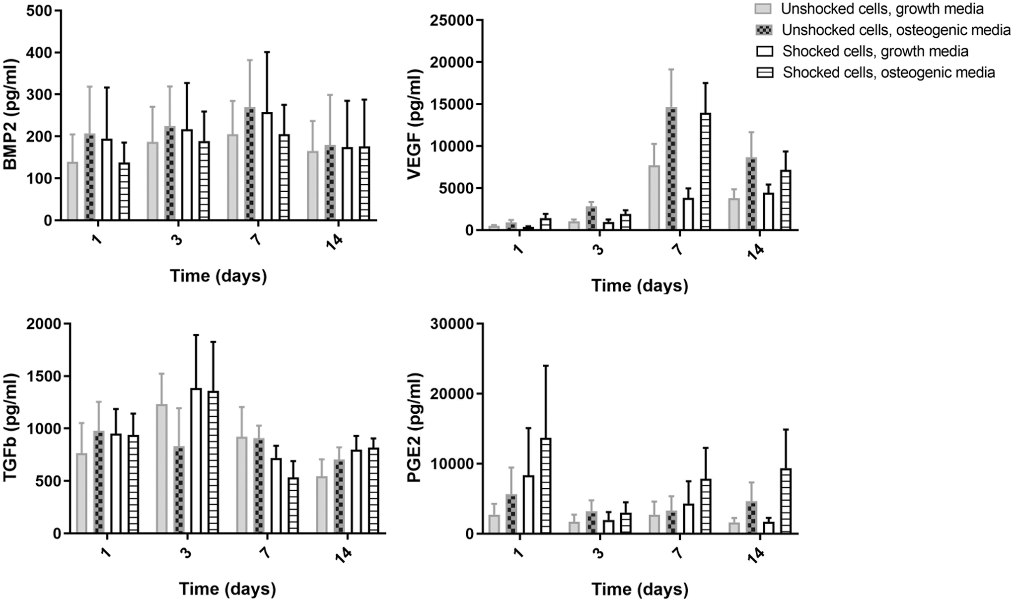
Protein expression of BMP2, VEGF, TGFβ, and PGE2. No interaction terms were found to be significant, therefore individual treatment groups are not labeled as significant. However, independent of the type of media used, ESW resulted in a statistically significant decrease in VEGF at day 3 and TGFβ at day 1 and 3. In contrast, time in culture for both unshocked cells in osteogenic medium (P < 0.005) and growth medium (P < 0.0005) resulted in an increase in VEGF compared with baseline. On day 3, osteogenic medium independent of ESW resulted in an increase in PGE2, ESW did not further increase PGE2 in our study. Bars indicate the mean + standard error of the mean. BMP2, bone morphogenic factor-2; VEGF, vascular endothelial growth factor; TGFβ, transforming growth factor beta; PGE2, prostaglandin-E2.
Effects of ESW on BMP2, VEGF, PGE2, TGFβ
Independent of the type of medium used, ESW resulted in 1.2-fold decrease in VEGF at day 3 (P < 0.05), and 1.7-fold decrease of TGFβ at day 1. In contrast, time in culture for unshocked cells in osteogenic medium (P < 0.005) or growth medium (P < 0.0005) resulted in an increase in VEGF. There was no change in PGE2 or BMP2 when ESW was evaluated independent of culture medium.
Intracellular ALPL
Independent of culture medium, ESW increased ALPL 1.4-fold on day 3 (P < 0.05). Independent of treatment group (shocked vs. unshocked), osteogenic medium resulted in a 1.5-fold increase in ALPL expression at day 3 (P < 0.05). Furthermore, ALPL expression resulted in a significant interaction term allowing groups to be evaluated independently on day 3 (Fig. 4). When treatment groups were evaluated independently at day 3, shocked cells in osteogenic medium had a statistically significant increase in ALPL compared with all other groups, including unshocked cells in osteogenic medium (P < 0.005).

Alkaline phosphatase from cellular lysates. A significant interaction was found for alkaline phosphatase at day 3. Therefore, treatment groups were evaluated independently. At day 3, shocked cells in osteogenic medium had a statistically significant increase in alkaline phosphatase compared with unshocked cells in growth medium (P < 0.005), shocked cells in growth medium (P < 0.005), and unshocked cells in osteogenic medium (P < 0.005). Differing letters indicate a statistically significant difference (P < 0.05). Bars indicate the mean + standard error of the mean.
rtPCR of ONT, Col3A1, RUNX2
Gene expression was analyzed on days 1, 3, 7, and 14 and summarized in Fig. 5. OCN not reported as expression was below the limit of detection of the assay. No significant differences were found in gene expression of ONT, Col3A1, RUNX2, or ALPL in any of the treatment groups. There was a small but statistically significant decrease in RUNX2 at day 14 in cells kept in osteogenic medium (independent of ESW) (P < 0.05).
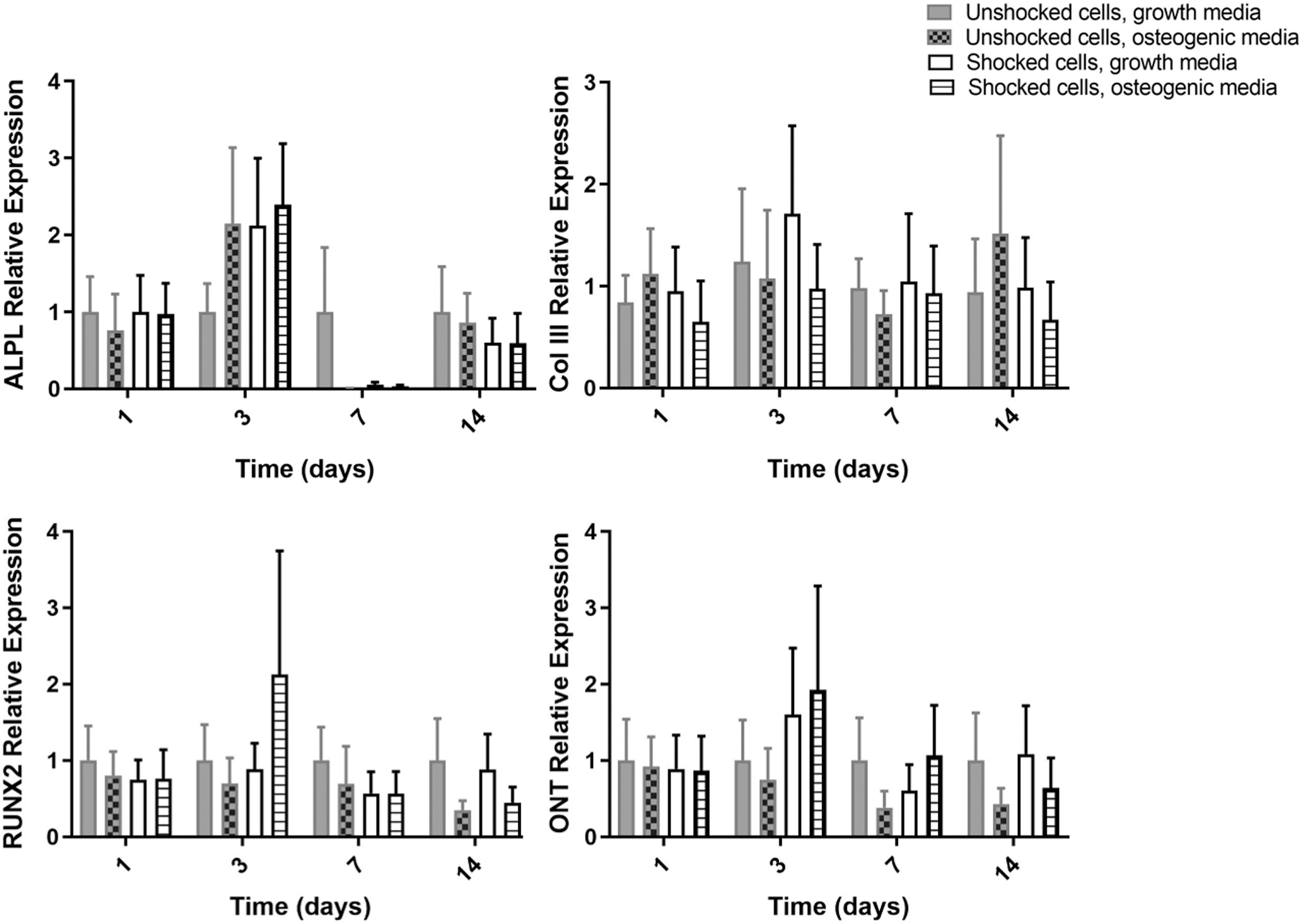
rtPCR. No significant interaction terms were found. Therefore, treatment groups could not be evaluated individually. No significant differences were identified in gene expression at day 1, 3, or 7. A small decrease in RUNX2 was identified in cells maintained in osteogenic medium independent of ESW exposure (P < 0.05). Bars indicate the mean + standard error of the mean.
rtPCR of ALPL
Unshocked cells in osteogenic medium had an increase in ALPL expression overtime during the culture period (P = 0.05) and unshocked cells in growth medium did not, indicating the osteogenic medium was effective in inducing osteogenesis. Independent of culture medium, ESW resulted in a 1.5-fold increase in ALPL expression at day 3, which trended toward significance (P = 0.1).
Trilineage differentiation
Unshocked BMDMSCs from all horses were able to undergo trilineage differentiation. ESW did not suppress the ability of BMDMSCs to undergo trilineage differentiation (Supplementary Fig. S1). No qualitative differences were noted in the ability of shocked cells versus unshocked cells to proceed through osteogenesis, chondrogenesis, and adipogenesis. Alizarin Red staining showed no suppression of osteogenic differentiation between shocked and unshocked BMDMSCs. However, no qualitative increase in osteogenesis was appreciated.
Discussion
Important findings from this study include that a single dose of ESW results in no significant negative effects on equine BMDMSC's ability to proliferate or differentiate. In addition, alkaline phosphatase protein expression increased 1.4-fold at 3 days following ESW. The increase in ALPL was transient and of less magnitude than reported in previous studies of human MSCs exposed to ESW [19], indicating a modest, transient, osteogenic effect, which was not upheld as cells divided in culture.
In people, ESW has proven efficacious in stimulating fracture repair when a nonunion is present [2,28]. Furthermore, studies examining the effect of ESW on human BMDMSCs suggest that ESW promotes osteogenesis of BMDMSCs [20,29]. Equine fracture repair remains challenging and the horse represents a valuable model for translational therapies [21,22]. Combination therapy of ESW and BMDMSCs would be a potentially attractive treatment to promote fracture healing.
In this study, ESW had no detrimental effects on proliferative ability or trilineage differentiation. No difference was detected in the morphology between shocked and unshocked cells in both osteogenic and growth medium. The results suggest that ESW may be used in combination with BMDMSCs without negative effects to the cells. This is consistent with previous studies of human MSCs exposed to ESW which found proliferation to be unaffected if 1,000 shocks or less were administered [19].
ESW did not widely affect the expression of genes associated with osteogenesis measured in this study. On day 14, osteogenic medium independent of ESW resulted in a statistically significant decrease in Runx2. However, the 2.4-fold decrease in relative expression late in the culture process was not reflected in morphology or protein expression and is not anticipated to cause a significant difference in osteogenic phenotype. The decrease in Runx2 expression contrasts with a previous study of equine BMDMSCs, which reports a 2.4-fold increase in Runx2 when cells were maintained in osteogenic medium [30]. However, in vitro osteogenesis assays of human BMDMSCs demonstrate small decreases in Runx2 expression at 7 days and only slight increases at day 14 and 21 [31]. Due to the variability in Runx2 expression, a recent study has suggested the ratio of Runx2/Sox9 as a better predictor of osteogenic potential [31]. Sox9 expression was not measured in this study and should be considered in future studies. Prior studies of human MSCs exposed to ESW resulted in increased expression of osteogenic genes, including ALPL, collagen type I, and OCN [19]. The current study resulted in no increase in collagen type I or OCN. In fact, OCN was below the detectable limit for all conditions. When evaluating the unshocked cells in osteogenic medium overtime there was an increase in ALPL expression demonstrating a successful osteogenic induction protocol. Although not statistically significant, ESW exposure resulted in a trend toward an increase in ALPL gene expression at Day 3 (P = 0.1) as did osteogenic medium independent of ESW exposure (P = 0.08). A trend in the increase in ALPL gene expression with no other increases in osteogenic gene expression may suggest a modest, transient, increase in osteogenesis as a result of ESW exposure but indicates there was no substantial, persistent, osteogenic effect at the dose of ESW used in this study.
Previous studies have reported an increase in osteogenic gene expression by human BMDMSCs exposed to ESW [20,29]. The contrast in results may be explained by species-specific responses to osteogenic stimuli [30]. The ESW dose was chosen from a prior publication where human BMDMSCs were exposed to ESW using a similar procedure of dissociation and exposure [20]. Clinically, this energy level coincides with commonly used settings for equine musculoskeletal treatment. A differing energy level or number of shocks may have yielded different results. However, a previous study in human BMDMSCs demonstrated that proliferation is negatively affected by doses of greater than 1000 impulses and determined the optimal in vitro dose of ESW for osteogenesis to be 500 impulses at the same energy level used in this study [19]. Concerns have been raised over measuring the effects of ESW on cultured cells with culture conditions having a potential effect on experimental effects [32]. Certainly, the limitation of in vitro studies must be recognized when interpreting results. However, in this study, the procedure of dissociation, ESW dose, and exposure mirrored previous studies to allow a reasonable comparison with prior literature [20].
Although ESW did not have substantial effects on osteogenic gene expression, growth factor and ALPL expression was affected. ESW exposure resulted in a significant decrease in VEGF at day 3. Previous reports have found osteogenic conditioned medium from equine BMDMSCs to be antiangiogenic [33]. Therefore, the transient decrease in VEGF may be suggestive of a transient osteogenic effect of ESW exposure. In agreement, on the same day (day 3), ALPL from cell lysates were increased. Increase in ALPL has been identified as predictive of in vivo osteogenic performance [34]. Although the increase only occurred on a single day for the ESW-exposed cells this could indicate a short-lived propensity for osteogenesis as a result of ESW exposure.
In previous studies, equine BMDMSCs did not produce measurable levels of PGE2 without stimulation [35]. In contrast, in this study, BMDMSCs produced low levels of PGE2 in culture. Although osteogenic medium independent of ESW resulted in an increase in PGE2, ESW did not further increase PGE2 excretion. In human BMDMSCs, PGE2 has been linked to increased osteogenesis as a result of ESW exposure [20]. The lack of increase in PGE2 as a result of ESW in our study is further evidence of a lack of a persistent, significant biological effect when equine BMDMSC are exposed to ESW.
Changes in gene and protein expression occurred at a single time point, 3 days following ESW exposure, but did not carry through to the 1-week and 2-week time points. Only a single ESW exposure was performed in this study. Multiple ESW exposures may have yielded different results. However, a single dose of ESW is common in both in vitro and in vivo studies [20,29]. In human studies treating fracture nonunion, a single administration of ESW is most often performed [2,28,36]. Furthermore, previous in vitro studies have found an effect from a single administration of ESW [19]. Therefore, although possible, it seems unlikely that multiple doses would lead to an increased effect.
When evaluating sources of equine MSCs, osteogenic differentiation was found to be the most prominent in BMDMSCs when compared with umbilical, tendon, and adipose-derived MSCs [37]. Evidence of equine BMDMSCs propensity for osteogenesis has also been demonstrated within in vivo studies; equine BMDMSCs when used with autologous platelet-enriched fibrin resulted in ectopic bone formation within the repair tissue [38]. The characteristic of equine BMDMSCs to be largely osteogenic may explain the lack of an additional response to ESW. In addition, BMDMSCs are a heterogeneous population, which have varying degrees of osteogenesis [39]. ESW stimulation of BMDMSCs may be dependent on the subpopulations of cells present.
In addition to BMDMSCs cellular characteristics, effects of in vitro culture must be considered. BMDMSCs proliferate rapidly in vitro. In this study, confluence doubled in 96 h. If the effects of ESW on MSCs is not transferred to the progeny of dividing cells, in vitro proliferation may cause dilution of the treated cells within the sample and subsequent dilution of the treatment effect. Despite the potential dilution, previous studies in human BMDMSCs have still exhibited a treatment effect [19].
In conclusion, a single ESW exposure had no detrimental effects on proliferation, trilineage capacity, or morphology of BMDMSCs. A significant but short-lived increase in ALPL protein expression and a trend toward increased ALPL gene expression was observed at day 3. However, no sustained effects on gene or protein expression lasted beyond day 3. In conclusion, the study suggests that a single administration of ESW at the dose used in this study does not negatively affect BMDMSCs and causes a significant increase in osteogenesis as evidenced by ALPL that is not sustained with cell division in vitro. These findings suggest that, clinically, it appears that ESW may be used concurrently with BMDMSCs without negative effects but higher frequency or ESW intensity may be needed for sustained osteogenic effects.
Footnotes
Acknowledgment
The authors would like to acknowledge Pulse Veterinary Technologies, LLC. for donation of the shockwave trode used in this study.
Author Disclosure Statement
L.G. and J.K. are shareholders in Advanced Regenerative Therapies. A.C. and J.P. have no disclosures.
Funding Information
This study was funded by the Research Council of the Colorado State University College of Veterinary Medicine and Biomedical Sciences. The shockwave trode used for this study was donated by Pulse Veterinary Technologies, LLC.
Supplementary Material
Supplementary Figure S1
References
Supplementary Material
Please find the following supplemental material available below.
For Open Access articles published under a Creative Commons License, all supplemental material carries the same license as the article it is associated with.
For non-Open Access articles published, all supplemental material carries a non-exclusive license, and permission requests for re-use of supplemental material or any part of supplemental material shall be sent directly to the copyright owner as specified in the copyright notice associated with the article.
