
Abstract
Human umbilical cord mesenchymal stem cells (hUC-MSCs) are a promising alternative source of mesenchymal stem cells (MSCs) that are enormously attractive for clinical use. This study was designed to investigate the effect of recombinant human bone morphogenetic protein-7 (rhBMP-7) and/or osteogenic media (OMD) on bone regeneration of hUC-MSCs seeded on nanohydroxyapatite/collagen/poly(
Introduction
Human umbilical cord mesenchymal stem cells (hUC-MSCs) derived from Wharton's jelly of the umbilical cord have been known as a promising alternative source of mesenchymal stem cells (MSCs) that are enormously attractive for clinical use [1]. hUC-MSCs are the only MSCs that have the characteristics of both prenatal and newborn stem cells [2 –6]. These cells are easily accessible without an additional invasive procedure, which have low cost and inexhaustible cell source. They had a high plasticity, developmental flexibility, and non-tumorigenesis [7,8] and minimal immunorejection [9] in transplantation, and had higher proliferative potential than human bone marrow mesenchymal stem cells (hBM-MSCs) or MSCs from other sources [10 –14], which differentiated into osteoblasts, adipocytes, chondrocytes, neurons, and endothelial cells under specific culture conditions [15 –20].
Although there have been a few studies on the use of hUC-MSCs in bone tissue engineering [20 –23], the osteogenic differentiation of hUC-MSCs in a three-dimensional environment has not been clearly elucidated.
Nanohydroxyapatite/collagen/poly(
Bone morphogenetic protein (BMP) belongs to the transforming growth factor β (TGF-β) superfamily and regulates cell proliferation and differentiation, leading to MSC differentiation into chondrocytes and osteoblasts to form bone [28 –30]. BMP-7, which has been known as an inducer of osteogenic differentiation, was able to individually stimulate osteoblastic (or chondrogenic) phenotypes in all kinds of mesenchymal precursor cell lines [31 –34]. However, whether recombinant human bone morphogenetic protein-7 (rhBMP-7) stimulates osteogenic differentiation of hUC-MSCs seeded on nHAC/PLA has not been demonstrated.
Osteogenic media (OMD), including dexamethasone, ascorbic acid, and β-glycerophosphate, are the gold standard procedure for MSC differentiation along the osteogenic lineage [15,16,23,27]. Previous studies showed that the combined use of OMD and BMP could enhance osteogenic differentiation of human MSCs [35,36], whereas some studies showed that BMP alone could not induce osteogenic differentiation of human MSCs [37 –39]. In this study, we aimed to investigate the effect of the combined use of rhBMP-7 and OMD on the proliferation and osteogenic differentiation of hUC-MSCs seeded on nHAC/PLA, and evaluate the effectiveness of hUC-MSC-based tissue-engineered bone to repair jaw bone defect in New Zealand rabbit.
Materials and Methods
Isolation of hUC-MSCs
hUC-MSCs were isolated using an explant culture method [40,41]. All surgical procedures and care administered to human samples were approved by the Medical Ethics Committee of Chinese People's Liberation Army (PLA) General Hospital and were performed according to institutional guidelines (ethics approval no. S2018-093-01). Simply, the umbilical cord tissues were obtained sterilely from full-term pregnancies (Maternity Department of Chinese PLA General Hospital, Beijing, China). After removal of the vascular, perivascular, and epithelial tissues, the remaining Wharton's Jelly tissue was minced into 1 cm3 small fragments, which were attached to a culture dish bottom and cultured in the serum-free medium (Military Medical Science Academy of the PLA, Beijing, China) at 37°C in 5% CO2. And then, the cells migrated from small fragments. The third passage cells were used to carry out a series of experiments.
Phenotype of hUC-MSCs
The cells were trypsinized and suspended in phosphate-buffered saline (PBS, pH 7.4) at a concentration of 5 × 10⁶ cells/mL, and a 100 μL sample was incubated with various fluorescein isothiocyanate-/phycoerythrin-labeled mouse anti-human antibodies for 45 min at room temperature. Cells were washed twice with PBS, centrifuged, and resuspended in 0.5 mL PBS. Control samples were incubated with PBS instead of antibody. A FACScan machine (Beckman Coulter, Fullerton, CA) was used to analyze antibody binding.
Multilineage differentiation potential of hUC-MSCs
Osteogenic differentiation was induced by OricellTM hUC-MSC osteogenic differentiation medium kit (HUXUC-90021; Cyagen Biosciences, Inc.). The cells were cultured in Oricell hUC-MSC osteogenic differentiation complete medium containing 175 mL hUC-MSC osteogenic differentiation basal medium, 20 mL fetal bovine serum, 2 mL penicillin–streptomycin, 2 mL glutamine, 400 μL ascorbate, 2 mL β-glycerophosphate, and 20 μL dexamethasone for 21 days, and examined for extracellular matrix (ECM) calcification using Alizarin red staining (Cyagen Biosciences, Inc.).
Adipogenic differentiation was induced by Oricell hUC-MSC adipogenic differentiation medium kit (HUXUC-90031; Cyagen Biosciences, Inc.). The cells were cultured in hUC-MSC adipogenic differentiation complete medium A containing 175 mL hUC-MSC adipogenic differentiation basal medium A, 20 mL fetal bovine serum, 2 mL penicillin–streptomycin, 2 mL glutamine, 400 μL insulin, 200 μL 3-isobutyl-1-methylanxthine (IBMX), 200 μL rosiglitazone, and 200 μL dexamethasone for 3 days, the media were then changed to hUC-MSC adipogenic differentiation complete medium B containing 175 mL hUC-MSCs adipogenic differentiation basal medium B, 20 mL fetal bovine serum, 2 mL penicillin–streptomycin, 2 mL glutamine, and 400 μL insulin for 1 day, and the media were then changed to hUC-MSCs adipogenic differentiation complete medium A for 3 days.
A and B took place by turn 3–5 times, and the cells were then induced in hUC-MSC adipogenic differentiation complete medium B for the last 4–7 days. After differentiation for 21 days, the cells demonstrated intracellular lipid accumulation using Oil red O staining.
Chondrogenic differentiation was induced by Oricell hUC-MSC chondrogenic differentiation medium kit (HUXUC-90041; Cyagen Biosciences, Inc.). The cells were counted after routine digestion, and 3–4 × 10⁵ cells were transferred to 15 mL centrifuge tubes, and were centrifuged at 250g for 4 min. The cells were then washed with premixed solution of hUC-MSC chondrogenic differentiation complete medium containing 194 mL hUC-MSC chondrogenic differentiation basal medium, 20 μL dexamethasone, 600 μL ascorbate, 2 mL ITS + supplement, 200 μL sodium pyruvate, and 200 μL proline two times for 5 min each at 150g, and the cells were centrifuged again using 0.5 mL hUC-MSC chondrogenic differentiation complete medium containing 1 mL premixed solution and 10 μL TGF-β3 at 150g for 5 min, but the supernatant was not abandoned.
The centrifugal pipe cover was loosened to facilitate gas exchange and was placed in an incubator at 5% CO2 and 37°C. When the cell cluster is gathered (usually 24 or 48 h), the bottom of the centrifuge tube is flicked away from the bottom of the centrifuge tube, and the spheroids were suspended in the liquid and induced. Culture medium was changed at 2- to 3-day intervals. After induction for 21 days, the cartilage balls were fixed and embedded by paraffin. The section was performed Alicin blue staining.
Seeding of hUC-MSCs and osteogenic differentiation protocol
nHAC/PLA (Beijing Allgens Medical Science and Technology Co., Ltd.) was made into blocks of 3.5 mm × 3.5 mm × 3.5 mm, 5 mm × 5 mm × 5 mm, and 10 mm × 4 mm × 3 mm, which were sterilized by cobalt 60. And then cells were seeded on culture plate and nHAC/PLA and induced toward osteogenic phenotype by OMD, namely Oricell hUC-MSCs osteogenic differentiation medium containing 175 mL hUC-MSC osteogenic differentiation basal medium, 20 mL serum-free medium instead of fetal bovine serum in the kit, 2 mL penicillin–streptomycin, 2 mL glutamine, 400 μL ascorbate, 2 mL β-glycerophosphate, and 20 μL dexamethasone. Other groups of cells were exposed to 100 ng/mL rhBMP-7 [42] alone, or in combination with OMD. The media were replaced on day 3–4 of incubation. The cells and constructs were ready for in vitro and in vivo studies.
Scanning electron microscopy
The cells were seeded on 10 mm × 4 mm × 3 mm nHAC/PLA in 24-well plate at a density of 1 × 10⁷ cells/cm2 and cultured in the 1 mL media with 100 ng/mL rhBMP-7. After culture for 2 and 7 days, the constructs were fixed by 2% paraformaldehyde and 2.5% glutaraldehyde (Sigma-Aldrich, St. Louis, MO) in 0.1 mol/L phosphate buffer. The samples were then rinsed in PBS, different ethanol concentrations, and a series of different hexamethyldisilazane concentrations, and were glued with conducing paste to appropriate mounting stabs, which were then coated with a several nanometer-thick layer of gold. Morphological characterization of cells and materials was done by means of a Hitachi S-520 scanning electron microscopy (SEM; Hitachi, Tokyo, Japan).
Cell counting kit-8 assays
The cells were seeded on 3.5 mm × 3.5 mm × 3.5 mm nHAC/PLA in 96-well plate at a density of 2 × 10⁴ cells/cm2 and cultured in the 100 μL media as per the abovementioned protocol. After culture for 1, 3, 5, 7 and 9 days, Cell Counting Kit (CCK-8; Dojindo, Japan) was used to examine the proliferation potential of hUC-MSCs seeded on nHAC/PLA.
Ca2+ concentration, PO4 3− concentration, alkaline phosphatase activity, osteocalcin secretion, and mineralized matrix formation measurement
The cells were seeded on 5 mm × 5 mm × 5 mm nHAC/PLA in 24-well plate at a density of 1 × 10⁶ cells/cm2 and cultured in the 1.5 mL media as per the abovementioned protocol. After culture for 7 and 14 days, the media were collected from the wells, respectively. The collected media were used to measure Ca2+ concentration, PO4 3− concentration, alkaline phosphatase (ALP) activity, and osteocalcin (OCN) secretion by Roche kit using automatic biochemical analyzer (Roche COBAS8000, Switzerland) in the Biochemistry Department of PLA General Hospital. The constructs cultured for 28 days were fixed and Alizarin red staining was performed to quantify mineralized matrix formation.
For mineralized matrix formation measurements, each construct was eluted for 30 min with 1 mL 10% acetic acid solution on the rocking bed. And then the absorbance value of the eluent was tested at 490 nm using a microplate reader ELX800 (Bio-Tek Company). Absorbance value of blank control (cell-free nHAC/PLA) was subtracted from the experimental group.
Jaw bone defect procedure and newly formed bone tissue labeling
Jaw bone defect model was performed in mature female New Zealand white rabbits (weight range of 2.50–3.00 kg; Laboratory Animal Center of the Academy of Military Medicine Sciences, China). All surgical procedures and care administered to the animals were approved by the Animal Care Committee of the PLA General Hospital and were performed according to institutional guidelines (ethics approval no. 2018-X14-87). New Zealand white rabbits in each group were subcutaneously injected with 4–5 mg/kg xylazine hydrochloride injection and anesthetized with 15 mg/kg of sodium pentobarbital by ear edge vein. A 10 mm incision was made and the tissue overlying the jaw bone over left incisor was dissected. A 10 mm × 4 mm × 3 mm defect was performed in the jaw bone using a surgical oscillating saw supplemented by copious sterile saline water irrigation (Fig. 4A).
The cells seeded on nHAC/PLA at 1 × 10⁷ cells/cm2 were cultured in the 1 mL media as per the abovementioned protocol for 7 days in vitro. And then, jaw bone defects were treated with hUC-MSCs + nHAC/PLA, hUC-MSCs + nHAC/PLA + OMD, hUC-MSCs + nHAC/PLA + rhBMP-7, and hUC-MSCs + nHAC/PLA + OMD + rhBMP-7 (Fig. 4B). Ten weeks after surgery, the newly formed bones were labeled by an intraperitoneal injection of tetracycline (30 mg/kg of body weight; Sigma-Aldrich) dissolved in physiologic saline. Ten days after injection, calcein (10 mg/kg of body weight; Sigma-Aldrich) dissolved in physiologic saline was administered intraperitoneally to the experimental rabbits. After 4 days, the rabbits were euthanized by an intravenous injection of pentobarbital sodium (Sigma-Aldrich) at 20 mg/kg body weight for a series of tests.
Microcomputed tomography
After surgery for 3 months, the jaw bone samples were resected and fixed in 10% formalin, and each sample was evaluated by the Quantum GX μCT System (PerkinElmer, Waltham, MA) with a source voltage of 70 kV, current of 114 μA, and 4.5 μm accuracy. Three-dimensional images of the defects were reconstructed from the scans by the Quantum GX μCT Workstation (PerkinElmer).
Bone mineral apposition rate measurement
The jaw bone samples fixed in 10% formalin were trimmed using waterproof polishing paper without demineralization and cut into 5 μm sections. The sections were then observed under the inverted fluorescence microscope (Leica-MPS30). Calcein was displayed in green and tetracycline was displayed in yellow within the section without demineralization. Bone mineral apposition rate of each section was determined by the tetracycline and calcein double labeling average interval/time period (10 days).
Histomorphometric analysis
The jaw bone samples fixed in 10% formalin were trimmed using waterproof polishing paper without demineralization and cut into 5 μm sections, which were stained by hematoxylin and eosin staining (H&E). The percentage of bone formation area within the section was used as an evaluation index of bone formation by using a Leica-Qwin 3.2 image analysis system (Leitz DMRD; Leica Microsystems, Inc., Bannockburn, IL). The percentage of bone formation area of each section was calculated to obtain an overall score for each sample.
Statistical analysis
All data were presented as the mean value ± standard deviation. Statistical analyses were performed using Statistical Package for the Social Sciences (SPSS13.0). Statistical significance was assessed by two-way analysis of variance (ANOVA) and three-factor repeated test ANOVA. t-Test was used for comparison. A confidence level of 95% (P < 0.05) was considered statistically significant.
Results
Culture and identification of hUC-MSCs
The hUC-MSCs derived from umbilical cord (Fig. 1A) were cultured using an explant culture method (Fig. 1B), having a typical fibroblastic morphology (Fig. 1C). They expressed CD73 (99.81%), CD90 (99.91%), and CD105 (99.75%) and unexpressed CD11a (0.09%) and CD45 (0.30%) (Fig. 1D). Under specific culture conditions, these cells differentiated into osteogenic, adipogenic, and chondrogenic lineages by Alizarin red, oil red O, and Alicin blue staining (Fig. 1E) after culture for 21 days.
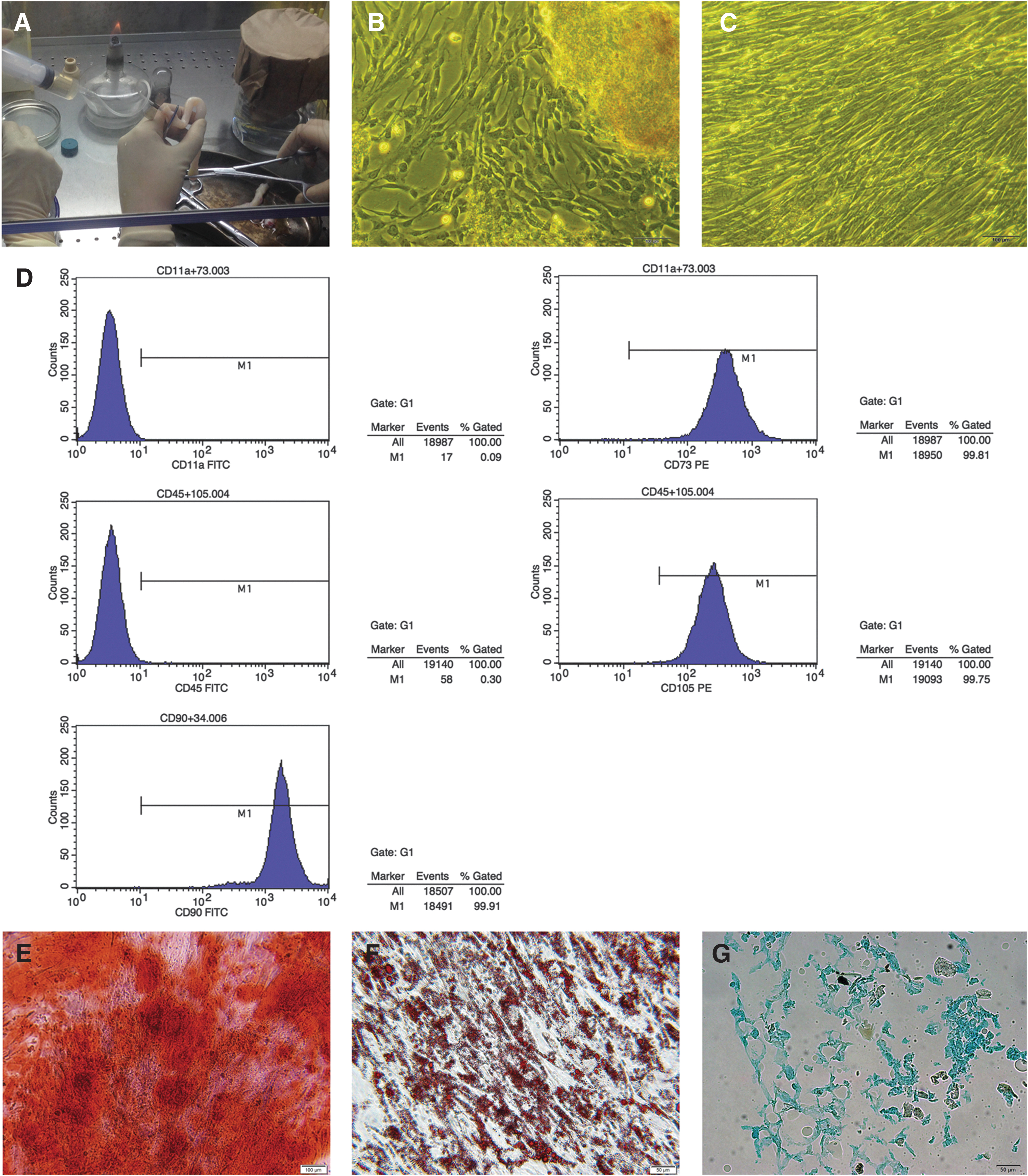
Culture and identification of hUC-MSCs.
SEM analysis
SEM showed that nHAC/PLA had a porous and hierarchical microstructure similar to natural cancellous bone (Fig. 2A, B). After culture for 2 days in media with 100 ng/mL rhBMP-7, the cells adhered, extended, and connected with each other, which produced a few ECMs on materials (Fig. 2C, D). After culture for 7 days, the cells produced large number of ECMs, in where the cells were covered by deposits (Fig. 2E, F).
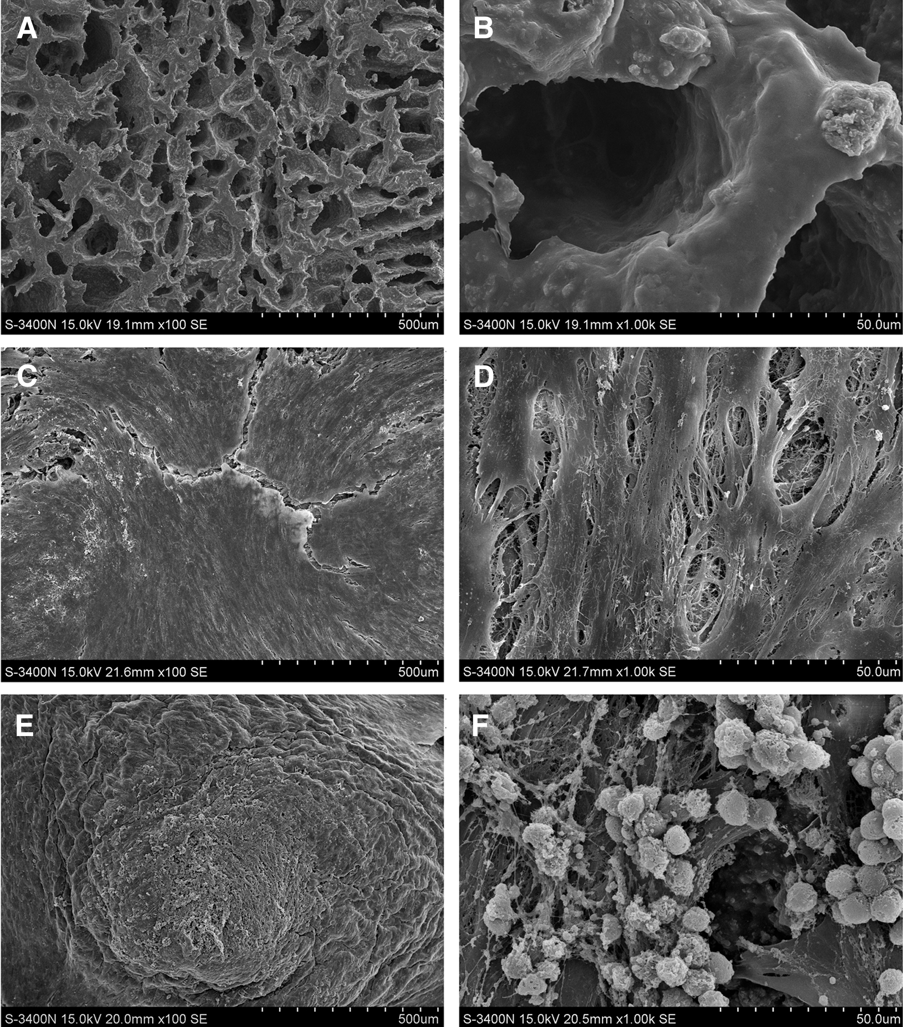
Scanning electron microscopy to observe the adhesion, proliferation, and differentiation of rhBMP-7-induced hUC-MSCs seeded on nHAC/PLA.
Proliferation potential of hUC-MSCs seeded on nHAC/PLA
Three-factor repeated test ANOVA showed that culture time (F = 585.065, P < 0.05) and OMD (F = 170.779, P < 0.05) had significant effect on the proliferation of hUC-MSCs seeded on nHAC/PLA, OMD + rhBMP-7 (F = 48.867, P < 0.05) had significant interaction effect on cell proliferation and rhBMP-7 (F = 1.381, P = 0.242) had no significant effect on cell proliferation. Partial Eta Squared (η 2) represents the contribution of each factor to total variation. η 2 was ranked as η 2 culture time(0.947) > η 2 OMD(0.568) > η 2 OMD + rhBMP-7(0.273) > η 2 rhBMP-7(0.011). The cell proliferation of hUC-MSCs + nHAC/PLA, hUC-MSCs + nHAC/PLA + OMD, hUC-MSCs + nHAC/PLA + rhBMP-7, and hUC-MSCs + nHAC/PLA + OMD + rhBMP-7 group separately reached the highest point at 9, 5, 7, and 7 days, respectively.
t-Test showed that the cell proliferation of hUC-MSCs + nHAC/PLA + OMD (t 1 = −5.515, P 1 < 0.05) and hUC-MSCs + nHAC/PLA + OMD + rhBMP-7 (t 1 = −3.774, P 1 < 0.05) group was significantly higher compared with hUC-MSCs + nHAC/PLA group. The cell proliferation of hUC-MSCs + nHAC/PLA + rhBMP-7 group was significantly lower than those of hUC-MSCs + nHAC/PLA (t 1 = 3.454, P 1 < 0.05) (t 9 = 4.496, P 9 < 0.05), hUC-MSCs + nHAC/PLA + OMD (t 1 = 14.424, P 1 < 0.05), and hUC-MSCs + nHAC/PLA + OMD + rhBMP-7 (t 1 = −10.294, P 1 < 0.05) group, respectively. The cell proliferation of hUC-MSCs + nHAC/PLA + OMD group was significantly lower compared with hUC-MSCs + nHAC/PLA (t 3 = 2.22, P 3 < 0.05) (t 5 = 2.425, P 5 < 0.05) (t 7 = 9.215, P 7 < 0.05) (t 9 = 19.243, P 9 < 0.05) and hUC-MSCs + nHAC/PLA + rhBMP-7 (t 3 = −2.276, P 3 < 0.05) (t 7 = −8.31, P 7 < 0.05) (t 9 = −4.592, P 9 < 0.05) group. The cell proliferation of hUC-MSCs + nHAC/PLA + OMD + rhBMP-7 group was significantly lower compared with UC-MSCs + nHAC/PLA (t 5 = 2.465, P 5 < 0.05) (t 7 = 4.778, P 7 < 0.05) (t 9 = 13.291, P 9 < 0.05) and hUC-MSCs + nHAC/PLA + rhBMP-7 (t 7 = 3.668, P 7 < 0.05) (t 9 = 2.746, P 9 < 0.05) group, but was significantly higher compared with hUC-MSCs + nHAC/PLA + OMD group (t 7 = −4.137, P 7 < 0.05) (t 9 = −4.724, P 9 < 0.05) (Fig. 3A).
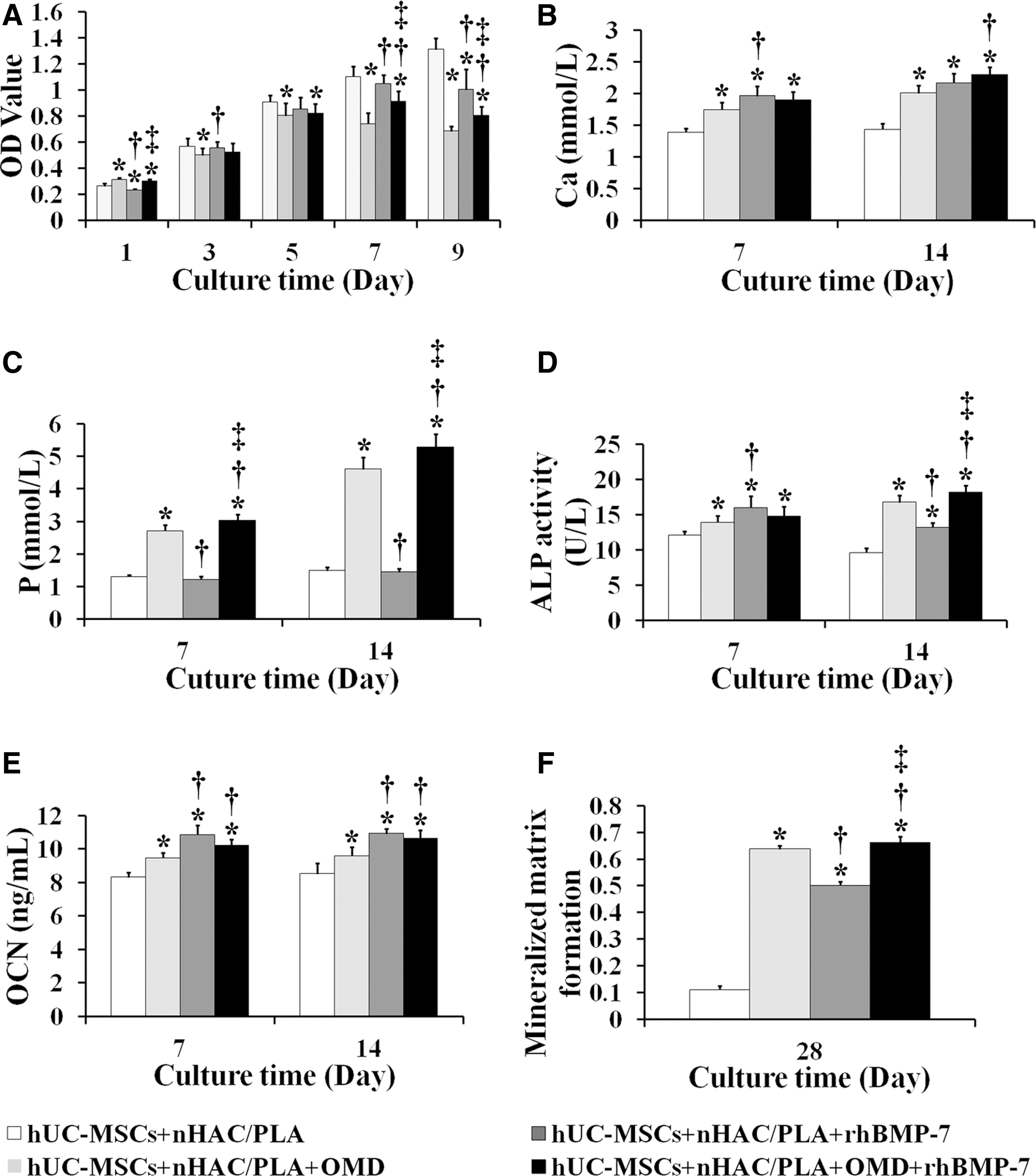
Proliferation and osteogenic differentiation potential of hUC-MSCs seeded on nHAC/PLA. Effect of rhBMP-7 and/or OMD on the proliferation (mean ± SD, n = 8)
Osteogenic differentiation potential of hUC-MSCs seeded on nHAC/PLA
Ca2+ concentration
Three-factor repeated test ANOVA showed that culture time (F = 39.334, P < 0.05), OMD (F = 46.866, P < 0.05), and rhBMP-7 (F = 143.253, P < 0.05) had significant effect on Ca2+ concentration of hUC-MSCs seeded on nHAC/PLA; OMD + rhBMP-7 (F = 35.676, P < 0.05) had significant interaction effect on Ca2+ concentration. η 2 was ranked as η 2 rhBMP-7(0.790) > η 2 OMD(0.552) > η 2 culture time(0.509) > η 2 OMD + rhBMP-7(0.484). The Ca2+ concentration of each group increased with the prolongation of culture time.
t-Test showed that the Ca2+ concentration of hUC-MSCs + nHAC/PLA + OMD (t 7 = −6.642, P 7 < 0.05) (t 14 = −6.946, P 14 < 0.05), hUC-MSCs + nHAC/PLA + rhBMP-7 (t 7 = −8.505, P 7 < 0.05) (t 14 = −8.137, P 14 < 0.05), and hUC-MSCs + nHAC/PLA + OMD + rhBMP-7 (t 7 = −8.414, P 7 < 0.05) (t 14 = −10.866, P 14 < 0.05) group was significantly higher compared with hUC-MSCs + nHAC/PLA group, respectively. The Ca2+ concentration of hUC-MSCs + nHAC/PLA+OMD group was significantly lower than those of hUC-MSCs + nHAC/PLA + rhBMP-7 (t 7 = −2.748, P 7 < 0.05) and hUC-MSCs + nHAC/PLA + OMD + rhBMP-7 (t 14 = −3.967, P 14 < 0.05) group (Fig. 3B).
PO4 3− concentration
Three-factor repeated test ANOVA showed that culture time (F = 317.120, P < 0.05), OMD (F = 1591.203, P < 0.05), and rhBMP-7 (F = 11.206, P < 0.05) had significant effect on PO4 3− concentration of hUC-MSCs seeded on nHAC/PLA; OMD + rhBMP-7 (F = 19.747, P < 0.05) had significant interaction effect on PO4 3− concentration. η 2 was ranked as η 2 OMD(0.975) > η 2 culture time(0.888) > η 2 OMD + rhBMP-7(0.331) > η 2 rhBMP-7(0.219). The PO4 3− concentration of each group increased with the prolongation of culture time.
t-Test showed that the PO4 3− concentration of hUC-MSCs + nHAC/PLA + OMD (t 7 = −18.991, P 7 < 0.05) (t 14 = −20.271, P 14 < 0.05) and hUC-MSCs + nHAC/PLA + OMD + rhBMP-7 (t 7 = −22.653, P 7 < 0.05) (t 14 = −22.278, P 14 < 0.05) group was significantly higher compared with hUC-MSCs + nHAC/PLA. The PO4 3− concentration of hUC-MSCs + nHAC/PLA + rhBMP-7 group was significantly lower than those of hUC-MSCs + nHAC/PLA + OMD (t 7 = 18.560, P 7 < 0.05) (t 14 = 20.473, P 14 < 0.05) and hUC-MSCs + nHAC/PLA + OMD + rhBMP-7 (t 7 = −21.98, P 7 < 0.05) (t 14 = −22.461, P 14 < 0.05) group. The PO4 3− concentration of hUC-MSCs + nHAC/PLA + OMD + rhBMP-7 group (t 7 = −3.107, P 7 < 0.05) (t 14 = −3.035, P 14 < 0.05) was significantly higher compared with hUC-MSCs + nHAC/PLA + OMD group (Fig. 3C).
ALP activity
Three-factor repeated test ANOVA showed that OMD (F = 113.403, P < 0.05) and rhBMP-7 (F = 67.333, P < 0.05) had significant effect on ALP activity of hUC-MSCs seeded on nHAC/PLA, and OMD + rhBMP-7 (F = 19.484, P < 0.05) had significant interaction effect on ALP activity. Culture time (F = 0.718, P = 0.402) had no significant effect on ALP activity. η 2 was ranked as η 2 OMD(0.739) > η 2 rhBMP-7(0.627) > η 2 OMD + rhBMP-7(0.328) > η 2 culture time(0.018). The ALP activity of hUC-MSCs + nHAC/PLA + OMD and hUC-MSCs + nHAC/PLA + OMD + rhBMP-7 groups increased, but hUC-MSCs + nHAC/PLA and hUC-MSCs + nHAC/PLA + rhBMP-7 groups reduced with the prolongation of culture time.
t-Test showed that the ALP activity of hUC-MSCs + nHAC/PLA + OMD (t 7 = −3.986, P 7 < 0.05) (t 14 = −15.299, P 14 < 0.05), hUC-MSCs + nHAC/PLA + rhBMP-7 (t 7 = −5.662, P 7 < 0.05) (t 14 = −10.019, P 14 < 0.05), and hUC-MSCs + nHAC/PLA + OMD + rhBMP-7 (t 7 = −4.327, P 7 < 0.05) (t 14 = −17.721, P 14 < 0.05) group was significantly higher compared with the hUC-MSCs + nHAC/PLA group. The ALP activity of hUC-MSCs + nHAC/PLA + OMD group was significantly lower compared with hUC-MSCs + nHAC/PLA + rhBMP-7 group (t 7 = −2.754, P 7 < 0.05). The ALP activity of hUC-MSCs + nHAC/PLA + OMD (t 14 = 7.534, P 14 < 0.05) and hUC-MSCs + nHAC/PLA + OMD + rhBMP-7 (t 14 = −10.193, P 14 < 0.05) group was significantly higher compared with hUC-MSCs + nHAC/PLA + rhBMP-7 group. The ALP activity of hUC-MSCs + nHAC/PLA + OMD (t 14 = −2.478, P 14 < 0.05) was significantly lower compared with hUC-MSCs + nHAC/PLA+ OMD + rhBMP-7 group (Fig. 3D).
OCN secretion
Three-factor repeated test ANOVA showed that OMD (F = 7.248, P < 0.05) and rhBMP-7 (F = 199.039, P < 0.05) had significant effect on OCN secretion of hUC-MSCs seeded on nHAC/PLA. OMD + rhBMP-7 (F = 44.557, P < 0.05) had significant interaction effect on OCN secretion. Culture time (F = 3.150, P = 0.084) had no significant effect on OCN secretion. η 2 was ranked as η 2 rhBMP-7(0.843) > η 2 OMD + rhBMP-7(0.546) > η 2 OMD(0.164) > η 2 culture time(0.078). The OCN secretion of each group increased with the prolongation of culture time.
t-Test showed that the OCN secretion of hUC-MSCs + nHAC/PLA + OMD (t 7 = −6.617, P 7 < 0.05) (t 14 = −4.275, P 14 < 0.05), hUC-MSCs + nHAC/PLA + rhBMP-7 (t 7 = −9.745, P 7 < 0.05) (t 14 = −12.83, P 14 < 0.05), and hUC-MSCs + nHAC/PLA + OMD + rhBMP-7 (t 7 = −9.762, P 7 < 0.05) (t 14 = −8.803, P 14 < 0.05) group was significantly higher compared with hUC-MSCs + nHAC/PLA group, respectively. The OCN secretion of hUC-MSCs + nHAC/PLA + OMD group was significantly lower than those of hUC-MSCs + nHAC/PLA + rhBMP-7 (t 7 = −4.892, P 7 < 0.05) (t 14 = −5.78, P 14 < 0.05) and hUC-MSCs + nHAC/PLA + OMD + rhBMP-7 (t 7 = −3.509, P 7 < 0.05) (t 14 = −3.786, P 14 < 0.05) group (Fig. 3E).
Mineralized matrix formation
Two-way ANOVA showed that OMD (F = 2697.481, P < 0.05) and rhBMP-7 (F = 985.539, P < 0.05) had significant effect on mineralized matrix formation of hUC-MSCs seeded on nHAC/PLA. OMD + rhBMP-7 (F = 763.243, P < 0.05) had significant interaction effect on mineralized matrix formation. η 2 was ranked as η 2 OMD(0.993) > η 2 rhBMP-7(0.98) > η 2 OMD + rhBMP-7(0.974).
t-Test showed that the mineralized matrix formation of hUC-MSCs + nHAC/PLA + OMD (t 28 = −68.350, P 28 < 0.05), hUC-MSCs + nHAC/PLA + rhBMP-7 (t 28 = −50.618, P 28 < 0.05), and hUC-MSCs + nHAC/PLA + OMD + rhBMP-7 (t 28 = −52.231, P 28 < 0.05) group was significantly higher compared with hUC-MSCs + nHAC/PLA group. The mineralized matrix formation of hUC-MSCs + nHAC/PLA + OMD (t 28 = 17.034, P 28 < 0.05) and hUC-MSCs + nHAC/PLA + OMD + rhBMP-7 (t 28 = −14.948, P 28 < 0.05) group was significantly higher compared with the hUC-MSCs + nHAC/PLA + rhBMP-7 group. hUC-MSCs + nHAC/PLA + OMD + rhBMP-7 group had significantly higher mineralized matrix formation than hUC-MSCs + nHAC/PLA + OMD group (t 28 = −2.318, P 28 < 0.05) (Fig. 3F).
Jaw bone defects repair capacity of hUC-MSCs seeded on nHAC/PLA
Three-dimensional micro-CT reconstruction was performed to detect the newly formed bone tissues 3 months after surgery. Mineralized tissue covering the defect could be observed in all groups (Fig. 4C–F).
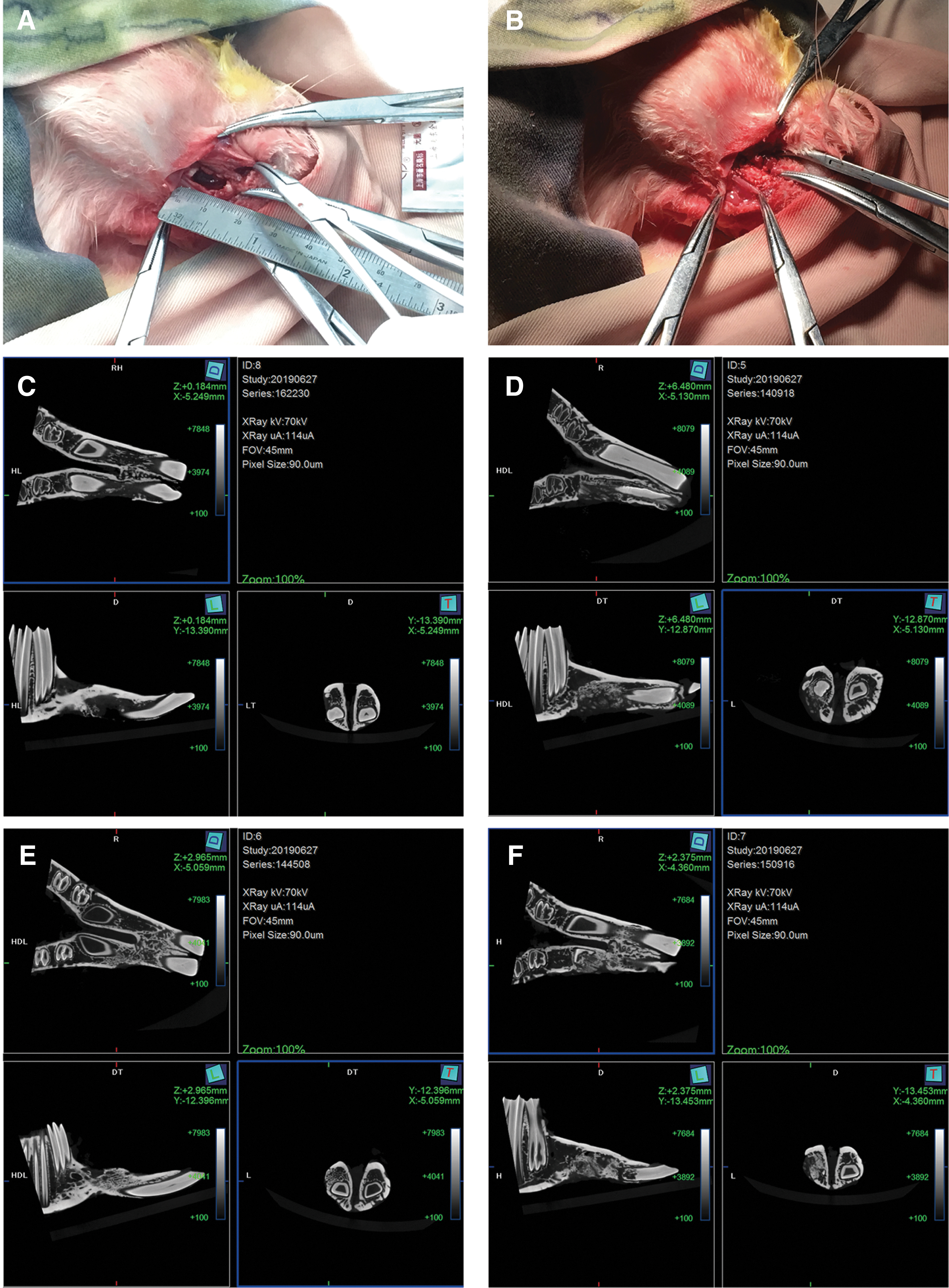
Schematic diagram of the surgery and representative microcomputed tomography reconstruction images of jaw bone defect repair after surgery for 3 months.
Within the section without demineralization, the calcein and tetracycline fluorescence were defected in all groups (Fig. 5A–D), Calcein was displayed in green and tetracycline was displayed in yellow. Two-way ANOVA showed that rhBMP-7 (F = 46.919, P < 0.05) had significant effect on the bone mineral apposition rate of hUC-MSCs seeded on nHAC/PLA. OMD + rhBMP-7 (F = 22.804, P < 0.05) had significant interaction effect on the bone mineral apposition rate. OMD (F = 2.065, P = 0.167) had no significant effect on the bone mineral apposition rate. η 2 was ranked as η 2 rhBMP-7(0.701) > η 2 OMD + rhBMP-7(0.533) > η 2 OMD(0.093).
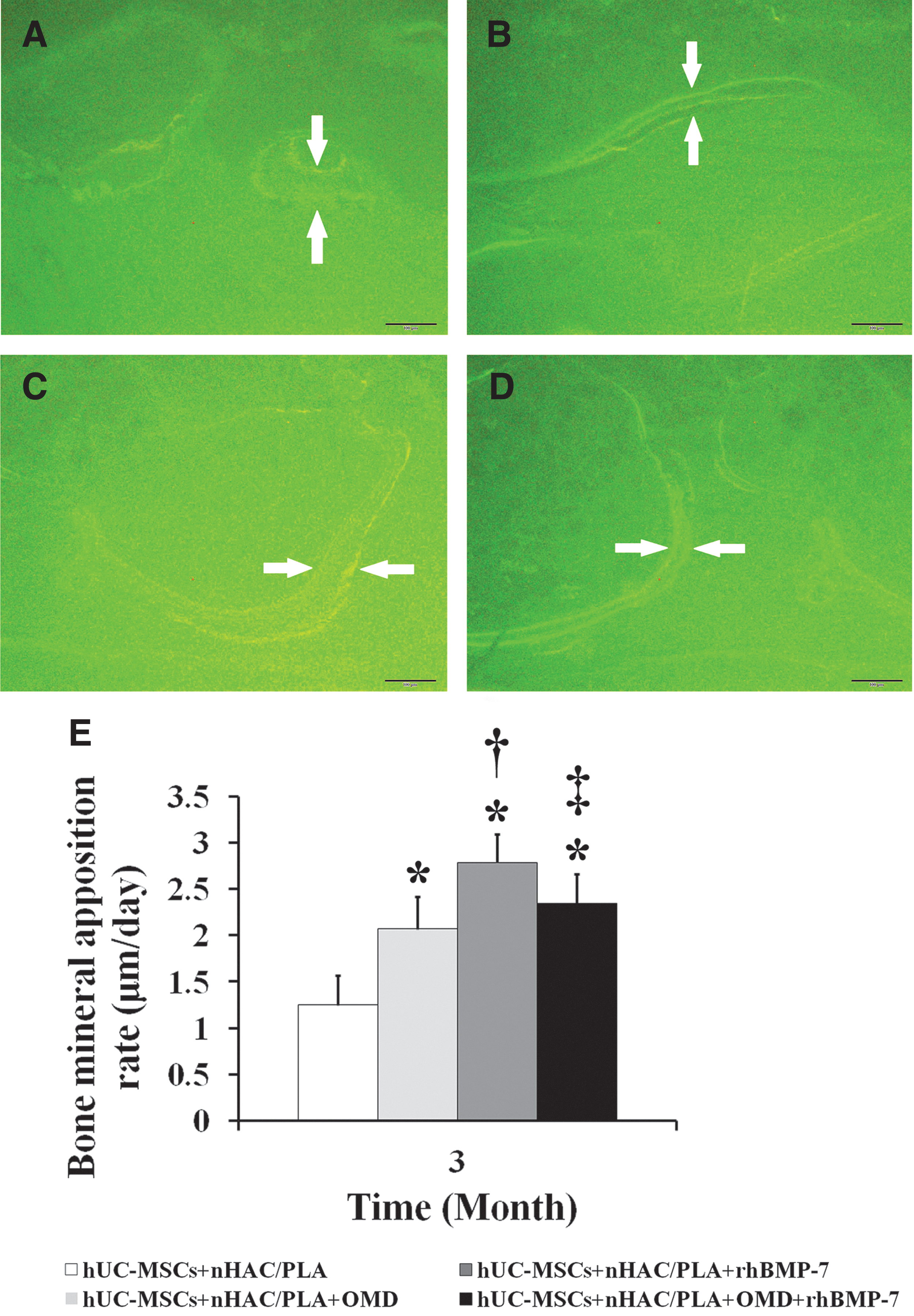
Bone mineral apposition rate of jaw bone defect repair after surgery for 3 months.
t-Test showed that the bone mineral apposition rate of hUC-MSCs + nHAC/PLA + OMD (t = −4.237, P < 0.05), hUC-MSCs + nHAC/PLA + rhBMP-7 (t = −8.460, P < 0.05) and hUC-MSCs + nHAC/PLA + OMD + rhBMP-7 group (t = −5.906, P < 0.05) was significantly higher compared with hUC-MSCs + nHAC/PLA group, respectively. The hUC-MSCs + nHAC/PLA + rhBMP-7 group had significantly higher bone mineral apposition rate than those of hUC-MSCs + nHAC/PLA + OMD (t = −3.799, P < 0.05) and hUC-MSCs + nHAC/PLA + OMD + rhBMP-7 (t = −2.455, P < 0.05) group (Fig. 5E).
H&E staining showed that new bone formation could be observed in all groups after surgery for 3 months (Fig. 6A–D). Most of the newly formed bones were perforated, some had been joined in sheets, and were well connected to the host bone. The newly formed bones had abundant blood vessels passing through. Bone edges were arranged in series of osteoblasts, and there were a lot of active osteoclasts in degraded nHAC/PLA.
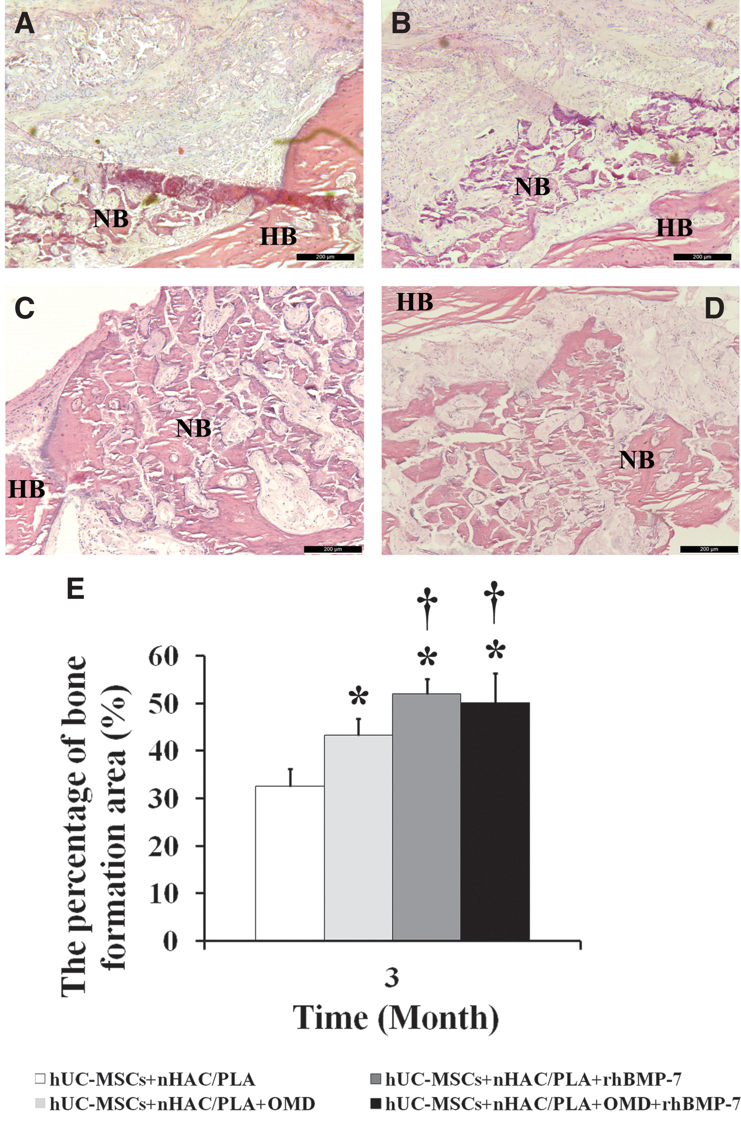
Histomorphometric analysis of the newly formed bone tissues after surgery for 3 months.
Two-way ANOVA showed that OMD (F = 6.241, P < 0.05) and rhBMP-7 (F = 56.128, P < 0.05) had significant effect on the percentages of the bone formation area of hUC-MSCs seeded on nHAC/PLA. OMD + rhBMP-7 (F = 12.956, P < 0.05) had significant interaction effect on the percentages of the bone formation area. η 2 was ranked as η 2 rhBMP-7(0.737) > η 2 OMD + rhBMP-7(0.393) > η 2 OMD(0.238). t-Test showed that the hUC-MSCs + nHAC/PLA + OMD (t = −5.215, P < 0.05), hUC-MSCs + nHAC/PLA + rhBMP-7 (t = −10.044, P < 0.05), and hUC-MSCs + nHAC/PLA + OMD + rhBMP-7 (t = −5.948, P < 0.05) group had significantly higher percentage of bone formation area than the hUC-MSCs + nHAC/PLA group. The percentage of bone formation area of hUC-MSCs + nHAC/PLA + rhBMP-7 (t = −4.6, P < 0.05) and hUC-MSCs + nHAC/PLA + OMD + rhBMP-7 groups (t = −2.334, P < 0.05) was significantly higher compared with hUC-MSCs + nHAC/PLA + OMD group. However, the highest percentage of bone formation area was presented in hUC-MSCs + nHAC/PLA + rhBMP-7 group (Fig. 6E).
Discussion
In the process of bone formation, osteoblasts undergo four stages: osteoblast proliferation, ECM maturation, ECM mineralization, and osteoblasts apoptosis [43]. In the in vitro culture system, the phenotypic development of osteoblasts is similar to the in vivo development and differentiation, and also undergo cell proliferation, ECM maturation, and matrix mineralization. In this study, our cultured cells derived from Wharton's jelly were MSCs, which is consistent with the results reported by other researches [13 –16]. SEM results displayed that nHAC/PLA had good biocompatibility and supported the adhesion and growth of hUC-MSCs.
During the whole growth period of hUC-MSCs combined with nHAC/PLA, OMD and OMD + rhBMP-7 inhibited cell proliferation, except for the initial stress response, rhBMP-7 had no effect on cell proliferation in the logarithmic phase, but inhibited cell proliferation in the nonlogarithmic phase. The study showed that BMP-7 downregulated the gene expression related to cell cycle, attenuating cell cycle progression and cell proliferation during early osteoblastic differentiation of human MSCs [44].
In this study, OMD, rhBMP-7, and OMD + rhBMP-7 also shortened cell cycle progression and attenuate cell proliferation, they separately reached the highest point of cell proliferation at 5, 7, and 7 days, which were earlier than the control group at 9 days. Hence, cell proliferation was inhibited. The inhibitory effect of OMD, rhBMP-7, and OMD + rhBMP-7 on cell proliferation was ranked as OMD > OMD + rhBMP-7 > rhBMP-7.
OMD is the standard procedure for the osteogenic differentiation of MSCs, including dexamethasone, ascorbic acid, and β-glycerophosphate. Dexamethasone is a synthetic glucocorticoid drug used in in vitro induction toward adipogenesis, osteogenesis, or chondrogenesis in an in vivo treatment for inflammation and autoimmune conditions [45]. The effect of dexamethasone on the proliferation of human MSCs is controversial. The study showed that high concentrations of dexamethasone inhibited the proliferation of hBM-MSCs and promoted apoptosis; low concentrations of dexamethasone promoted cell proliferation [45]. Different concentrations of dexamethasone also had different effects on the proliferation and apoptosis of hUC-MSCs [46].
Ascorbic acid in osteogenic differentiation of MSCs mainly promotes type I collagen secretion [47], whereas the accumulation of collagen partly leads to the cessation of cell proliferation [48]. β-Glycerophosphate served as a phosphate source for bone mineralization; the higher PO4 3− concentration affected cell proliferation [47]. In our study, OMD inhibited the proliferation of hUC-MSCs seeded on nHAC/PLA, which may be related to the added dexamethasone, ascorbic acid, and β-glycerophosphate. However, the addition of rhBMP-7 reversed the inhibitory effect of OMD on cell proliferation, the relevant mechanisms needed to be further explored.
Human MSCs can be differentiated into osteoblasts in vitro, which requires an OMD, including dexamethasone, ascorbic acid and β-glycerophosphate. In this study, culture time had no significant effect on ALP activity and OCN secretion. It indicated that our isolated hUC-MSCs required a longer period of time to differentiate into osteoblasts in consistent with previous study [49]. OMD, rhBMP-7 and OMD + rhBMP-7 enhanced osteogenic differentiation of hUC-MSCs seeded on nHAC/PLA. OMD, which is golden standard of osteogenic differentiation medium of MSCs, contains dexamethasone, ascorbic acid and β-glycerophosphate. Dexamethasone induced MSC differentiation into osteoblasts by activating WNT/β-catenin signaling-dependent Runx2 expression. BMP signaling regulates Runx2 activity. Runx2–BMP interaction plays an important role in dexamethasone-induced osteogenic differentiation of MSCs [50].
Ascorbic acid promoted osteogenic differentiation of MSCs by increasing collagen type 1 secretion and forming the ECM, and then activating Col1/α2β1 integrin-mediated intracellular signaling. Osteoblasts must get in touch with a collagen-containing ECM before they can differentiate [51]. β-Glycerophosphate serves as a phosphate source for bone mineral and induces osteogenic gene expression by extracellular related kinase phosphorylation [47]. Therefore, OMD, by the effects of these substances on intracellular signaling cascades, leads to osteogenic differentiation of MSCs.
The study showed that BMP-7 upregulated osteogenic gene expression in early osteoblastic differentiation of human MSCs, whereas BMP-7-induced osteogenic differentiation was independent of endogenous BMP-2 expression [44]. The addition of rhBMP-7 enhanced osteogenic differentiation of murine MSCs and produced a reciprocal expression profile in the endogenous BMP expression, and osteogenic differentiation was regulated by a complex network of multiple BMPs.
Interestingly, during the proliferation and osteogenic differentiation of hUC-MSCs seeded on nHAC/PLA, when the culture with OMD had the lowest cell proliferation, its inhibitory effect on cell proliferation was strongest, when the culture with rhBMP7 had the highest OCN secretion, its promoting effect on OCN secretion also was largest. However, when the culture with OMD + rhBMP-7 had the highest PO3- 4 concentration and mineralized matrix formation, its promoting effect on PO4 3− concentration and mineralized matrix formation was not the highest, but OMD. The promoting effect of OMD, rhBMP-7, and OMD + rhBMP-7 on PO4 3− concentration and mineralized matrix formation was ranked as OMD > OMD + rhBMP-7 > rhBMP-7 and OMD > rhBMP-7 > OMD + rhBMP-7. It suggested that the effect of the combined use of OMD and rhBMP-7 on PO4 3− concentration and mineralized matrix formation was not as good as that of OMD alone.
The addition of rhBMP-7 did not enhance the effect of OMD on PO4 3− concentration and mineralized matrix formation, although the combination of OMD and rhBMP7 had a significant effect on PO4 3− concentration and mineralized matrix formation. In the process of bone formation, Ca2+ and PO4 3− are needed to form hydroxyapatite that is deposited in the ECM for mineralization [52]. In this study, not only did β-glycerophosphate in OMD provide PO4 3− but also nHAC/PLA that was degraded by the proliferation and differentiation of hUC-MSCs growing on it; therefore, the culture with OMD or OMD + rhBMP-7 had significantly higher PO4 3− concentration compared with rhBMP-7 and untreated control group.
Previous study showed that cells treated with high concentrations (≥2 mM) of β-glycerophosphate could produce mineralization or nonapatitic mineralization when they were incapable of osteogenic differentiation, which could be detected by Von Kossa or Alizarin red staining [53]. This might be one of the reasons why the culture with OMD or OMD + rhBMP-7 had higher mineralized matrix formation than that with rhBMP-7.
When hUC-MSCs seeded on nHAC/PLA were induced by OMD and/or rhBMP-7 for 7 days in vitro, the cultures were implanted into jaw bone defects in rabbit. The micro-CT, bone mineral apposition rate, and H&E staining showed that hUC-MSCs + nHAC/PLA + rhBMP-7 group had higher bone formation than other groups. These results indicated that the combined use of rhBMP-7 and OMD had no ideal synergistic effect on bone regeneration of hUC-MSCs seeded on nHAC/PLA in jaw bone defect.
Our in vitro results demonstrated that the culture with rhBMP-7 had higher cell proliferation, Ca2+ concentration, and OCN secretion, but had lower PO4 3− concentration and mineralized matrix formation. During cell proliferation period, the active cell growth and cell activity are fundamental to development of the bone cell phenotype [48]. That the culture with rhBMP-7 had the higher proliferation may be one of the advantages of having the most in vivo bone formation.
Although the culture with OMD or OMD + rhBMP-7 had higher PO4 3− concentration and mineralized matrix formation, a confluent cell monolayer required at least a 3-week period of continuous treatment of dexamethasone, β-glycerophosphate, and ascorbic acid for osteogenic differentiation [54], whereas osteogenic gene expression of human MSCs was upregulated when the cells were treated with BMP-7 for 24 or 120 h in vitro [44]. A long, continuous, and high concentration of β-glycerophosphate had led to many false-positive results about mineralized matrix formation in osteogenic differentiation of MSCs [55].
In addition, the formation of hydroxyapatite for mineralization requires appropriate concentrations of Ca2+ and PO4 3− outside the matrix vesicles [52]. The bone-specific Ca/P ratio of an oriented and organized Ca/P deposition, not unorganized dystrophic Ca/P deposition, is 1.67 [55]. In this study, Ca/P ratio of the culture with rhBMP-7 was 1.614 at 7 days and 1.5 at 14 days, but 0.643 and 0.436 in that with OMD and 0.624 and 0.435 in that with OMD + rhBMP-7 at 7 and 14 days. It indicated that the culture with rhBMP-7 had the closest bone-specific Ca/P rate.
Human MSCs harvested from reaming debris combined with β-tricalcium phosphate induced by BMP-7 alone had superior bone formation when implanted ectopically in severe combined immunodeficiency mice [32]. Furthermore, BMP-7 downregulated the gene expression related to chemokine/cytokine activity during early osteoblastic differentiation of human MSCs, which was confirmed by cytokine secretion measurement in culture media [44]. BMP-7-induced primary human aortic smooth muscle cell had the higher anti-inflammatory activities [56]. These reports suggested that BMP-7 may play an important role in maintaining vascular integrity. The study showed that hUC-MSCs had higher endothelial differentiation potential than BM-MSCs [11]. Therefore, the interaction between a variety of products secreted in vivo by hUC-MSCs seeded on nHAC/PLA due to the induction of rhBMP-7 in vitro and jaw bone microenvironment may be essential for sufficient and timely bone regeneration of hUC-MSCs, which needs further research and investigation.
Conclusively, our study suggested that OMD, rhBMP-7, and OMD + rhBMP-7 all enhanced osteogenic differentiation of hUC-MSCs seeded on nHAC/PLA. The largest bone mineral apposition rate and bone formation were presented in the cultures with rhBMP-7. The combined use of rhBMP-7 and OMD may have no ideal synergistic effect on bone regeneration of hUC-MSCs seeded on nHAC/PLA in rabbit jaw bone defect.
Footnotes
Acknowledgments
We are very grateful to the staff and faculty of Institute of Stomatology, Chinese PLA General Hospital, Beijing Key Laboratory of Chronic Heart Failure Precision Medicine, and Department of Chemistry, Jinan University.
Author Disclosure Statement
No competing financial interests exist.
Funding Information
This work was approved by the committee of experimentation of Chinese People's Liberation Army General Hospital. This work was supported by People's Liberation Army General Hospital Clinical Research Support Fund (no. 2016FC-CXYY-2007, 2015FC-CXYY-1003) and National Natural Science Fund (no. 81271180).