
Abstract
Dental-tissue-derived stem cells have been used for tissue engineering owing to their ease of isolation and efficacy in in vitro and in vivo proliferation and differentiation. Nanohydroxyapatite/chitosan/gelatin (nHA/CG) three-dimensional porous scaffolds are promising for bone tissue engineering, especially jaw bone regeneration, because of their structural and functional similarity to natural bone. In our previous study, the efficiency of scaffolds with stem cell complexes in osteogenesis was confirmed in vivo in immunocompromised mice. However, studies on the bone regeneration efficiency of stem cell-seeded nHA/CG scaffolds using large animal jaw bone defect models have not been conducted. This study evaluated the bone regeneration potential of the nHA/CG scaffolds with transplanted human periodontal ligament stem cells (hPDLSCs) in critical-sized jaw bone defects in minipigs. The hPDLSCs isolated from periodontal ligaments of discarded teeth (postorthodontic purposes) were seeded onto the nHA/CG scaffolds. The scaffold was successfully synthesized according to our previous studies. Forty-eight critical-sized jaw bone defects were created in 12 minipigs. The defects were randomly assigned to one of three groups [scaffolds with seeded hPDLSCs (hPDLSCs/nHA/CG), only scaffold (nHA/CG), and a negative control group, ie, no cells and scaffolds implanted into defects] to investigate jaw bone regeneration. The bone regeneration capacities of the three groups were assessed for up to 12 weeks. The results showed that the hPDLSCs adhered well to the nHA/CG scaffold in vitro, and the cell-nHA/CG composites significantly increased new bone formation and generated large bones with normal architectures and vascularization in vivo compared to the nHA/CG and control groups. Immunohistochemistry staining showed that runt-related transcription factor 2 (Runx2) was highly expressed in the bone marrow formed in the hPDLSCs/nHA/CG group. This study provides strong evidence for future clinical applications of the nHA/CG scaffolds transplanted with hPDLSCs to regenerate the bone in large jaw bone defects.
Introduction
Large segmental jaw bone defects caused by trauma, infections, tumor resection, or nonunions in severe fractures are a difficult clinical problem. However, alternative therapy to conventional methods such as bone grafting, the gold standard in bone defect therapies, and prostheses is associated with limited availability, donor site morbidity, and a risk of immune-rejection. Bone tissue engineering provides an exciting method to repair bone defects in oral orthopedics. This technology enables the regeneration of tissues using scaffolds, growth factors, and biological factors, with stem cells playing a leading role in tissue regeneration. The tissue engineering repair strategy involves creating bone-like tissue constructs that resemble the natural architecture and function of bones, by combining scaffolds with stem cells capable of inducing osteogenesis [1 –4].
Dental-tissue-derived stem cells are a good choice as the cell source for bone tissue engineering. Periodontal ligaments (PDL) contain heterogeneous cell populations that can differentiate into bone-generating cells (osteoblasts) [5]. Various stem cells have been used in tissue engineering and preclinical studies because of their ease of isolation and efficacy in proliferation and differentiation in vitro and in vivo [6 –9]. Human periodontal ligament stem cells (hPDLSCs) are a type of the dental mesenchymal stem cell (MSC)-like population. These cells express the MSC markers STRO-1 and CD44/CD146.
The osteogenic differentiation of isolated and cultured human mesenchymal stem cells (hMSCs) derived from adult bone marrow has been well characterized [10]. The use of MSCs is an advantageous alternative therapeutic option in bone regeneration, periodontics, and tendon tissue regeneration [5,11 –13]. Various studies have demonstrated the ability of MSCs to exert a profound inhibitory effect on the proliferation of T cells, B cells, dendritic cells, and natural killer cells in vitro and in vivo. These findings suggest that MSCs can be used to reduce transplant rejection. The viability of transplanted MSCs in large animal models has also been established [14 –17]. Therefore, the potential use of stem cells for bone regeneration and tissue repair in clinical applications is very promising [18 –21].
Tissue engineering utilizes scaffolds as biomimetic strategies to induce certain functions in seed cells, such as cell attachment, migration, and proliferation, which affect the efficacy of regenerative treatments [22 –26]. Scaffolds are carriers for seeded autogenous cells; they not only substitute for the extracellular matrix (ECM) but also can be used to induce the formation of desired tissue following the growth of cells from surrounding areas post-transplantations.
In our previous study, we produced a nanohydroxyapatite/chitosan/gelatin (nHA/CG) bone scaffold by freeze-drying, in an effort to generate a good biocompatible bone graft substitute. The nHA/CG scaffold significantly increased the seed cell proliferation and osteogenic differentiation in vitro and in vivo [27,28]. The composition of scaffolds affects their biological and mechanical properties. nHA is often used to produce nanocomposite scaffolds to improve their biodegradability and mechanical properties. Chitosan and gelatin are common organic components used in bone scaffolds [26,29 –33]. These components also contribute to the adhesion and differentiation of stem cells.
Bone tissue engineering provides an option to supplement existing treatment regimens for the regeneration of large maxillofacial defects. Although we have demonstrated that the nHA/CG scaffold is biocompatible and osteoconductive in vivo in a small animal model for 12 weeks [27,28], large animal model studies have to be performed, as they are crucial for further investigations of clinical applications [25]. Good comparability with human system is a prerequisite for testing bone-substituting materials in an animal study. Minipigs are similar to humans in terms of their anatomy, physiology, and immune system [34]. Moreover, minipigs were the animal of choice for this experiment because their bone regeneration rate (1.2–1.5 μm/day) is comparable to that of humans (1.0–1.5 μm/day) [35]. Owing to these factors, the minipig jaw bone defect model is an appropriate large animal model that enables evaluation of jaw bone regeneration.
In this study, the therapeutic capacity of the nHA/CG scaffold loaded with hPDLSCs to repair large defects was assessed in a large animal model. This study provides a rationale for future applications of this technique in clinical studies.
Materials and Methods
Composition of the scaffold
All the synthetic procedures were performed as described in our previous study and are shown under the Supplementary Data [28]. Briefly, the three-dimensional (3D) porous scaffold (8 mm diameter × 5 mm thick) components were nHA, chitosan, and gelatin. The relative ratio (wt/vol %) of the components (nHA, chitosan, and gelatin) was 3:1:1 [27,28]. The nHA content significantly influenced the scaffold density and enhanced its bend strength. Chitosan was used, as it is a biodegradable material with a broad potential in tissue engineering applications [36]. Gelatin was used because it is inexpensive and biodegradable [37]. The scaffolds were prepared by freeze-drying, as previously described [28] (the precise method of synthesis of the scaffold is presented in the Supplemental Data). Scanning electron microscopy (SEM) images at different magnifications of the nHA/CG scaffold are shown in Fig. 1. The scaffold showed a very high porosity in the SEM images (200 × , 500 × ), and the pore walls were seen to have a rough structure at higher magnifications (5000 × , 50000 × ).
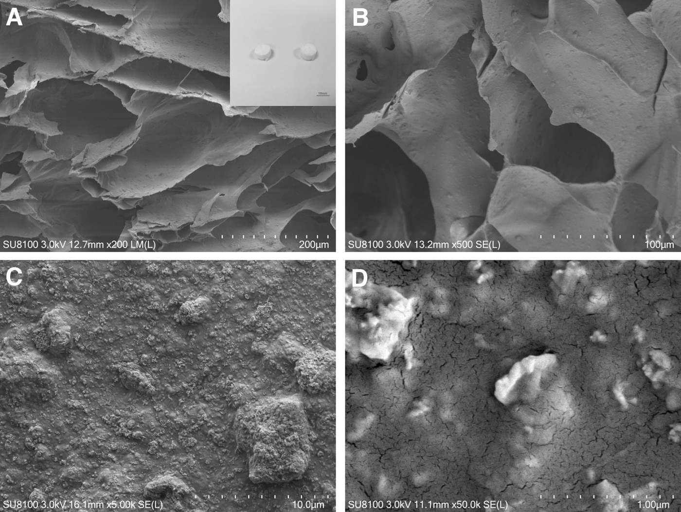
SEM images of the scaffold. Microstructure of the scaffold visualized with SEM.
Isolation and multipotent differentiation of human PDLSCs
Teeth with healthy PDLs were obtained after tooth extractions for orthodontic purposes. All the teeth samples were processed within 2 h of extraction. The PDL was gently separated from the middle third of the tooth root surface. The PDLs obtained were then digested in 2 mL of Type I collagenase (3 mg/mL; Worthington Biochemical, Lakewood, NJ) for 1 h at 37°C. The cells were cultured in basal medium consisting of Minimum Essential Medium-alpha (α-MEM) (Gibco, Gibco, Gaithersburg, MD), containing 20% fetal bovine serum (FBS) (Gibco, Life Technologies, Grand Island, NY), 100 U/mL of penicillin and 100 mg/mL of streptomycin, 2 mM glutamine (Amresco, Solon, OH), and 100 μM L-ascorbic acid phosphate (Sigma-Aldrich), at 37°C in 5% CO2. When 70%–80% confluency was achieved, the hPDLSCs were passaged into T-75 culture flasks at 6 × 10³ cells per cm2 in the growth medium, as previously described. The medium was changed every 2 to 3 days. Cells at passage 3 were used for the experiments. Alkaline phosphatase activity assays were performed on cells cultured in an osteogenic induction media for 7 days. The osteogenic differentiation medium consisted of α-MEM, 10% FBS, 10−7 M dexamethasone, 10 mM β-glycerophosphate, 100 μM phosphor-ascorbic acid, and 20 mM 4-(2-hydroxyethyl)-1-piperazine ethanesulfonic acid buffer. The hPDLSCs were plated in α-MEM supplemented with 5% fetal calf serum, 100 mM L-ascorbate-2-phosphate, 1 mM sodium pyruvate, 50 μg/mL streptomycin, 50 U/mL penicillin G, 2 mM
Expression of osteogenic differentiation marker genes in osteogenesis-induced hPDLSCs
The hPDLSCs were seeded in triplicate in six-well plates at a density of 3 × 10⁴ cells/mL and grown to 80% confluence. The cells were then induced with osteogenic media. At days 2 and 7, RNA extraction and cDNA synthesis were carried out. Quantitative reverse transcription-polymerase chain reaction (qRT-PCR) for the BMP2 and BMP4 genes was also carried out. The primers used were 5′-GCC AAA CAC AAA CAG CGG AA-3′ (forward) and 5′-GGG AGC CAC AAT CCA GTC AT-3′(reverse) for BMP2 and 5′-TGG GCA CCT CAT CAC ACG AC-3′(forward) and 5′-CCA TAG TTT GGC TGC TTC TC-3′(reverse) for BMP4. hPDLSCs are MSCs that show a classical fibroblast-like phenotype, with the ability to differentiate into osteogenic cells. qRT-PCR analysis was used to quantify the osteogenic differentiation of the hPDLSCs in vitro.
Seeding the hPDLSCs onto the nHA/CG scaffolds in vitro and transplantation in vivo
Before cell seeding, the scaffolds were soaked in α-MEM, containing 10% FBS, for 24 h to facilitate hPDLSC attachment, and then placed into a 24-well culture plate. The hPDLSCs were collected with a solution of 0.05% trypsin/ethylene diamine tetraacetic acid (EDTA) (Invitrogen). They were then slowly and evenly seeded onto the presoaked nHA/CG scaffolds (using 3 × 10⁶ cells). The cells adhered to the scaffold after 24 h of incubation. A certain volume of culture medium was slowly added to the plate wells to submerge the samples continuously. For the in vivo study, the composites were implanted into jaw defects of minipigs created after the hPDLSCs were incubated for 24 h on the scaffolds.
Animal studies
We used 12 healthy female minipigs weighing 30–35 kg and aged ∼10 months. The Ethics and Research Committee at the Nanjin University, Jiangsu Province, China, approved this study (no. IACUC-2003078). The animals were preanesthetized with a combination of acepromazine (0.25 mg/kg), buprenorphine (0.01 mg/kg), and medetomidine (35 μg/kg) injected in the neck area of the pigs intramuscularly. In this study, fentanyl, midazolam, and propofol were administered through continuous intravenous infusion for the anesthesia, along with controlled ventilation. Conventional dental infiltration anesthesia (articaine 40 mg and 1% epinephrine) was administered at the surgical sites. Two nonhealing defects without the periosteum of ∼1.0 cm width × 1.2 cm length × 0.6 cm depth (Fig. 2) were created in one quadrant of the maxillary bone of each minipig, and the 48 defects were randomly divided into three groups. The nHA/CG scaffold was seeded with 3 × 10⁶ cells/mL of the hPDLSCs (hPDLSCs/nHA/CG group, N = 16) (Fig. 2); the nHA/CG scaffold without cells (nHA/CG group, N = 16) was randomly placed into the defect area, and the negative control group (N = 16) was left empty. After 12 weeks of operation, the minipigs were euthanized by anesthesia overdose. The samples were then analyzed with micro-computed tomography (micro-CT) and histology.

Implantation of scaffold loaded with the hPDLSCs.
3D cone beam CT images and direct measurements of the new bone
Cone beam CT (CBCT) images were taken using a NewTom VG scanner (QR Srl, Verona, Italy) according to the manufacturer's protocol, with a voxel size of 0.20 mm. The gray values of the new bone sections were quantitatively calculated using the density measurement tool in the Mimics software, version 20.01.
micro-CT measurements
At 12 weeks after the experiments began, the minipigs were sacrificed. The bone specimens were then scanned using micro-CT (μ-CT80; Scanco Medical, Switzerland) at an energy of 7 kV and an intensity of 114 μA. micro-CT was performed by slice increment of the defects at 62.4 μm thickness intervals. The density measurement tool in the Mimics software was used to calculate the ratio of bone volume/tissue volume (BV/TV), trabecular thickness (
Histological assessment (hematoxylin and eosin and Masson) and immunohistochemistry staining
After CT scanning, the specimens were fixed in 10% formalin for 48 h, decalcified with 10% EDTA for 10 weeks, embedded in paraffin blocks, and sectioned to 5 μm thickness. All the sections were then stained with hematoxylin and eosin (HE) and Masson. The immunohistochemistry staining was done according to the manufacturer's instructions, using the runt-related transcription factor 2 (Runx2) rabbit monoclonal antibody [EPR14334] ab192256 and the CD34 rabbit monoclonal antibody [Servicebio] GB13013.
Statistical analysis
Statistical analyses were performed using the Statistical Package for Social Sciences software package (SPSS 22.0, Chicago, IL). Data obtained from the histomorphometric examinations were expressed as mean ± standard deviation and analyzed by one-way analysis of variance. Tukey's multiple comparison test was used to identify differences. A P value of <0.05 was considered statistically significant.
Results
Isolation and characterization of the hPDLSCs
The hPDLSCs isolated from healthy human donor teeth exhibited a high degree of proliferation and multipotent differentiation, with most cells appearing spindle shaped upon microscopic examination. The cells showed a long cytoplasmic process shape with a good degree of contact with neighboring cells (Fig. 3A). Moreover, after culturing the cells in osteogenic induction media for 7 days and in adipogenic induction media for 4 weeks, the PDL cell-derived colonies formed alkaline phosphate (Fig. 3B) and lipid vacuoles (Fig. 3C). Histological assessments (HE staining) and SEM, as shown in Fig. 3D–F, revealed that the hPDLSCs showed good adherence to the nanostructured scaffold.

Multipotent differentiation of the hPDLSCs and expression of osteogenic differentiation marker genes in vitro. SEM images of the hPDLSCs attached on the scaffold.
Osteogenic gene expression during hPDLSCs osteogenesis
The bone-associated gene (BMP2 and BMP4) expression in cells cultured in the osteogenic media and in α-MEM was analyzed with qRT-PCR. At days 2 and 7, the expression of osteogenic differentiation markers in the hPDLSCs cultured in the osteogenic media was significantly higher (P < 0.05) than that in the hPDLSCs cultured in α-MEM postanalysis, as assessed with qRT-PCR (Fig. 3G, H).
CBCT analysis
To evaluate the potential of bone regeneration using the nHA/CG scaffold containing hPDLSCs in large animals, critical-sized defects were created in the jaw bone of minipigs. Twelve weeks after the operation, new bone development in each group was assessed with CBCT imaging. The gray values of newly formed bone in each group were quantitatively calculated [Fig. 4C(a)]. The gray values of animals with the nHA/CG scaffolds seeded with the hPDLSCs were significantly higher than those of the control (untreated) (P < 0.0001) and nHA/CG groups (P < 0.0001).
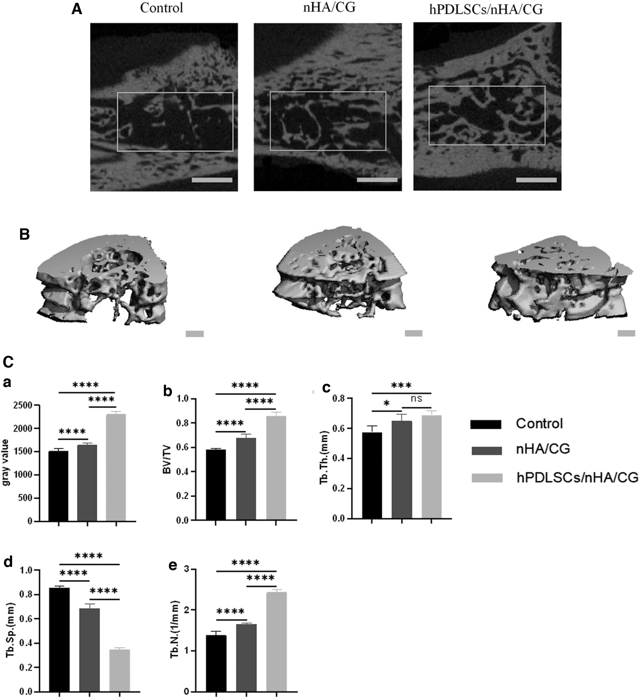
The 2D and 3D images of new bone formation by micro-CT and quantitative analysis after 12 weeks.
micro-CT analysis of the volume of newly formed bone in minipigs
Ex vivo micro-CT imaging showed the regeneration levels among the examined groups.
In the negative control group, only a small amount of new bone was formed along the defect margin. The defects in the nHA/CG group showed more new bone formation after 12 weeks at the edges of the defects than those of the negative control group. In both the hPDLSCs/nHA/CG and nHA/CG groups, examination of all the specimens indicated that the residual scaffolds had been degraded. However, the hPDLSCs/nHA/CG group showed the greatest amount of new bone formation, followed by the nHA/CG and negative control groups. The cross-sections and representative 3D reconstructions of the defects at 12 weeks are presented in Fig. 4A. The reconstructed images displayed trabecular bone details. Quantitative analysis of the micro-CT data revealed that the negative control group exhibited a significantly lower (P < 0.05) bone regeneration potential than the hPDLSC/nHA/CG and nHA/CG groups. The BV/TV ratio was significantly higher in the hPDLSCs/nHA/CG group than in the nHA/CG (P < 0.0001) and negative control groups (P < 0.0001). The thickness of the trabecular bone formed around the defect (
Histological evaluation of the new bone formation
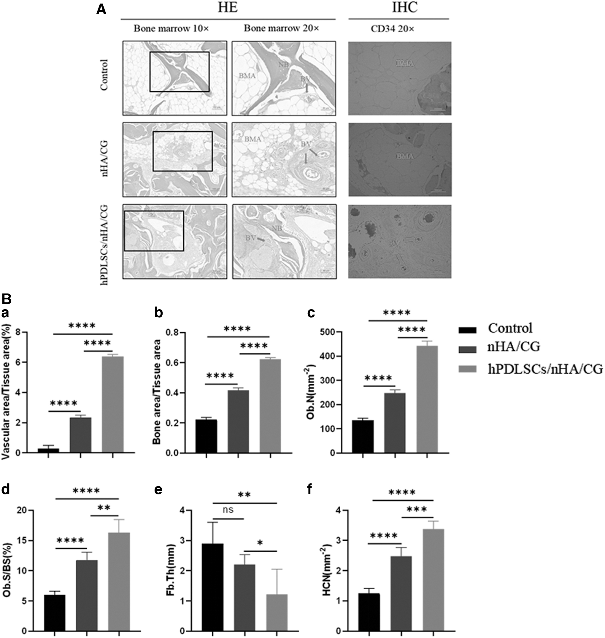
Histological images were analyzed to observe the microstructure of tissues in the defect area. The representative histological images of each group, at low and high magnification, are depicted in Fig. 5. The hPDLSCs/nHA/CG group consistently had significantly higher amounts of new bone to repair the defects than the nHA/CG and negative control groups. In the negative control group, bone formation was irregular in most sections; instead of bone formation, large regions filled with fibrous tissues were observed in bone defects. We assessed the collagen content and bone maturity in the defect area in the treated animals through Masson's trichrome and HE staining. The hPDLSCs/nHA/CG group exhibited significantly more new bone formation than the other groups, thus confirming the micro-CT results. Osteoblasts were visible on the newly formed bone, indicating an active bone regeneration process. Representative immunohistochemical images of the defects are shown in Fig. 5A. Strong expression of Runx2 was observed in areas of bone marrow within the defect regions in the hPDLSCs/nHA/CG group, while the nHA/CG and negative control groups showed milder expression of these osteogenic markers. These findings imply the potential functional role of hPDLSCs in bone tissue regeneration. Some cell populations with osteogenic markers were found in the bone marrow of the hPDLSCs/nHA/CG group. Runx2 was selected as a marker to observe differentiated osteoblast cells in the bone marrow, and the results showed that there were more Runx2-positive cell populations in the bone marrow of the hPDLSCs/nHA/CG group minipigs than in that of other groups. In addition, excessive bone marrow deposits were found in the negative control group. Collectively, these results suggest that biomaterials with hPDLSCs could be effective promoters of an active environment for bone formation.

Representative histological sections of the bone defects of each group at 12 weeks after surgery
The HE staining assays showed that the hPDLSCs/nHA/CG group had a few bone marrow adipocytes and increased angiogenesis (Fig. 6A). The analysis of HE staining and the expression of CD34 with immunohistochemical staining in areas of the bone marrow revealed that the scaffolds seeded with hPDLSCs promoted the formation of blood vessels. The histological quantitative analysis is shown in Fig. 6B. The formation of teratomas or other tumors in the bone or other organs was absent within the 12 weeks of our study (Fig. 7).
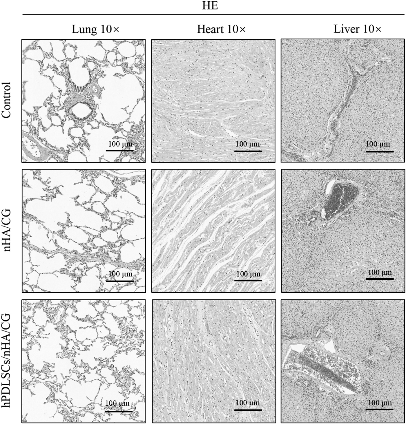
HE-stained histological images of the lung, heart, and liver sections from all the groups at 3 months postsurgery. Scale bar: 100 μm.
Discussion
The application of biomaterials for human bone repair requires extensive preclinical safety testing using laboratory animals. The use of miniature pigs in studies is advantageous because this animal has characteristics similar to those of humans, and these characteristics bring the experiences of human clinical trials closer at the preclinical stage. Furthermore, minipigs have close similarities to humans in terms of their bone structure [16,38 –41]. Our study aimed to achieve bone repair by using biomimetic 3D porous scaffolds seeded with hPDLSCs, to examine the effect of the combination of hPDLSCs and the scaffold on bone regeneration. Porous scaffolds have usually been combined with osteogenic factors such as cells or growth factors, in previous studies [42 –45]. In this study, an nHA/CG scaffold was evaluated for its efficacy in bone repair in large animals, based on a previous study [27,28]. In addition, we evaluated the role of the hPDLSCs in jaw bone repair. The nHA/CG scaffolds improved the osteogenic differentiation of human induced pluripotent stem cells (hiPSCs) from hematopoietic growth factors in vitro and in vivo according to our previous study [28]. The hiPSCs seeded on scaffolds generated large bones after the scaffolds were transplanted in vivo for 12 weeks. However, the development of preclinical models using large animals requires the use of well-characterized cell lines. Human PDL contains a population of multipotent postnatal stem cells that can be isolated and expanded in vitro, providing a unique reservoir of stem cells from an accessible tissue resource [5]. Several studies have shown that hMSCs migrate to injured areas, survive for a long period, and repair the tissues [18,46 –50]. However, the osteogenic capacity of MSCs from minipigs is unknown. Heino et al. found that minipig MSCs had a significantly lower ability, than hMSCs, to form differentiated and functional osteoblasts [39].
Our study used a 3D porous scaffold to repair large bone defects in minipigs. The nHA/CG scaffold was found to possess an excellent bone regeneration potential. High porosities in scaffolds, greater than 93%, benefit cell migration and tissue growth [51,52]. The scaffolds in our study exhibited uniform pores with sizes in the range of 100–300 μm and are, therefore, suitable for initiating bone tissue regeneration. The nHA used was similar to that found in natural bones and promoted the disorganized formation of new bones [53].
Chitosan-based scaffolds are used in periodontal and craniofacial regenerative therapies because these scaffolds support the initial attachment and proliferation of osteoblasts [54]. To mimic both the physical architecture and the chemical composition of the natural bone ECM, we developed a method to fabricate an nHA/CG composite scaffold in this study. The scaffold developed was seeded with hPDLSCs for enhanced bone regeneration, and it significantly promoted in vivo bone formation in critical-sized jaw bone defects. The ECM activates intracellular signaling that alters cell shape and cytoskeletal dynamics to modulate cell growth and viability, and directs the cell migration and cell fate [55].
Nanostructured scaffolds based on nanomaterials capable of closely resembling the native ECM structure have been designed. These nanomaterials are similar to the native bone niche [56,57]. Our study showed that the porous nHA/CG scaffold had excellent biocompatibility and was suitable for hPDLSC attachment and proliferation. The scaffold showed an appropriate degradation rate to satisfy the time of bone regeneration. A suitable biodegradation cycle to match the cell and tissue growth rates without toxic degradation products is a requirement that an ideal scaffold for bone tissue engineering must fulfill. Therefore, biodegradability is an important index to evaluate whether a bone repair material can be applied in clinical treatment.
Bone repair is a complex physiological process initiated by multiple events, such as angiogenesis and the bone marrow environment. Accumulating evidence suggests that improvements in angiogenesis play an important role in repairing bone defects [58 –60]. Our results showed that the scaffolds loaded with hPDLSCs promoted the formation of blood sinuses in the bone marrow. Vascular pericytes have shown osteogenic potential in vitro and in vivo [61]. Neovascularization in the skeletal system and osteogenesis appear to be coupled, suggesting an interaction between the endothelial and osteoblastic cells [62].
The bone regeneration process is stimulated by a suitable bone marrow environment, as previously stated. In our study, scaffolds with hPDLSCs facilitated the formation of this environment. These results were confirmed by the higher level of Runx2 observed with immunohistochemical staining. Runx2 is a DNA-binding transcription factor with a well-known role in endochondral and intramembranous bone development [63]. It directs the differentiation of MSCs into preosteoblasts and further into immature osteoblasts [64]. The expression of Runx2 is initially detected in preosteoblasts, increases in immature osteoblasts, and then decreases during osteoblast maturation [65]. Flattened bone lining cells are thought to be quiescent osteoblasts that form the endosteum on trabecular and endosteal surfaces and underlie the periosteum on the mineralized surfaces of the bones. Runx2 and other factors control the differentiation of MSCs into osteoblasts or adipocytes [66]. The increased expression of Runx2 in the hPDLSCs/nHA/CG group suggests the involvement of a signaling pathway in the formation of mineralized ECM as induced by hPDLSCs. In addition, Runx2 plays an important role in maintaining the homeostasis of bone tissues not only in the bone mass but also in the bone marrow environment [67]. When MSCs are stimulated, they proliferate and differentiate in response to local cues provided by the organ they are recruited in [68]. Yang et al. showed that osteogenesis was promoted in bone defect areas when adipogenesis was inhibited in bone marrow stem cells (BMSCs). Adipocytes and osteoblasts are generally derived from BMSCs and share an inverse relationship [69]. Johann et al. found that fatty degenerative osteolysis of the medullary spongiosa develops in the areas of the jaw bone that remains incompletely healed; these areas are pathohistologically characterized by fibrosis and adipocyte degeneration. The in vivo investigations in our study indicated fatty degeneration of the medulla in the large bone defects in the negative control group, since large bone defects may affect the connectivity among osteoblasts. Scaffolds support cell adhesion and movement. Thus, communication between cells was promoted. Further investigation is ongoing regarding the exact signaling pathway of Runx2 expression in the bone marrow during the osteogenesis-inducing process of the hPDLSCs seeded on nHA/CG scaffolds.
Conclusion
Our study showed that nanostructured 3D porous scaffolds, in combination with hPDLSCs, presented good results for the regeneration of critical-sized jaw bone defects at 12 weeks in minipigs, with no inflammation. The hPDLSCs/nHA/CG group had a significantly higher extent of new bone formation, with the normal architecture of natural bones, blood vessels, and a bone marrow environment that stimulated bone formation. No ectopic bones or tumors were formed in the bone or other thoracic organs within 12 weeks after the transplantation of the scaffolds loaded with the hPDLSCs. Therefore, the nHA/CG scaffold loaded with hPDLSCs may be a promising treatment for large bone defects.
Footnotes
Author Disclosure Statement
No competing financial interests exist.
Funding Information
This study was supported by the National Natural Science Foundation of China (No.81700939 to YQ J), Natural Science Foundation of Jiangsu Province (BK20200150), Jiangsu Provincial Medical Youth Talent (No. QNRC2016116), the Medical Science and Technology Development Foundation, Nanjing Department of Health (No. YKK16163, YKK16160), and Six Talent Peaks Project in Jiangsu Province (YY-100).
Supplementary Material
Supplementary Data
References
Supplementary Material
Please find the following supplemental material available below.
For Open Access articles published under a Creative Commons License, all supplemental material carries the same license as the article it is associated with.
For non-Open Access articles published, all supplemental material carries a non-exclusive license, and permission requests for re-use of supplemental material or any part of supplemental material shall be sent directly to the copyright owner as specified in the copyright notice associated with the article.