
Abstract
Osteoarthritis is a frequently occurring joint disorder in veterinary practice. Current treatments are focused on pain and inflammation; however, these are not able to reverse the pathological condition. Mesenchymal stem cells (MSCs) could provide an interesting alternative because of their immunomodulatory properties. The objective of this study was to evaluate the potential of a single intravenous (IV) injection of xenogeneic equine peripheral blood-derived MSCs (epbMSCs) as treatment for articular pain and lameness. Patients with chronic articular pain were injected intravenously with epbMSCs. They were evaluated at three time points (baseline and two follow-ups) by a veterinarian based on an orthopedic joint assessment and an owner canine brief pain inventory scoring. Thirty-five dogs were included in the safety and efficacy evaluation of the study. Results showed that the epbMSC therapy was well tolerated, with no treatment-related adverse events and no increase in articular heat or pain. A significant improvement in lameness, range of motion, joint effusion, pain severity, and interference scores was found 6 weeks post-treatment compared with baseline. This study demonstrates that future research on IV administration of epbMSCs is warranted to further explore its possible beneficial effects in dogs with chronic articular pain and lameness.
Clinical Trial gov ID: EC_2018_002
Introduction
Chronic articular pain or osteoarthritis (OA) is one of the most frequently occurring joint disorders in veterinary practice as a consequence of metabolic disturbance and inflammatory responses in the joints [1]. It is prevalent in ∼20% of the dogs older than 1 year and characterized by progressive degeneration and remodeling of the synovial joints, leading eventually to chronic pain, discomfort, swelling of the joint, and lameness [2,3].
Weight management, tailored exercise, and medical treatments for OA such as nonsteroidal anti-inflammatory drugs (NSAIDs) and corticosteroids are mainly focused on pain relief and treatment of the inflammatory reaction, but are not able to slow down disease progression or reverse the pathological condition [4]. Persistent high doses of certain older-generation NSAIDs are associated with gastrointestinal, renal, and hepatic abnormalities. Corticosteroids provide a rather short pain relief and could possibly induce further cartilage damage [4,5]. Therefore, there is a very high demand for effective and long-term treatment options in canine OA.
Mesenchymal stem cells (MSCs) have been proposed as a potential alternative because of their immunomodulatory properties that could inhibit the inflammation process of OA, slow down its progression in a very short term, and even potentially cause reversion of the sustained damage [6 –10]. Several canine studies have investigated their safety and efficacy in the treatment of OA and shown very interesting results [11 –14].
The majority of these canine studies have used autologous MSCs derived from adipose tissue or bone marrow (BM), which were administered by an intra-articular injection into the affected joint. However, because OA often affects multiple joints in a dog, this kind of MSC therapy is very expensive and time-consuming. An intra-articular injection is an invasive procedure, which requires sedation, experience, and a targeted diagnosis.
Therefore, the systemic administration of MSCs through an intravenous (IV) injection would offer substantial benefits in therapy application [15]. Previous reports describing anti-inflammatory effects after systemic MSC administration [16,17] and the ongoing research to improve MSC homing capacities create perspective for IV MSC therapy in the treatment of canine OA [18].
As mentioned above, current canine studies mainly investigate the use of autologous MSCs in the treatment of OA. However, the use of allogeneic or xenogeneic MSCs would be a more favorable option as they offer a stringent selection of healthy and high-quality stem cell donors. They allow production of a ready-to-use product, avoiding the invasive harvesting and time-consuming cultivation of MSCs from each individual patient [7,19 –21].
Because of the relative low culture capacity of canine MSCs compared with equine or human MSCs, the use of xenogeneic equine MSCs would be preferred above allogeneic canine MSCs, especially for commercial applications [22,23], in particular, when MSCs could be derived from equine peripheral blood, which allows multiple MSC collections per year with minimal discomfort or morbidity for the donor animal.
To the best of our knowledge, IV use of xenogeneic epbMSCs in dogs has not been described. The objective of this preliminary study was to subjectively evaluate the clinical response of dogs suffering from naturally occurring articular pain (and unresponsive to current standard therapies) to a single IV injection of epbMSCs.
Materials and Methods
Dogs
In this prospective study, a total of 50 canine patients were treated with the investigational product (IVP) in multiple veterinary practices (n = 10). Patient inclusion was restricted by the following inclusion criteria: joint pain in one or multiple joints for several days/weeks, nonresponsiveness to conservative therapies, confirmed lameness, confirmed pain by anamnesis, a confirmed joint condition by radiography (RX) or other imaging modalities, and completion of Canine Brief Pain Inventory (CBPI) questionnaires, including pain severity score (PSS) ≥3 and pain interference score (PIS) ≥3.
Patients with following conditions and treatments were excluded from the study: sprains, pregnancy, other diseases that could influence the clinical study, PSS <3 and PIS <3, changes in dog's regular medical treatment (eg, NSAIDs, food supplements, physiotherapy), and corticosteroid administration within the washout period or an ongoing corticosteroid treatment. Data were collected at the day of injection (Day 0) and at two evaluation points during the study (3 and 6 weeks post-treatment).
Evaluations were performed by a veterinarian with at least 5 years of practical experience in the field of canine orthopedics. A general veterinary physical examination (temperature, respiratory rate, pulse rate, capillary filling time, body condition, body weight, behavior, and attitude) was performed at each follow-up time point by the examining vet. During the follow-up time points, the occurrence of adverse events (uncommon behavior and posture) was recorded and reported by the examining veterinarian.
Additional clinical and physical examination could be scheduled when requested by the dog owner. The dog was supervised by the examining veterinarian during a period of 30 min post-treatment. Owners, who were well informed, were tasked to report the occurrence of potential adverse events in between evaluation points. All regular medical treatments were continued during the study.
This animal study was approved by the local ethics committee of Global Stem Cell Technology (approval number EC: 2018_002). This ethics committee is approved by the Flemish government with permit LA1700607 and consists of a majority of independent animal welfare experts. The research of the present study was in accordance with national and international animal welfare regulations (Directive 2001/82/EC as amended, Belgian animal welfare legislation (KB 29/05/2013), Directive 2010/63/EU and EMEA/CVMP/816/00-Final). A written informed consent was signed by all the owners.
Isolation and cultivation of epbMSCs (IVP)
According to previously described methods, epbMSCs were isolated from venous blood collected from the vena jugularis of one donor horse (ethical approval: EC_2012_001 and 2016_003). Before cultivation of epbMSCs, serum was tested for the presence of multiple transmissible diseases, as described by Broeckx et al., according to the principles of European Pharmacopoeia 5.2.5 (management of extraneous agents in immunological veterinary medicinal products) [24].
Subsequently, the stem cells were cultivated in a good manufacturing practice (GMP)-certified production site (BE/GMP/2018/123) according to GMP guidelines. Blood was centrifuged at 1,000 g for 20 min at room temperature. The buffy coat was collected and diluted with phosphate-buffered saline (PBS). The cell suspension was gently layered on a Percoll gradient (density 1.080 g/mL; GE Healthcare). Subsequently, it was centrifuged at 600 g for 15 min at room temperature.
The interphase was collected and washed three times with PBS by centrifuging at 200 g for 10 min. Next, cells were seeded in a T75 flask in culture medium consisting of low-glucose Dulbecco's modified Eagle's medium (DMEM) (Invitrogen) supplemented with 20% fecal calf serum (GIBCO), dexamethasone, and a solution of antimycotics and antibiotics (Invitrogen).
The medium was refreshed twice weekly, and the culture was maintained at 37°C and 5% CO2. At 70% confluence, cells were trypsinized with 0.25% trypsin-EDTA passage (P)0 and further cultured. At P5, cells were characterized on viability, morphology, presence of cell surface markers, and population doubling time. Evaluation of the presence (cluster of differentiation (CD)29, CD44, and CD90) and absence (major histocompatibility complex (MHC) II, CD45, and monocyte markers) of specific cell surface markers was accomplished using flow cytometry, as previously described [23].
Cell viability was assessed using trypan blue. The presence of a spindle-shaped morphology was assessed using microscopy. In addition, trilineage differentiation was performed, as described by Depuydt et al. [25]. Afterward, the cells were further cultivated until P10. Again, cells were characterized (shorter panel: presence of CD44 and CD90 and absence of major histocompatibility complex II), and viability, population doubling time, and sterility were tested.
The acceptance criteria for all intermediate cell stock and batch release testing cannot be disclosed as they are considered proprietary company information. Nevertheless, the intermediate cell stock and batches used in the current study passed all internal release criteria. Cells were trypsinized and resuspended at a final concentration of 300,000 cells/mL in DMEM low glucose with 10% dimethyl sulfoxide (DMSO). epbMSCs were stored and transported at −80°C in cryovials until further use.
Sterility of the final product was confirmed by testing the absence of aerobic bacteria, anaerobic bacteria, fungi, endotoxins, and mycoplasma. Dogs were treated with epbMSCs from batches originating from the same cell stock at P5.
Study design
All included canine patients were injected with one vial of the IVP containing 1 mL of epbMSC suspension (DMEM +10% DMSO) independent of body weight. The vial was thawed in the palm of a hand and injected intravenously. Subsequently, the dogs were clinically evaluated by the same experienced veterinarian at three evaluation points (Day 0: baseline evaluation on the day of treatment administration, Follow-up 1: 3 weeks post-treatment, and Follow-up 2: 6 weeks post-treatment) and thoroughly observed by a well-informed owner at all times.
At the evaluation points, the effect of the treatment was investigated and scored by an orthopedic examination, lameness evaluation, range of motion (ROM) determination (subjective scoring+goniometry measurement), and an evaluation of the impact on the general clinical condition [26 –29] (Table 1). The goniometer, to determine ROM in degrees (Table 2), was applied by placing the center over the axis of the limb and the transparent arms aligned with the anatomic landmarks on the limb, and % change in ROM was reported for better visualization.
An Overview of the Scoring System Used During the Orthopedic and General Clinical Examination
ROM, range of motion.
Goniometry Measurements of Normal Median Canine Range of Motion [30]
In addition, the measured ROM values were transformed into a scoring system (Table 1), including the absence/presence of crepitus, to show a more general idea of the quality of the joint. ROM was compared with goniometry measurements of normal median canine ROM according to Jaegger et al. [30]. Furthermore, pain severity, pain interference, and quality of life were scored by the owners using the Canine Brief Pain Inventory (CBPI) at each of the follow-up points, according to Brown [31].
The CBPI consists of 11 questions with a scoring system ranging from 0 to 10. The first four questions are used for scoring the severity of pain, the next six questions are used to determine pain interference, and the last question gauges the overall quality of life of the dog (Table 3). Finally, at all three time points, the veterinarian subjectively evaluated and scored articular heat, articular pain, and joint effusion by palpation of the affected joints (Table 4).
The Canine Brief Pain Inventory
PIS, pain interference score; PSS, pain severity score.
An Overview of The Subjective Joint Assessment Scoring System
Statistical analysis
Due to a lack of comparable data before the study, no power calculation could be performed upfront. Data were imported into spreadsheet software and analyzed using IBM SPSS Statistics for Windows, version 5, software for statistical analysis.
A nonparametric test, the related samples Friedman's two-way analysis of variance by ranks with time as a factor, was performed for all the evaluated scores: lameness, ROM, clinical condition, articular heat, articular pain, joint effusion, PSS, and PIS. For the absolute ROM, a repeated-measures Analysis of Variance with time (Day 0, Follow-up 1, and Follow-up 2) as a within-subject effect was used. Normal distribution of studentized residuals was checked using Kolmogorov–Smirnov tests and Q-Q plots. Bonferroni correction for multiple comparisons was performed.
Results
Dogs
For 5 of the 50 treated canine patients, data could not be obtained for the Follow-up 1 and/or Follow-up 2 time point(s), leading to missing data over the whole study period. Furthermore, nine dogs did not comply with the preset inclusion and exclusion criteria. Finally, one dog was euthanized before the end of the study due to stomach rupture caused by gastric dilatation volvulus. The remaining 35 dogs that complied with the preset inclusion and exclusion criteria and had a sufficient follow-up data set were included in the data analysis of this study.
These dogs suffered from pain in one or more of the following joints: elbow, stifle, or hip. Of these 35 included patients, 18 dogs had 1 affected joint (elbow: 12, stifle: 4, and hip: 2), 11 dogs had bilateral affected joints (elbow: 1, stifle: 3, hip: 6, and shoulder: 1), and 6 dogs had multiple bilateral affected joints (elbow+hip: 1, stifle+hip: 3, elbow+stifle: 1, and elbow+stifle+hip: 1) (Table 5). The most affected joint during baseline measurement was used to evaluate efficacy.
Overview of Included Patients
m.d.: missing data; M: male; F: female; MC: male castrated; FC: female castrated; CV: Canis vulgaris; 0: no joint affected; 1: unilateral joint affected; 2: bilateral joints affected.
There were no findings during the general physical examination by the veterinarian. No suspected adverse drug reactions or serious adverse events related to the treatment were recorded during this study.
Orthopedic examination
An overview of the lameness assessment at all three follow-up points is illustrated in Fig. 1 (Supplementary Table S1). At the start of the study (Day 0), 23% of dogs had a score of 5 of 5, 26% of dogs had a score of 3 or 4 of 5, 17% of dogs had a score of 2 of 5, and 9% of dogs had a score of 1 of 5. Three weeks after treatment administration (Follow-up 1), lameness scores were significantly decreased, with 44% of dogs showing a lameness score of 1 or 2 of 5 (26% score 1 and 18% score 2) compared with 26% of dogs with a score of 1 or 2 at baseline (Day 0) (P < 0.05).
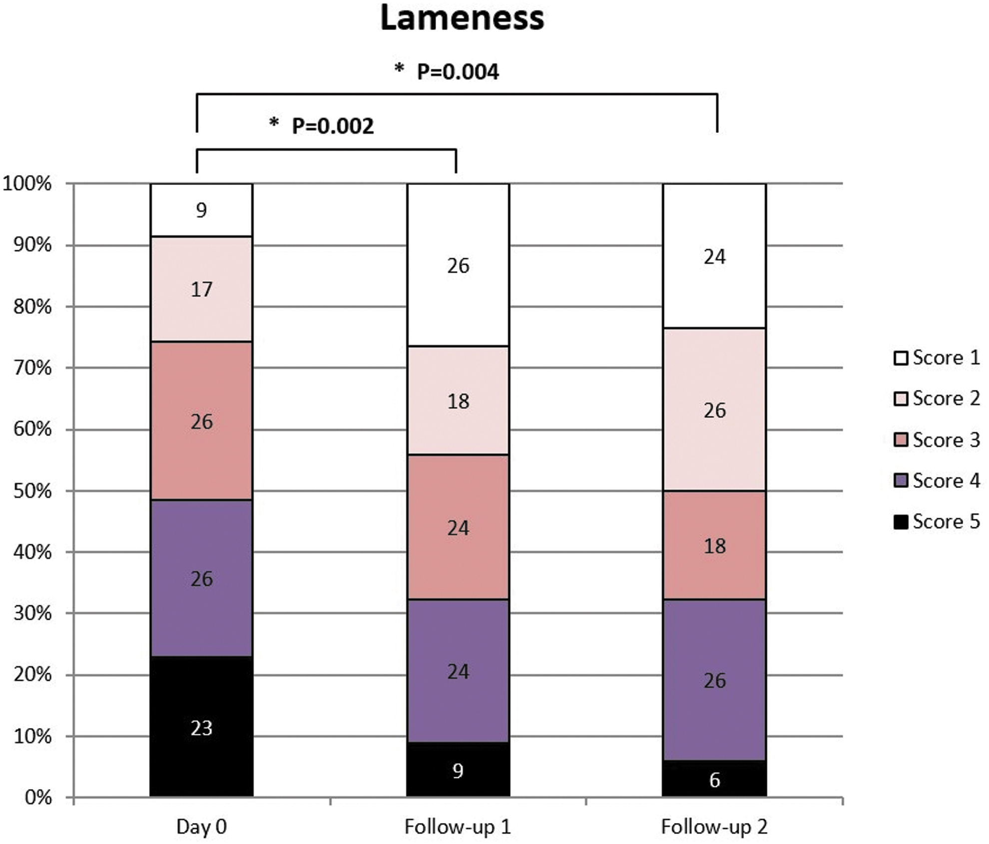
Overview of the lameness assessment for all three follow-up points. The percentage of patients of each score is presented for the three time points. At follow-ups 1 and 2, lameness scores were significantly decreased compared with Day 0. No significant differences in lameness scores could be detected between Follow-up 1 and Follow-up 2. *p < 0.05.
Six weeks after treatment administration (Follow-up 2), 50% of dogs had a lameness score of 1 or 2 of 5 (24% score 1 and 26% score 2), which is significantly better compared with baseline conditions (P < 0.05). No significant differences in lameness scores could be detected between Follow-up 1 and Follow-up 2 (P = 0.484). Furthermore, assessment of lameness of individual patients showed a decrease of lameness over a period of 6 weeks in 62% of cases. The remaining 38% of dogs showed a stable (26%) or worsened (12%) degree of lameness 6 weeks post-treatment.
A second parameter tested during the orthopedic examination was the ROM of the affected joints. Results of the scoring are presented in Fig. 2. A significant impact on the ROM was seen when comparing the second follow-up with baseline conditions (Day 0) (P < 0.05). Fifty percent of dogs had a score of 0 or 1 at Follow-up 2 compared with 33% on Day 0.
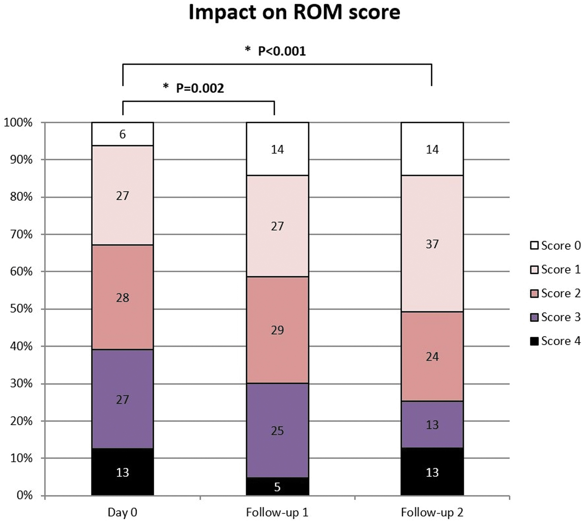
Overview of the ROM score for all three follow-up points. The percentage of patients for each score is presented for the three time points. The impact on ROM decreased significantly between Day 0 and both follow-ups. No significant difference was seen between Follow-up 1 and Follow-up 2. *p < 0.05. ROM, range of motion.
A significant difference was also found between Day 0 and Follow-up 1 (P < 0.05). Follow-up 1 and Follow-up 2 did not differ significantly (P = 0.455). Additionally, an evaluation of ROM per joint 6 weeks post-treatment showed an improvement of ROM in 34% of the joints. The ROM remained unchanged or worsened in, respectively, 61% and 5% of the joints.
The ROM was also evaluated by measuring the % change in degrees using a goniometer. Three weeks after treatment (Follow-up 1), the ROM in degrees was significantly higher (P < 0.05) with a median increase of 9.0 [95% confidence interval (CI): (3.5 to 16.0)] compared with baseline. After 6 weeks (Follow-up 2), the ROM in degrees was significantly higher (P < 0.05) with a median increase of 8.5 [95% CI: (2.5 to 13.5)] compared with baseline, but the two follow-up periods did not differ significantly (P = 0.964).
Considering the impact on clinical condition, no significant difference was found between all three time points.
Canine brief pain inventory
During the three visits of the patients at the veterinary practice (Day 0, Follow-up 1, and Follow-up 2), the owners completed the CBPI, assessing the PSS, PIS, and quality of life (Supplementary Table S3). This questionnaire revealed a significant decrease in PSS between Day 0 and Follow-up 1 [median = −1; 95% CI: (−1.5 to −0.5), P < 0.05], but not between Day 0 and Follow-up 2 [median = −1; 95% CI: (−1.0 to 0.0), P = 0.073] and between the two follow-up periods.
Concerning PIS, a significant decrease was found between Day 0 and Follow-up 1 [median = −1.4; 95% CI: (−2.4 to −0.9), P < 0.05] and between Day 0 and Follow-up 2 [median = −2.15; 95% CI: (−3.1 to −1.4), P < 0.05], but not between Follow-up 1 and Follow-up 2 [median = −0.5; 95% CI: (−1.0 to 0.1), P < 0.05].
Finally, an improvement in quality of life was observed in 66% of dogs over the 6-week study period after treatment administration. The quality of life remained the same or decreased during the 6-week study period in, respectively, 23% and 11% of cases.
Joint assessment
Finally, the joint assessment was performed after treatment administration. No significant differences could be found for heat sensation scores. Articular pain decreased significantly between Day 0 and Follow-up 1 [median = −1.0; 95% CI: (−1.5 to −0.5), P < 0.05]. No significant differences were found between Day 0 and Follow-up 2 (P = 0.073) and between both follow-up periods (P = 0.429).
A decrease in joint effusion scores was found after treatment administration. The joint effusion score decreased significantly between Day 0 and Follow-up 1 [median = −1.0; 95% CI: (−1.5 to −1.0), P < 0.05] and between Day 0 and Follow-up 2 [median = −1.0; 95% CI: (−1.5 to −1.0), P < 0.05], but the two follow-up periods did not differ significantly (P = 1.000) (Fig. 3).
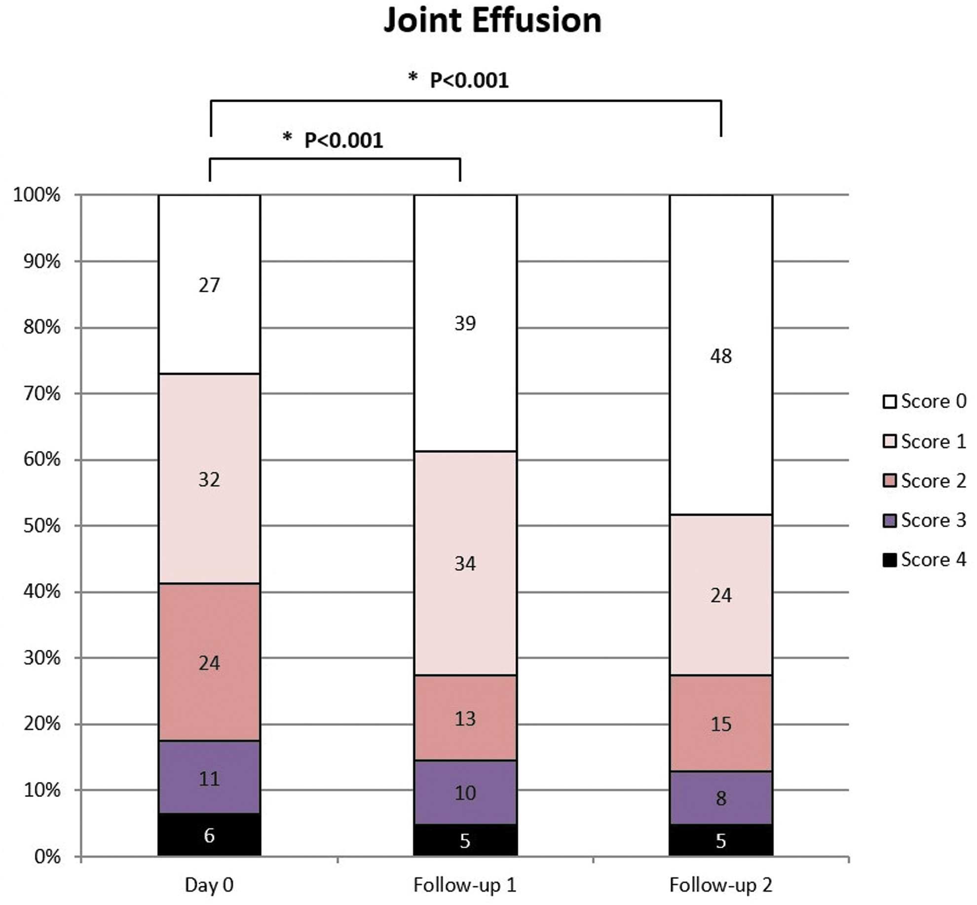
Overview of the joint effusion scores for all three follow-up points. The percentage of patients for each score is presented for the three time points. The joint effusion score decreased significantly between Day 0 and Follow-up 1 and between Day 0 and Follow-up 2, but the two follow-up periods did not differ significantly. *p < 0.05.
An evaluation of joint effusion per joint showed a decreased amount of effusion in 38% of joints 6 weeks after epbMSC administration. An equal or increased amount of joint fluid was detected in, respectively, 56% and 6% of the joints.
Discussion
To the best of our knowledge, this is the first study investigating the clinical response of dogs with articular pain to xenogeneic epbMSCs injected intravenously. The stem cell therapy appeared to be well tolerated as there were no suspected adverse drug reactions or serious adverse events related to the treatment administration reported during this study. Furthermore, joint assessment showed no increase in articular heat sensation. Significant decreases in articular pain, lameness, joint effusion, and impact on motion range were detected after treatment administration.
Previous studies have investigated the safety and efficacy of autologous, allogeneic, and xenogeneic MSCs in the treatment of OA, with moderate to very promising results [11 –15,28]. However, our research group is the first to describe the xenogeneic use of equine MSCs in dogs as a possible treatment option for chronic articular pain. Previously, the use of intra-articular administration of equine chondrogenic induced MSCs in the treatment of OA was described by our group, reducing lameness and pain in treated dogs according to the owner's evaluation [28].
In the current study, native MSCs were injected intravenously instead of intra-articularly, which might have a substantial systemic anti-inflammatory effect. This systemic anti-inflammatory effect might lead to reduced pain and inflammation in affected joints without the MSCs' local presence [16,17,32]. Furthermore, MSCs have been shown to be able to migrate to inflammatory regions, contributing to an additional local anti-inflammatory effect [18].
This type of administration also offers the advantage of intravenous injection of a ready-to-use formulation. IV injection of MSCs in the treatment of canine OA was previously described by Olsen et al., who used allogeneic MSCs and found an improvement in the clinical client-specific outcome measure–activity score. No difference in other outcome measures and owner questionnaires could be found after treatment.
Compared with the study by Olsen et al., our study was able to present a wider array of significantly different outcome measures [15]. Although these outcome measures were mostly subjective, they indicate that the use of a single IV injection of epbMSCs may be a promising and safe treatment for joint pain and lameness in dogs.
The safety of epbMSC injection was currently only evaluated based on clinical parameters. However, the xenogeneic administration of MSCs in previous studies has been shown to induce an increase in CD4+ cells [33]. Immunological assays such as the mixed lymphocyte reaction determining immunogenicity of MSCs could be performed in the future.
Moreover, as allogeneic and xenogeneic MHC-mismatched BM MSCs have been shown to induce a cell-mediated immune response after intra-articular injection in horses, activation of the cellular immune system after IV injection of epbMSCs in dogs should be analyzed in future studies [33 –35]. It should be mentioned that our study group tested the MHC II levels of MSCs by flow cytometry, using a 2% cutoff level.
MSCs can express different levels of MHC II, depending on the species and source of MSCs, which is one of the most important extracellular markers causing a cellular immune response of the host [36]. Nevertheless, the abovementioned studies are necessary to gain more insights into the immunogenicity profile of this canine stem cell therapy.
In the current study, epbMSCs have a clinical effect until a minimum of 6 weeks post-treatment. Although current results are interesting, they should be interpreted with caution due to several study limitations. The major limitation of the current study was the lack of a control group, randomization, and blinding. The lack of blinding could have caused a placebo effect. The data we obtained from the veterinarians specified the location of pain; however, the exact diagnosis was not requested.
In addition, the instructions given to the recruiting veterinarians were to enroll nonresponsive patients based on the known history of the dog; however, the nonresponsive period was not defined and was at the discretion of the veterinarian. Furthermore, no records were made from conventional treatment history. This lack of information combined with the heterogeneous group of patients makes data interpretation challenging.
In addition, this study was performed without objective scoring criteria such as gait analysis based on force or pressure plate since these tools are not standard or available in veterinary practices, which could have caused additional bias. Therefore, a future study with experimental animals using an OA model would make it possible to examine the potential histological effects. In addition, future studies should use MSC batches derived from multiple donor horses to investigate the potential variability across donor horses.
Despite the abovementioned limitations, the main objective of the current study was to evaluate the research potential of epbMSCs injected intravenously in dogs with chronic articular pain. Since epbMSCs seem to positively influence chronic articular pain without adverse events, future research on this topic is warranted. Randomized, blinded, placebo-controlled clinical trials in canine patients will give more insights into the short- and long-term efficacy and safety of epbMSCs for treatment of canine OA.
Conclusions
In conclusion, the results of this study demonstrate that a single IV injection of epbMSCs appears to be well tolerated by canine patients suffering from joint pain and lameness. Although no final conclusions can be made on efficacy, joint pain and lameness did not aggravate after epbMSC injection and there was even a tendency toward improvement.
These beneficial results encourage further controlled, randomized, and blinded research on this topic to provide more insights into the precise mode of action, safety, efficacy profile, and optimal dosage of xenogeneic epbMSCs for IV administration in dogs.
Footnotes
Acknowledgments
Gratitude is directed toward J.H.S. and J.S. as head of the department of Medical Imaging and Orthopedics of Domestic Animals, Faculty of Veterinary Medicine, Ghent University, who enabled this study. In addition, authors would like to specially thank G.P. and Y.S. who guided and supported this article.
Author Disclosure Statement
Authors J.H.S., L.H., E.D., and G.P. are employed with Boehringer Ingelheim. The content of this article contains a stem cell product under development owned by Boehringer Ingelheim. The remaining authors declare that the research was conducted in the absence of any commercial or financial relationships that could be construed as potential conflicts of interest.
Funding Information
The authors declare that this study was sponsored by Boehringer Ingelheim. The sponsor had the following involvement with the study: study design, data collection and analysis, decision to publish, and preparation of the manuscript.
Supplementary Material
Supplementary Table S1
Supplementary Table S2
Supplementary Table S3
Supplementary Table S4
References
Supplementary Material
Please find the following supplemental material available below.
For Open Access articles published under a Creative Commons License, all supplemental material carries the same license as the article it is associated with.
For non-Open Access articles published, all supplemental material carries a non-exclusive license, and permission requests for re-use of supplemental material or any part of supplemental material shall be sent directly to the copyright owner as specified in the copyright notice associated with the article.