




Abstract
The therapeutic potential of autologous stem cell transplantation for heart repair diminishes in the elderly due to stem cell aging. Rejuvenating aged stem cells to enhance their protective effects on injured cardiomyocytes is crucial for aging patients with heart failure. In this study, we aimed to investigate whether neuron-derived neurotrophic factor (NDNF) over-expression improves the protective effect of aged stem cells for injured cardiomyocytes and explore the underlying mechanism. Human bone marrow was collected from both young and old patients, and bone marrow mesenchymal stem cells (BMSCs) were cultured. Lentivirus expression vectors carrying NDNF genes were used to transfect aged BMSCs. Fatal hypoxia-induced injury in H9C2 cells served as an in vitro ischemia model. The conditioned medium from different BMSC groups was applied to assess the beneficial effects on hypoxia-induced damage in myocardial H9C2 cells. Results revealed that the conditioned medium of NDNF over-expressed old BMSCs increased H9C2 cell viability and reduced oxidative stress and apoptosis levels under fatal hypoxia. NDNF over-expressed old BMSCs exhibited an antiapoptotic role by upregulating the antiapoptotic gene Bcl-2 and downregulating the proapoptotic genes Bax. Additionally, the protective effects were mediated through the elevation of phosphorylated AKT. Our data support the promise of NDNF as a potential target to enhance the protective effects of autologous aged BMSCs on ischemic cardiomyocytes and then improve the curative effects of stem cell for ischemic heart injury in aged patients.
Introduction
Cardiovascular diseases are the leading cause of death and disability around the world. 1 Cardiac ischemia, caused by vascular diseases such as coronary atherosclerosis, usually induces massive cardiomyocytes death. The existing therapeutic treatments are based on reperfusion strategies. Although they have the potential to save myocardial infarction to a certain extent, the dead and dying cardiomyocytes in adults are irreversible. And the cardiac function and quality of life were severely affected because of the excessive cardiomyocyte loss. Therapeutic strategies focus on cardioprotection and cardiac repair, such as stem cell therapy, have grown into a new strategy for the treatment. 2
The use of stem cell-based therapy has been quickly launched. Cardiomyocyte apoptosis and cell death were reduced, and cardiovascular regeneration was enhanced by MSCs treatment. 3 Many clinical trials have demonstrated the safety and effect in improving heart function of stem cell therapy. 4 Stem cell transplantation effectively boosts cardiac function, but its efficacy wanes in older patients due to the diminished regenerative capacity of aging bone marrow mesenchymal stem cell (BMSC) therapy. Simultaneously, allogeneic stem cell transplantation is less potent due to alterations in cell surface antigens and heightened susceptibility to adaptive immune rejection. 5 Strategies for rejuvenating aged MSCs to optimize autologous MSC-based cardioprotection and myocardial therapy are needed.
Neuron-derived neurotrophic factor (NDNF) serves various biological functions aligned with stem cell rejuvenation goals, encompassing the stimulation of cell proliferation, inhibition of apoptosis, and promotion of angiogenesis. 6 Our previous study demonstrated that NDNF over-expression rejuvenates aged human BMSCs, enhancing angiogenesis postischemic injury. 7 However, the direct protective effects and the mechanism of NDNF over-expression in aged human BMSCs on damaged cardiomyocytes remain unclear. Cardiomyocyte survival is crucial for cardiac function, with reactive oxygen species (ROS)-mediated oxidative stress identified as a key process in cardiac ischemia and ischemia/reperfusion-induced injury. ROS overproduction may induce cellular damage and cardiomyocyte apoptotic death. This study utilized an in vitro hypoxic environment to establish an ischemia injury model in H9C2 cells, assessing the protective effects and mechanisms of NDNF over-expressed aged BMSCs on myocardial ischemia injury.
Materials and Methods
Culture and overexpression of NDNF in aged bone marrow mesenchymal stem cells
Our research received approval from the research ethics board of Shanxi Medical University. With written informed consent, human bone marrow was collected from young (20–30 years old) and old (over 60 years old) patients during bone marrow aspirate and biopsy. BMSCs were cultured and passaged using established methods. 7
BMSCs were transfected with lentivirus expression vectors carrying NDNF genes (Lenti-Puro-EF1α-NDNF-Homo-IRES-eGFP, Cyagen Biosciences Inc., Santa Clara, CA) following the manufacturer’s instructions. Successful NDNF over-expression was confirmed by detecting NDNF mRNA and protein levels. Subsequently, BMSCs and their supernatants were collected for further experiments.
Myocardial ischemic model in vitro under the protection of BMSCs conditioned culture medium
After reaching 80% confluence, NDNF-transduced old BMSCs (Old+NDNF), empty vector-transduced old BMSCs (Old), and empty vector-transduced young BMSCs (Young) were further cultured with 2% FBS-DMEM/F12 medium for an additional 48 h. The harvested conditioned medium was then used for subsequent analysis. H9C2 cardiomyocytes from the Cell Bank of the Chinese Academy of Sciences were cultured in normoxia (21% O2, 5% CO2) with 10% FBS-DMEM until reaching approximately 80% confluence. Subsequently, H9C2 cells were treated with various conditioned media from BMSCs, harvested, and exposed to a hypoxic environment of 0.1% O2 and 5% CO2 to induce an in vitro myocardial ischemic injury model.
Cell viability assay
H9C2 cells (8000 cells/well) were placed into 96-well plates and incubated for 24 h. Following ischemic injury under the protection of BMSCs conditioned medium, the CCK-8 assay was conducted using the cell counting kit-8 assay kit (MedChemExpress, Shanghai, China) according to the manufacturer’s instructions. OD values were collected at 450 nm on a microplate reader (Eppendorf, Hamburg, Germany).
Detection of ROS production in H9C2
H9C2 cells (8 × 10⁴ cells/well) were placed into 24-well plates and grown for 24 h. To determine the level of ROS produced in H9C2 cells following ischemic injury, dihydroethidium (DHE) staining was employed. H9C2 cells in each group were incubated with DHE(5 μM, MedChemExpress, Shanghai, China) at 37°C in dark for 30 min. After staining with DAPI and mounting, pictures were taken by the NIKON fluorescence microscope. The average DHE staining area percentage was calculated for quantitative analysis.
TUNEL staining
The level of apoptosis was measured using the one-step TUNEL apoptosis detection kit (Beyotime Biotechnology, Shanghai, China), following the manufacturer’s instructions.
Western blotting
H9C2 cells were collected on ice using protein lysis buffer, supplemented with protease and phosphatase inhibitors. Equal protein amounts were loaded onto SDS gel, transferred to PVDF membranes, and, after blocking, incubated with primary antibodies. Subsequently, HRP-conjugated secondary antibodies were applied, and signals were detected using enhanced ECL Western blotting detection reagent. Antibodies utilized: NDNF (1:1000, ab175602, Abcam, Cambridge, England); p-AKT (1:1000, 4060, Cell Signaling, Boston, MA, USA); AKT (1:1000, 4691, Cell Signaling, Boston, MA, USA); Bax (1:200, sc-20067, Santa Cruz Biotechnology, Heidelberg, Germany); Bcl-2 (1:500, 68103, Proteintech, Wuhan, China); GAPDH (1:1000, TA-08, ZSGB-BIO, Beijing, China).
Total RNA extraction and RT-PCR
Total RNA was extracted with TRIZOL reagent (Invitrogen, Waltham, MA, America). The RNA concentration was assessed using a spectrophotometer (Eppendorf, Hamburg, Germany), and reverse transcription of cDNA was carried out using by high capacity cDNA reverse transcription kit (ThermoFisher, Waltham, MA, America) according to the manufacturer’s instructions. Primers used were as follows:
NDNF–forward primer: 5′-CCTTTGGAGTGGAAGCTGAG-3′; NDNF–reverse primer: 5′-GTAGACATGACGCCCCAGTT-3′; GAPDH–forward primer: 5′-GGAGCGAGATCCCTCCAAAAT-3′; GAPDH–reverse primer: 5′-GGCTGTTGTCATACTTCTCATGG -3′.
Statistical analyses
Values were presented as mean±SEM and analyzed using GraphPad Prism software. One-way analysis of variance with Tukey post-hoc tests was employed for comparisons among the three groups. Student’s t-test was used for analyzing differences between two groups. Statistical significance was set at P < 0.05.
Results
The conditioned medium of NDNF over-expressed aged BMSCs increased the H9C2 cell survival under ischemic injury
Efficiency of NDNF transfection into aged BMSCs was assessed 72 h post transfection. NDNF mRNA expression significantly increased in old + NDNF compared with empty-vector transfected BMSCs (old) (Fig. 1A). Protein expression of NDNF in BMSCs, as measured by Western blotting (Fig. 1B), showed a substantial elevation in NDNF-transfected old BMSCs, confirming successful transfection. To verify the protective role of NDNF over-expressed old BMSCs on cardiomyocytes, H9C2 cells were treated with different conditioned media from BMSCs under lethal hypoxia. Cell survival after ischemic injury was assessed using a CCK8 assay. H9C2 cell viability with conditioned media from old + NDNF BMSCs was higher than that from old BMSCs (Fig. 1C).

The conditioned medium of NDNF over-expressed aged BMSCs increased the H9C2 cell survival under ischemic injury.
Effects of NDNF over-expressed old BMSCs on hypoxia-induced oxidative stress and apoptosis in H9C2
To explore the approach of NDNF over-expressed old BMSCs on cardiomyocytes protection, we assessed superoxide generation in H9C2 cells through DHE staining, calculating fluorescence intensity as a measure of oxidative stress. The young BMSC-treated group exhibited the lowest DHE-positive area in H9C2 cells, while the old group showed the highest. Notably, the oxidative stress level in H9C2 cells treated with old + NDNF BMSCs was significantly lower than in those treated with old BMSCs (Fig. 2).

The conditioned medium of NDNF over-expressed aged BMSCs down-regulated the hypoxia-induced oxidative stress of H9C2 cell.
Next, we evaluated apoptosis in H9C2 cells using TUNEL staining and detecting apoptosis-related proteins. Quantification of TUNEL+H9C2 cells revealed the lowest area of apoptotic cells in the young BMSC-treated group. Importantly, fewer apoptotic cells were observed in H9C2 cells treated with old + NDNF BMSCs compared with those treated with old BMSCs (Fig. 3A, 3B). Consistent with this result, Western blotting showed markedly reduced expression of the pro-apoptosis-related protein Bax and higher expression of the anti-apoptosis-related protein Bcl-2 in old + NDNF BMSCs treated H9C2 cells compared with the old group (Fig. 3C, 3D).
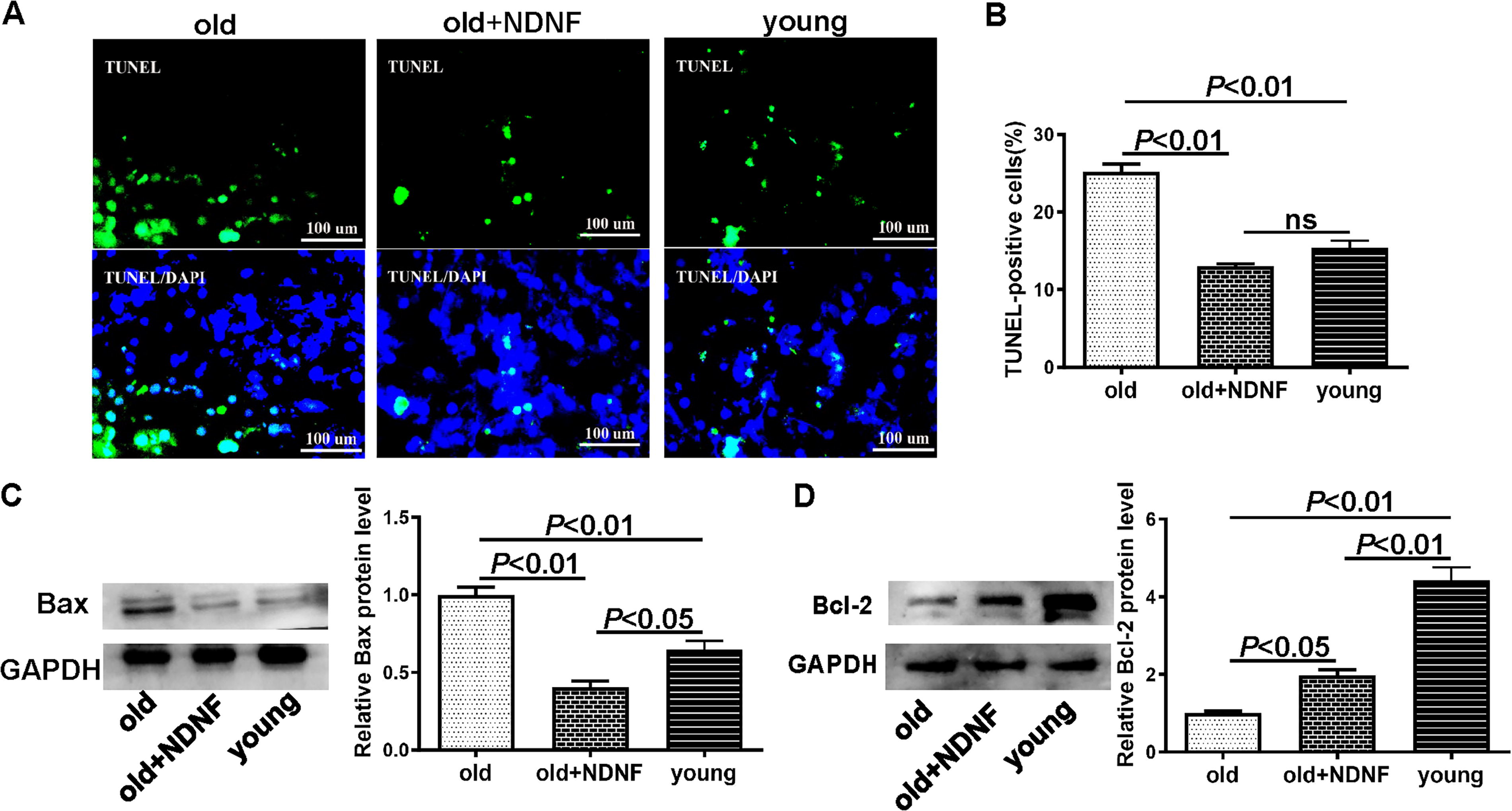
The conditioned medium of NDNF over-expressed aged BMSCs down-regulated the hypoxia-induced apoptosis of H9C2 cell.
NDNF over-expressed old BMSCs activated p-AKT pathway in H9C2
The expression of phosphorylated AKT (p-AKT) was significantly lower in H9C2 under the protection of conditioned medium from old BMSCs than that from young BMSCs, while its expression was significantly increased in H9C2 under the protection of conditioned medium from old + NDNF BMSCs than that from old BMSCs (Fig. 4A). At the same time, the total AKT expression was similar among all three groups (Fig. 4B).
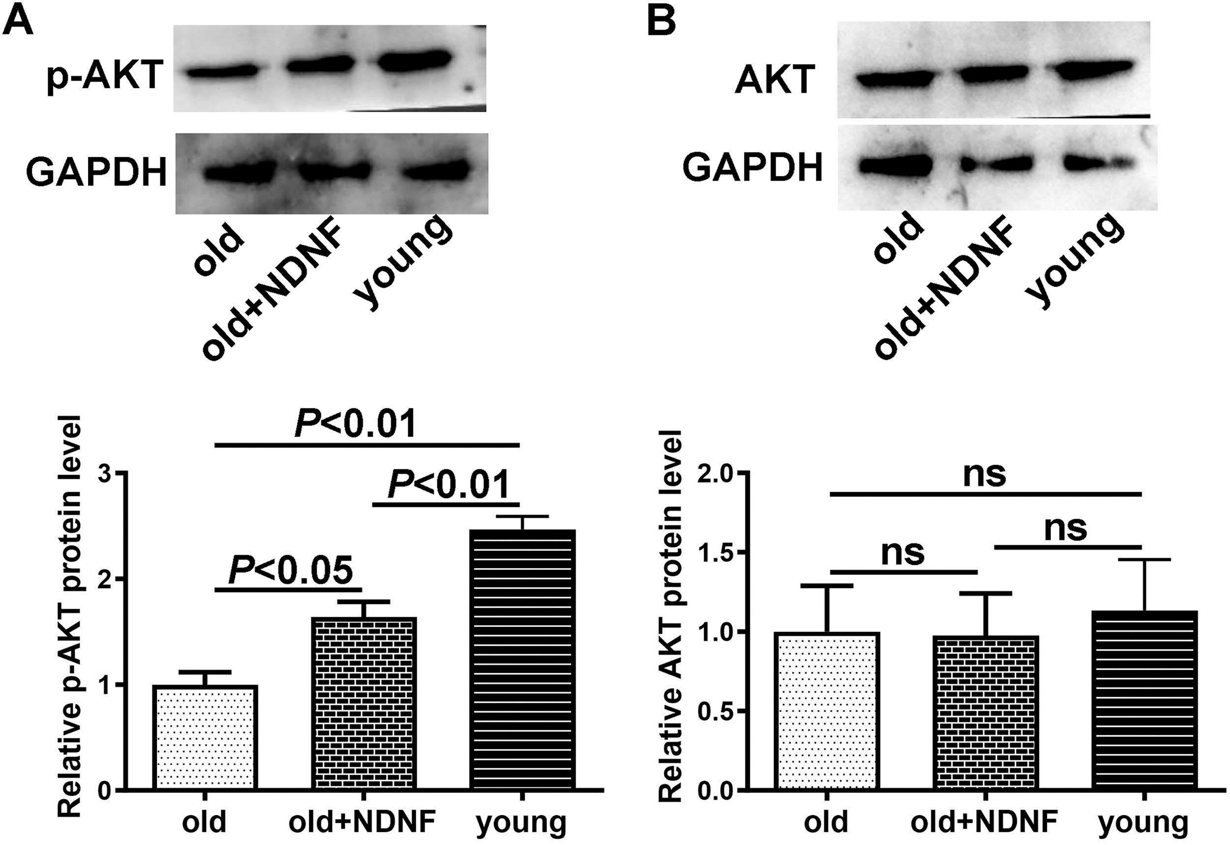
The protection of NDNF over-expressed old BMSCs on ischemic H9C2 was through activation of p-AKT.
Discussion
Ischemic heart disease poses a great threat to human health because of its high morbidity and mortality. Myocardial infarction leads to inflammatory outburst, oxidative stress, cardiomyocytes death, the ventricular remodeling and other pathological changes, and even heart failure. 8 The main problem is that the damaged cardiac tissue can’t regenerate and cardiac function is difficult to recover. 9
Stem cell therapy has arisen as a promising strategy for the management of ischemic heart disease, for the protective capacity to injured cardiomyocyte. Studies have confirmed that bone marrow-derived mesenchymal stem cells can improve cardiac function and reduce myocardial infarct size in ischemic heart disease and confirmed the effectiveness of autologous and allogeneic mesenchymal stem cells. 10–11 However, physical function gradually deteriorates with age. Aged BMSCs have reduced capacity for self-renewal, proliferation, and pluripotent differentiation. So, they are unable to effectively exert their therapeutic effects on ischemic heart disease. 12 The allogeneic cell transplantation will be rejected and lost therapeutic efficacy in the late stage of transplantation. 13 Therefore, promoting the rejuvenation of aged BMSCs and improving their ability to protect injured cardiomyocytes is particularly important for ischemic heart disease in elderly patients.
NDNF is a neuron-derived neurotrophic factor. It can stimulate cell growth and inhibit apoptosis. Experiments in mice have demonstrated that endothelial cell secretion of NDNF can enhance the function and increase the survival of endothelial cells in mice with ischemic limb injury, and systematically increasing NDNF levels in mice can improve cardiac function, increase angiogenesis after MI. 14 We previously demonstrated the over-expression of NDNF can rejuvenate aged hBMSC and implantation of NDNF over-expressed old hBMSCs following MI into the cardiac border area of mice decreased scar size and improved angiogenesis. 7 However, if the NDNF over-expressed old hBMSCs has the direct protective effects on damaged cardiomyocytes and the underling mechanism is still unclear. In this study, it was demonstrated that NDNF over-expressed old hBMSCs indeed has the direct protective effects on damaged cardiomyocytes. The results showed that the conditioned medium of NDNF over-expressed aged BMSCs increased the H9C2 cell survival under ischemic injury.
Oxidative damage is the important underlying mechanism or etiology for myocardial ischemia injury. Large amounts of ROS are produced during myocardial ischemia injury. The oxidative damage causes excessive myocardial cell apoptosis and then impairs the cardiac function. So, the ROS production and associated apoptosis of cardiomyocytes are the two vital pathological factors in ischemia-induced cardiomyocyte injury and also the major factors that determine the survival of injured cardiomyocytes. 15 The protective effects of BMSC are related with the secretion of immunomodulatory and antiapoptotic factors. 16
Our findings in the current study indicated that there was a significantly higher level of oxidative stress, as detected by DHE staining, in H9C2 cells treated with old BMSCs compared with those treated with young BMSCs. Furthermore, H9C2 apoptosis after old BMSCs treatment was significantly more than after young BMSCs treatment. This is one of the reasons that the therapeutic effects of stem cell transplantation for cardiac repair are less pronounced in aged patients. And more importantly, our results showed that the oxidative stress level was significantly lower and apoptosis was significantly less in ischemic H9C2 treated with old + NDNF BMSCs than that of old BMSCs. In other words, our results suggest that overexpression of NDNF may improve the regenerative effects of transplanted aged BMSCs in response to ischemic heart injury by modulating levels of oxidative stress and subsequent apoptosis of damaged cardiomyocytes.
AKT is an important factor for cell survival. Our data showed that NDNF over-expressed BMSCs induced activation of the phosphorylated AKT (p-AKT) pathway, suggesting that the protective effects of NDNF over-expressed BMSCs are mediated by this pathway. This result is consistent with several existing studies. The activation of the AKT signaling pathway has been demonstrated to confer protection against apoptosis of cardiomyocytes in mice with diabetes. 17 Furthermore, activation of the IGF-1/PI3K/AKT signaling pathway exerts a cardioprotective effect by reducing apoptosis and inflammatory stress in cardiomyocytes. 18 However, additional research is required to determine the exact mechanisms underlying the protective effects of NDNF-overexpressing BMSCs on H9C2.
However, there are some limitations of the cell model we used in this article. H9C2 cells, derived from rat embryonic heart tissue, possess a unique combination of cardiomyocyte and skeletal muscle properties, making them a valuable tool for various research studies in biophysics, biochemistry, and pathophysiology. 19 However, it is important to note that differences exist between H9C2 cells and in vivo heart tissue, including variations in extracellular matrix composition, cell-cell interactions, and mechanical forces. Additionally, disparities may arise when comparing cardiomyocytes from different species and tissue sources. These variations may impact the cellular behavior and impede the precise replication of physiological and pathological phenomena in vivo. 20 We will further the investigation with cardiac organoids derived from human stem cells in the following research, which is a better model and may offer a more effective means of replicating physiological and pathological states within the human body.
Conclusion
In summary, NDNF could enhance aged hBMSC protective effects against ischemic injury of cardiomyocytes by suppressing the oxidative stress and apoptosis. Moreover, the protective function was potentiated by the activation of AKT pathway. NDNF could be a potential target to enhance the protective effects of autologous aged BMSCs on ischemic cardiomyocytes, and it could be a promising target to improve the curative effects of stem cell for ischemic heart injury in aged patients.
Footnotes
Ethics Statement
This research was approved by the research ethics board of Shanxi Medical University (2017LL058).
Author Disclosure Statement
The author reports no conflicts of interest in this work.
Funding Information
This study was supported by the National Natural Science Foundation of China (81702239), Fund Program for the Scientific Activities of Selected Returned Overseas Professionals in Shanxi Province (20230021), Research Project Supported by Shanxi Scholarship Council of China (2021-078), China postdoctoral science foundation(2021M691994), the Natural Science Foundation of Shanxi Province (20210302123296, 20210302124571, 202103021224243), National Clinical Key Specialty Construction Fund (Y2022ZD001).