




Abstract
The human stomach breaks down and transports food by coordinated radial contractions of the gastric walls. The radial contractions periodically propagate through the stomach and constitute the peristaltic contractions, also called the gastric motility. The force, amplitude, and frequency of peristaltic contractions are relevant to massaging and transporting the food contents in the gastric lumen. However, existing gastric simulators have not faithfully replicated gastric motility. Herein, we report a soft robotic gastric simulator (SoGut) that emulates peristaltic contractions in an anatomically realistic way. SoGut incorporates an array of circular air chambers that generate radial contractions. The design and fabrication of SoGut leverages principles from the soft robotics field, which features compliance and adaptability. We studied the force and amplitude of the contractions when the lumen of SoGut was empty or filled with contents of different viscosity. We examined the contracting force using manometry. SoGut exhibited a similar range of contracting force as the human stomach reported in the literature. Besides, we investigated the amplitude of the contractions through videofluoroscopy where the contraction ratio was derived. The contraction ratio as a function of inflation pressure is found to match the observations of in vivo situations. We demonstrated that SoGut can achieve in vitro peristaltic contractions by coordinating the inflation sequence of multiple air chambers. It exhibited the functions to massage and transport the food contents. SoGut can simulate the physiological motions of the human stomach to advance research of digestion.
Introduction
Soft actuators and robots can emulate or enhance the functionalities of the human body.1–3 A number of soft robotic devices have been developed for assisting or mimicking the abilities of hands,4,5 legs,6,7 and heart.8,9 However, robots that are dedicated to replicating the peristaltic contractions of the human stomach are still primitive in the field of food science. 10
The human stomach exhibits peristaltic contractions that are a series of wave-like contractions (Fig. 1a).11,12 The amplitude, frequency, and force of these contractions are relevant to the normal physiological and pathological conditions of the stomach. Similar to the human heart, the stomach is a muscular organ and has a pacemaker region to coordinate the muscle contractions. In a healthy stomach, the pacemaker region at the upper part of the stomach stimulates a contraction every 20 s. The resultant contraction propagates through the body and antrum of the stomach for a period of 60 s. The contracting force in the antrum plays a key to grinding the food. 10 The value of the contracting force varies from 0.2 to 1.0 N. The amplitude of contraction is also varied for the different parts of the stomach and digestion phases. 13 Stomach anatomy is affected by many factors such as the body position, food contents, and surrounding organs. The peristaltic contractions serve two main functions. The first is massaging the food to break down and mix it with gastric juice. The second is transporting the food from the stomach to the intestines when the food is ground small enough to pass through the pylorus.
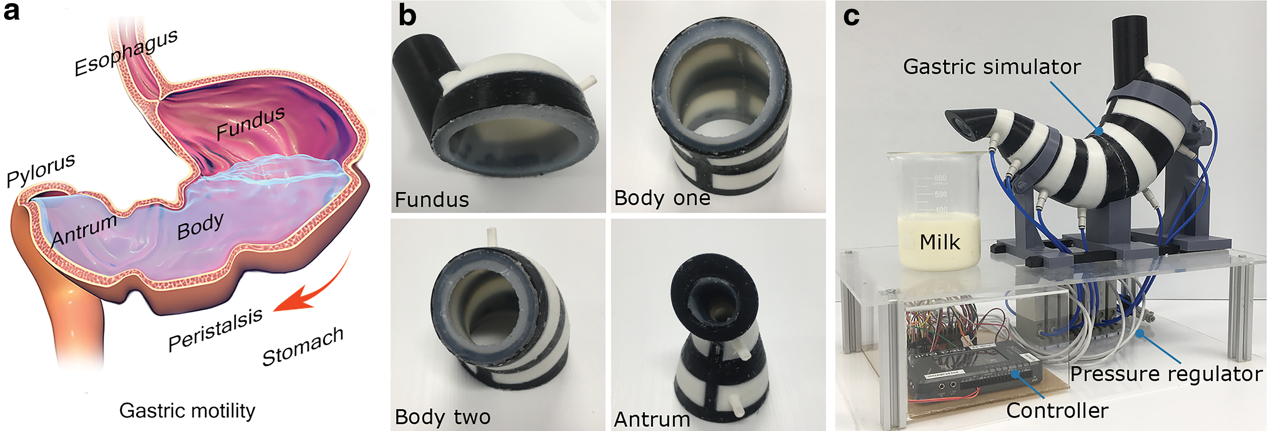
The soft robotic gastric simulator (SoGut).
Gastric simulators that closely emulate the peristaltic contractions are in demand.10,14,15 This class of simulator has two main practical applications: (1) to advance the investigation of the in vivo digestion process of food and drugs16,17 and (2) to promote the development of new technologies such as stomach pacemakers18,19 and microrobots that are working in the human stomach.20,21
Several gastric simulators have been developed to reproduce the change of the food in the human stomach for food science and nutrition.22–27 However, the existing devices lack the realistic gastric anatomy and motility. For example, the dynamic gastric model has a cone-shaped elastic membrane used to imitate the body of the stomach.22,23 Pressurized water pushes the membrane to realize the contraction. A barrel and piston are combined to mimic the breakdown functions in the antrum. Another gastric simulator named TNO intestinal model-1 consists of two adjoining glass units with flexible chambers inside.24,25 Pressurized water surrounds the chambers and forces the food contents to move back and forth in the two glass units, leading to the mixture of the food contents. Human gastric simulator is designed to produce the peristaltic contractions of the stomach. 26 It consists of a latex lining with a set of rollers placed around the lining. Rollers move against and push the lining inward. Although it generates the contraction with the similar frequency and force to the human stomach, it overlooks the amplitude of contractions and the stomach shape. Another simulator is a silicone stomach model wrapped with ropes for validation of surgical endoluminal robots. 27 The ropes that are pulled by the motors compress the model surface, but the amplitude and force of contractions are not investigated. To summarize, the research on physical gastric simulators done so far is multidisciplinary and has different research motives. Those works for evaluations of new drugs, foods, and surgical plans have resulted in process simulators,22–25 mechanical simulators12,26 and a biomimetic robot, 27 respectively. The comparison of the existing gastric simulators is summarized in Table 1. The corresponding illustrations are listed in Supplementary Table S1.
The Features of Existing Gastric Simulators
Gastric anatomy refers to the tubular shapes of the simulators that contain key regions of fundus, body, and antrum. The peristaltic contractions are a series of wave-like radial contractions generated by the simulators. The physiological process refers to the simulation that involves chemicals such as gastric juice. The sign “—” means the number is not available.
DGM, dynamic gastric model; HGS, human gastric simulator; SoGut, soft robotic gastric simulator; TIM-1, TNO intestinal model.
We previously analyzed the technical requirements and proposed conceptualization of a robotic gastric simulator. 28 We summarized the key features of the peristaltic contractions and shape of the stomach. As a step further toward a functional gastric simulator, we developed a soft ring-shaped actuator that generates radial contractions, where its quasi-static performance under the inflation pressure was modeled and validated. 29 However, by itself, it cannot emulate the gastric anatomy and peristaltic contractions.
In this study, we report a soft robotic gastric simulator (SoGut) that mimics the gastric peristaltic contractions in an anatomically realistic manner. SoGut embodies the anatomy and motility that agree with the observation of the human stomach. The novelty of this study lies in the systematic approach to designing and examining the SoGut, which has substantially advanced our early study on the soft ring actuator. 29 The other part of novelty is that SoGut mimics the in vitro contracting force, contraction ratios, and peristaltic motions of the human stomach, which the previous gastric simulators are short of. SoGut can serve as an important new tool to investigate digestion. This study provides engineers and roboticists with a holistic approach to developing bioinspired and biomimetic systems using soft robotic techniques. The rest of the article is organized as follows. The Description of SoGut section describes the design and fabrication of SoGut. Experiments section elaborates the experimental setup for examining the contraction ratio, contracting force, and peristaltic motions. Results are analyzed in Results and Discussions section. Conclusion section concludes the article with ideas for the future developments.
The Description of SoGut
Design and working principle
SoGut comprises four segments that embody the gastric anatomy as shown in Figure 1a and b. The segments are embedded with circular air chambers that can generate radial contractions. To demonstrate the radial contractions, each air chamber was inflated in an increment of 1 kPa (Supplementary Movie S1). It was observed that air chambers #1 and #7 can be operated within the pressure range of 0–6 kPa and air chambers #2 to #6 at the range of 0–9 kPa.
SoGut consists of a skeleton, a membrane, and shells with a shape akin to the human stomach (Fig. 2a). The skeleton provides a framework combining the shells and membrane to form air chambers. The key dimensions are labeled on the structure of SoGut bereft of shells (Fig. 2b) with their values given in Table 2. The skeleton and membrane constitute seven open air chambers that are arranged along a curved axis. The shells that are attached to the skeleton enclose air chambers. The geometry and dimensions of SoGut are in accordance with the shape of the human stomach in the literature.11,13

The design and working principle of SoGut.
The Dimensions of Soft Robotic Gastric Simulator
The circular air chambers of SoGut are activated with the inflation pressure to achieve radial contractions. Figure 2c shows the cross-sectional view of SoGut along the curved axis at the rest state. The membrane covering each air chamber #1 to #7 is the deformable area and the skeleton and shell are the rigid areas. The diameter of air chambers at the rest state is denoted as Di, where i is the index of air chamber. Figure 2d shows the radial contractions of SoGut under the inflation pressure. The diameter of the inflated air chamber is denoted as di. The T-structure firmly secures the edges of the membrane within the slots of the skeleton. With the shells surrounding the skeleton and membranes, air chambers are fully closed and activated under the inflation pressure.
Materials and fabrication procedures
The fabrication of SoGut involves three-dimensional (3D) printed molds (Prusa i3; Prusa Research, Czech Republic), elastomer (Ecoflex 00-30; Smooth-on, Inc., USA), silicone adhesive (Sil-Poxy; Smooth-on, Inc.), cyanoacrylate adhesives (Loctite 401; Henkel, Australia), and wax (Beeswax; Jacquard, New Zealand). All computer-aided design (CAD) files of the molds were developed in SolidWorks (Education Edition 2018; Dassault Systemes, France). The molds were 3D printed.
SoGut comprises four segments that are robotic fundus, body one, body two, and antrum corresponding to the anatomy of the human stomach as shown in Figure 3. The fabrication of each segment involves 3D-printed parts, wax, and Ecoflex. 3D printed parts include cores, skeletons, solid shells, and shells. The difference between the solid shells and the shells is that the latter have a cavity as part of air chambers. The molten wax is used as a placeholder to fit in the gap between the skeletons and solid shells. Ecoflex is used to form the membrane. Silicone adhesive was used to seal the seams between the molds when the uncured elastomer was poured inside the assembly. Cyanoacrylate and silicone adhesives were applied to attach the four robotic segments together.

Fabrication procedure of SoGut. The fabrication procedure of robotic fundus
The first step of making the robotic fundus involves two cores, a solid shell and a skeleton as shown in the first column of Figure 3a. Then, the Ecoflex (uncured elastomer) was poured into the assembled molds. The elastomer cured, embedded into the skeleton, and formed the membrane in the second column of Figure 3a. At last, a shell covered the gap between the membrane and the skeleton to make up air chambers.
For the robotic body one, first, the core, skeleton, and solid shells were prepared in Figure 3b. The molten wax was carefully fitted on the beam of the skeleton to fill the gap between the skeleton and solid shells. Second, uncured elastomer was poured into the gap of the assembled molds. Silicone adhesive was used to seal the seam to prevent the leakage. After the curation, the solid shell and wax were removed, forming the circular gap between the skeleton and the membrane. Third, the shells covered the gaps leading to the circular air chambers.
The similar procedures were applied to robotic body two and antrum (Fig. 3c, d). Once the robotic fundus, body one, body two, and antrum were ready, they were connected consecutively using silicone and cyanoacrylate adhesive. The assembly was the prototype of SoGut.
To make the prototype portable, SoGut was mounted on a platform as in Figure 1c. The platform was laser cut from an acrylic board to support the prototype. The pneumatic control system was equipped to regulate the inflation pressure. It involves a custom circuit, seven electro-pneumatic regulators (ITV00 series; SMC, Japan), and a controller (NI myRIO-1900; National Instruments, USA) loaded with customized LabVIEW scripts.
Finite element model of the membrane
We visualized the deformation through finite element analysis to assist the design. A finite element model of the membrane was constructed to visualize the radial contractions, with SolidWorks (Education Edition 2018; Dassault Systemes). The membrane of SoGut was modeled using the hyperelastic neo-Hookean model. This model was chosen because it is suitable for predicting the performance of the incompressible elastomeric material. The neo-Hookean strain energy density function, defined as
where
In the simulation, we applied air chambers #2 to #7 with the inflation pressure on the membrane, the same value as the physical experiment in Supplementary Movie S1. At the meantime, the T-structures were constrained. The skeleton, shells, and pneumatic tubes were neglected, considering they have little impact on the contractions. The contractions of an idealized ring-shaped actuator with uniform thickness have been simulated in the previous study. 29 However, the simulation in this study is based on SoGut, a device made of a series of ring actuators with varying dimensions. Whereas the previous simulation is used for the validation of the theoretical modeling, the simulation of SoGut serves to visualize the deformations and guide the fabrication process. The simulations of this finite element analysis are in Supplementary Movie S2.
Experiments
This study was designed to develop a SoGut that can emulate the gastric peristaltic contractions in an anatomically realistic way. To achieve this aim, we laid out three objectives. The first was to characterize the contracting force when the lumen of SoGut was empty or filled with food contents of different viscosity. The second was to investigate the amplitude of the contractions when SoGut interacts with food contents of different viscosity. The third is to examine the capability of the proposed simulator to mimic in vivo peristaltic contractions.
Food preparation
Three types of liquids with varying consistencies were selected as the food contents to test the performance of SoGut in Supplementary Figure S1. They were (1) honey-like fluid, (2) pudding-like fluid, and (3) ground congee. The selections were made because they are akin to the contents in the human stomach. The experiments with three foods in this study are intended to show the SoGut's capability. Honey-like fluid and pudding-like fluid were prepared with a commercial food thickener (Altrix Rapid Thickener; Douglas Nutrition Ltd., USA). The food thickener is commonly used to prepare food for dysphagic patients. Following the user instruction of the product, we mixed 10 and 20 scoops of food thickener powder into 1.0 L water, respectively, to obtain honey-like and pudding-like fluids. For the closer mimicry to the stomach contents, the third we chose was 1.0 L of mixed congee (Black Bean and Sesame; Taisun, Ltd., Taiwan). The congee was ground by a smoothie maker (300W Smoothie Maker; Living & Co., New Zealand) for 5 s to mimic the mastication process.
To quantitatively evaluate the difference between these selected food contents, we examined their viscosity. The viscosity test on the selected food contents complied with the National Dysphagia Diet guideline. 31 We obtained the data through an MCR 301 rheometer (Anton Paar GmbH) at the temperature of 25°C and the shear rate of 50 s−1. The viscosity of the honey-like fluid, pudding-like fluid, and ground congee are 0.30, 1.36, and 5.31 Pa/s, respectively.
Experimental setup for the contracting force
To characterize the contracting force at air chambers #6 and #7, we utilized manometry as shown in Figure 4. It is a common technique that measures the pressure in the esophagus and stomach. The manometry catheter (P33-15205CC152; Sandhill Scientific, USA) was placed in the lumen of SoGut. The manometry catheter is equipped with five evenly spaced pressure sensors at its distal end. When measuring the contacting pressure at air chamber #6, we aligned one of the pressure sensors with air chamber and then collected the contacting pressure during the inflation process. We placed the sensor above the midpoint of air chamber. Since the sensor and air chamber were in the lumen of SoGut, the configuration was achieved according to the dimensions of SoGut and the manometry catheter. The contracting force is the product of the measured pressure and the areas where the sensor underwent the pressure exerted by the inflated air chamber. The inflation process increased at an increment of 1 kPa to the pressure of 9 kPa. The inflation process was repeated five times to assess consistency and reproducibility. This measurement method on air chamber #6 was conducted at four test settings that are empty and three selected food contents, respectively. The same procedure to measure the contracting force was applied to air chamber #7. The sampling frequency of the pressure sensor was 30 Hz (S98-200C; Sandhill Scientific, USA).

Experimental setup for the contracting force measurement. The manometry system contains two parts: the manometry catheter and the data acquisition system. The catheter has the five pressure sensors that are evenly spaced at the distal end of the catheter. One of the pressure sensors is placed on the midpoint of air chambers #6 and #7, respectively, to measure the contacting pressure. Color images are available online.
Experimental setup for the contraction ratio
To characterize the contraction ratio of SoGut, we used videofluoroscopy to observe the radial contraction of air chambers under the inflation pressure in Figure 5. Videofluoroscopy is a common method to observe the human esophagus and stomach. Although the contractions and peristalsis vary from person to person, healthy adults show general features,
28
which we intend to have SoGut emulate. We filled 1.0 L of the barium-laced contents in the lumen to observe the contractions of SoGut through the videofluoroscopy video. With the focus on the radial contractions, we examined the contraction ratio of air chambers #2 to #6. Air chambers #1 and #7 were left out, because air chamber #1 achieves the tonic contraction, rather than radial contraction. Air chamber #7 is prescribed with only two states that are fully open and fully closed. To characterize the contraction ratio as a function of pressure, we inflated air chambers in a step of 1 kPa over the prescribed pressure ranges. Each test setting was repeated three times, where SoGut was filled with three contents of different viscosity, respectively. Videofluoroscopy recorded the X-ray video at 30 frames per second and a resolution of 720

Experimental setup for the contraction ratio measurement. Videofluoroscopy involves an X-ray generator, an X-ray detector, and a computer that displays and stores the X-ray video. SoGut was placed between the X-ray generator and detector. The inset shows one frame of the X-ray video obtained. Color images are available online.
Inflation sequence to replicate the peristaltic contractions
To replicate the peristaltic contractions, we coordinated the inflation sequence for multiple air chambers according to the literature on the peristaltic contractions in the human stomach.10,11 The inflation started from air chamber #2 and proceeded to the next neighboring chamber at 10 s intervals until air chamber #6. We repeated four successive propagating contractions while recording X-ray videos of SoGut through videofluoroscopy. A customized MATLAB script was used to derive the diameters of air chambers from the videos and calculate the contraction ratio as a function of time.
Results and Discussions
Characterization of contracting force
The peristaltic contractions of the stomach, particularly in the antrum, play a significant role in breaking down, mixing, and transporting the food contents. 17 Thus, air chambers #6 and #7 at the bottom of SoGut were examined to replicate the antral force. The antral force generated by the contractions is reported to lie within the range of 0.2–1.0 N, although different values were given in the literature, depending on the measurement methods.23,32,33
We examined the contracting force at air chambers #6 and #7 through manometry when SoGut was empty or filled with contents. The pressure sensors at the end of the manometry catheter measured the contacting pressure between the food contents and the catheter (Fig. 6a). When the air chamber is inflated, there is a difference in the pressure between the inside and the outside of air chamber. The difference leads to the deformation of the membrane of the air chamber. The pressure sensor on the catheter (red part) measured the contacting pressure at the air chamber #6 and #7. The contracting force was regarded as the interaction of membrane of the air chamber and the food contents, and was created by inflating the pressurized air into the rest-state air chamber. The contracting force acting on the food contents was calculated as the product of the contacting pressure measured by the sensor Pi and the corresponding area of air chamber #i pressing on the food

Characterization of the contracting force.
The contracting force is plotted as a function of inflation pressure (Fig. 6b–e). The experimental results show that when SoGut was empty or filled with different food contents, the contracting force was within the range of antral force as the research reported.23,32,33 The contracting force increased at different rates when the inflation pressure increased at each test setting. The discrepancy between air chambers of #6 and #7 is mainly caused by the different geometry and dimensions. The contracting force at the empty state was examined as shown in Figure 6b, which served as a baseline to qualitatively compare the contracting force when interacting with different food contents. It is noticed that the food contents have an impact on the contracting force. It is associated with the viscoelasticity of the food contents. The energy of the contraction was partially dissipated by the contents due to the viscosity and to some extent stored in the contents due to the elasticity. The viscoelasticity of the pudding (Fig. 6d) had the energy of the contraction dissipated in the contents or stored in the contents, leading to the smaller contracting force compared with that at the empty state (Fig. 6b). In the same way, the contracting force with the honey, which is of the smallest viscoelasticity, exhibited the largest contracting force as shown in Figure 6c. The result of the case of pudding indicates that the contracting force became larger at the air chamber #6 than that of the air chamber #7. It is probably associated with other factors of contents such as friction that describes the interactions between the contents and membrane surface. It is worth noting that in the literature the contraction forces observed in humans vary with the measurement methods and patients' conditions. To our best knowledge, there is little literature that shows the force specific to a spot in the stomach. With the aid of SoGut, we can simulate the contracting force specific to a spot in accordance to specific medical records of various pathological states.
Characterization of contraction ratio
The amplitudes of the peristaltic contractions are the key to grinding and propelling the food contents in the stomach. The amplitude is commonly represented as the contraction ratio, which is the normalization of the diameters of the contractions to its initial values.13,34 The contraction ratio in the human stomach varies within the range of 0–0.9. The variation is mainly affected by the digestion phases, the food contents, and the stomach conditions. Similarly, we adopted the contraction ratio to characterize the amplitude of the contractions in SoGut.
In this study, we examined the contraction ratio when SoGut was filled with food contents of different viscosity using videofluoroscopy. Air chambers #2 to #6 were inflated in an increment of 1 kPa, while the deformation of air chambers of SoGut was recorded concomitantly using videofluoroscopy. We repeated this experiment three times to assess the repeatability of the measurements.
The frames extracted from the videofluoroscopy videos demonstrated the profiles of contraction of air chambers (Fig. 7a–c). These frames were processed to derive the diameter di of the corresponding inflated air chamber. The contraction ratio is calculated as the normalization of the change in the diameters of air chambers under inflation pressure to the initial diameters, namely ε i =(D i −d i ) i /d i , i = 2,3,4,5,6. The contraction ratio of each air chamber is plotted as a function of pressure when SoGut was filled with honey-like fluid, pudding-like fluid, and ground congee (Fig. 7d–f). Air chambers #5 and #6 achieved the contraction ratio up to 0.9, consistent with that of the human stomach.13,35 Air chambers #2 to #4 exhibited a smaller contraction ratio compared with air chambers #5 and #6. It could be attributed to the gravity of the food contents. Besides, the discrepancies of the trends of the respective air chambers indicate the contraction ratio is affected by the viscosity of the food content given the inflation pressure applied. It is also noticed that some of the contraction ratios are zero at a pressure of about 4 kPa. The recorded zero-contraction under a certain pressure is probably associated to the irregularly deformed membrane. The barium-laced contents remain in the gaps of the irregular shape, leading to the stain on the profiles in the videos. The stain results in the imperfect representation of the actual deformed membrane, which contains the recorded zero-contraction under a certain pressure. Despite this inherent drawback of the measurement method, videofluoroscopy offers a feasible way to the contractions. This experimental result suggests that we could regulate the contraction ratio by changing the inflation pressure when SoGut is filled with a given food content.

Characterization of the contraction ratio.
Replication of the peristaltic contractions
After characterizing the contracting force and contraction ratio of SoGut, we coordinated the inflation sequence of the air chambers to emulate the in vivo peristaltic contractions. The gastric peristaltic contractions in the normal physiological stomach are different from the pathological one in terms of the frequency, force, amplitude, and propagating directions. In this study, we focused on replicating the normal physiological peristalsis. For the healthy human stomach, the contraction starts at the pacemaker region every 20 s and takes around 60 s to propagate to the antrum of the stomach, which means three contractions can be observed every minute.10,11 We prescribed the inflation sequence of the multiple air chambers of SoGut according to the healthy stomach. We observed the in vitro peristaltic contractions of SoGut through videofluoroscopy when it was filled with the barium-laced honey-like fluid (Supplementary Movie S3).
The barium-laced fluid was transported by the peristaltic contractions of SoGut (Fig. 8a). The prescribed inflation sequence and the associated contraction ratio of air chambers are plotted over time (Fig. 8b–f). It shows the one contraction that propagated down to the bottom of SoGut took ∼60 s, and the gaps between two contractions at the same air chamber were 20 s. The in vitro peristaltic contractions of SoGut demonstrate the similar pattern to that of the normal physiological stomach. SoGut massaged and transported the food contents simultaneously akin to the functions of human stomach. However, the lack of the direct observation of the specific cases in the literature limits the comparisons with the observation of the human stomach. Nevertheless, SoGut shows the capability of emulating the peristaltic motions of the average healthy adults. The results indicate that SoGut can simulate the peristaltic contractions with the pressure-regulated force, amplitude, and frequency, to serve as a reproducible and reliable test environment for digestion research.

The replication of in vivo peristaltic contractions in SoGut.
Discussion
The performance of SoGut has been examined by characterizing the force and amplitude of the radial contractions as a function of the inflation pressure. The contracting force in the antrum of SoGut was within the similar range of the human stomach. The contraction ratio that evaluates the amplitude was observed to be in good agreement with the literature reported. It is found that the contraction ratios of air chambers were related to the inflation pressure and the viscosity of the contents. We proceeded to demonstrate that SoGut can achieve in vitro peristaltic contractions by coordinating the inflations of multiple air chambers in the temporospatial domain. The peristaltic contractions concurrently achieved the massaging and transporting function, akin to the situation in the stomach. These results suggest that SoGut can offer a quantitative and reproducible simulation environment.
SoGut has substantially advanced the previous study on soft ring actuators that radially contracts. The early study 28 specifies the technical requirements for a robotic gastric simulator and another study 29 describes the design and theoretical model of a single soft ring actuator that has a uniform thickness. Although these studies laid a foundation of our understanding for a bioinspired and biomimetic gastric simulator, they are entirely different from this article that presents the system design, manufacture, and validation against the medical evidence.
SoGut represents a fundamental advance in replicating the in vivo peristaltic contractions. Compared with the existing gastric simulators, the key contribution of SoGut is the capability of generating the force and amplitude of radial contractions in an anatomically realistic way. Besides, the geometry of SoGut is scalable to customized dimensions due to the adaptable 3D printed components. The pneumatically actuated system is controllable for users to obtain the desired contractions. Users can also coordinate the inflation sequence so that SoGut can simulate both healthy and diseased gastric motility in a repeatable and quantitative manner. Equipped with these features, SoGut has practical applications in both food science and medicine. Although the digestion process is a combination of the physiological and mechanical process, SoGut can serve as a reliable platform to examine the impact of the motility on the food and drug digestion taking place in the stomach. In addition, it can be incorporated with the development of other gastric-related technologies, such as the artificial pacemaker of the stomach, and microrobots that work in the internal organs.
Conclusion
Existing gastric simulators lack the replication of gastric anatomy and peristaltic contractions, which limit researches in the food science, nutrition, and digestion. However, the soft robotic techniques proposed in this study provide an alternative way to emulate in vivo gastric peristaltic contractions. This advance of gastric simulators, in turn, introduces peristaltic contractions into the soft robotics field, where the bending and elongation movements are commonly investigated.
In this study, we have reported a SoGut that emulates peristaltic contractions in an anatomically realistic manner. The design and examination of SoGut has advanced substantially our early study on the single soft ring actuator. Compared with the existing gastric simulators, SoGut has the capability of mimicking the in vitro contracting force, contraction ratios, and peristaltic motions of the human stomach. It can serve as an important new tool to investigate digestion and promote novel technologies for the human stomach.
However, there are some limitations worth noting. Although SoGut can simulate the gastric peristaltic contractions, the impact of the food contents on the contraction needs further study. Besides, although videofluoroscopy offers a projection of the contractions, we still lack a direct observation of the motion of the food contents and air chambers, for which endoscopy can be used in future studies. The lack of measurement of the actual contacting area also limits the evaluation of the contracting force, for which the ultrasound imaging can be applied. Moreover, although the contraction can propagate continuously through the stomach due to the soft material continuum, the limited number of air chambers in SoGut makes the contraction restricted at discrete spots and peristalsis less smooth. This could be solved by increasing the number of air chambers in the next version of SoGut.
Future study should extend the application of SoGut by mimicking pathological situations. SoGut should be devised to study diseased events happening within the stomach of targeted populations, such as young children and elderly people. In particular, the pharmaceutical industry can apply SoGut to examine how the gastric motility affects the efficacy of drugs in babies and young children under more realistic conditions.
Footnotes
Acknowledgment
Y.D. acknowledges his doctoral scholarship from China Scholarship Council.
Author Disclosure Statement
No competing financial interests exist.
Funding Information
Research was supported by Medical Technologies Centre of Research Excellence (MedTech CoRE) and The Riddet Institute, a centre of research excellence, New Zealand.

References
Supplementary Material
Please find the following supplemental material available below.
For Open Access articles published under a Creative Commons License, all supplemental material carries the same license as the article it is associated with.
For non-Open Access articles published, all supplemental material carries a non-exclusive license, and permission requests for re-use of supplemental material or any part of supplemental material shall be sent directly to the copyright owner as specified in the copyright notice associated with the article.
