
Abstract
Abstract
Background:
Pneumoperitoneum usually is caused by hollow viscus perforation with associated peritonitis. Severe pneumoperitoneum secondary to infection of a hematoma with gas-forming organisms is exceedingly rare.
Methods:
Case report and literature review.
Results:
A 43-year-old man with a history of abdominoperineal resection for rectal cancer developed abdominal distention, fever, and elevated white blood cell count after lysis of adhesions with bowel resection for recurrent small bowel obstruction. Abdominal radiography and a computed tomography scan demonstrated a large amount of free air in the peritoneal cavity. Contrary to expectations, reexploration revealed no signs of viscus perforation or anastomotic leak, but instead a large pelvic hematoma with an odor was identified and evacuated. Cultures from the hematoma yielded anaerobic gram-negative bacilli (not Bacteroides fragilis). The patient recovered uneventfully.
Conclusion:
Infected hematoma should be recognized as a cause of pneumoperitoneum after surgery. Awareness of this rare condition may prevent unnecessary surgical explorations in doubtful situations.
Case Report
A 43-year-old man with history of rectal cancer, for which he had received neoadjuvant chemoradiation followed by abdominoperineal resection a year earlier, presented with high-grade small-bowel obstruction. He failed conservative management and was brought to the operating room for an exploratory laparotomy. The distal ileum was densely stuck into the pelvis with enormous dilation of the proximal small intestine. After extensive lysis of the adhesions, an ileocecectomy with ileocolonic anastomosis was performed because of concerns about the viability of the released bowel segment. Postoperatively, the patient was started on total parenteral nutrition via a right subclavian central venous catheter. He was able to tolerate a clear liquid diet in postoperative day (POD) 7 after removal of the nasogastric tube.
Despite being stable hemodynamically, the patient's hematocrit dropped progressively from 32.0% on POD 1 to 21.1% on POD 9. He also developed jaundice with elevated liver function enzymes. The total bilirubin concentration reached as high as 9.4 mg/dL (normal = 0–1.4 mg/dL), direct bilirubin 8.1 mg/dL (normal 0–0.5 mg/dL), aspartate transaminase 133 U/L (normal 0–45 U/L), alanine aminotransferase 150 U/L (normal 0–50 U/L), and alkaline phosphatase 173 U/L (normal 0–136 U/L). Hemolysis and hepatitis work-up were both negative. An abdominal ultrasound scan was negative for gallstones and cholecystitis, and magnetic resonance cholangiography likewise was negative for bile duct obstruction.
On POD 10, the patient developed a fever of 101.7°F. He also had an elevated white blood cell count of 14.4 × 10⁹/L. Despite a grossly distended abdomen, he denied any significant abdominal pain or nausea/vomiting. He had mild tenderness in the lower abdomen on palpation. There was no rebound tenderness. The wound was clean, and the ostomy was functional. As part of a fever evaluation, a chest radiograph was done, and unexpectedly, a huge amount of free air was identified under the diaphragm (Fig. 1). The diaphragm was tented superiorly, indicating substantial pressure generated by the volume of the pneumoperitoneum. Because of the discrepancy between the clinical and radiographic findings, an abdominal computed tomography (CT) scan was done that confirmed the presence of free air (Fig. 2). In addition, a large gas-containing hematoma was identified in the pelvis.

Enormous pneumoperitoneum. Diaphragm was tented superiorly by the high gas pressure in the peritoneal cavity (arrows).
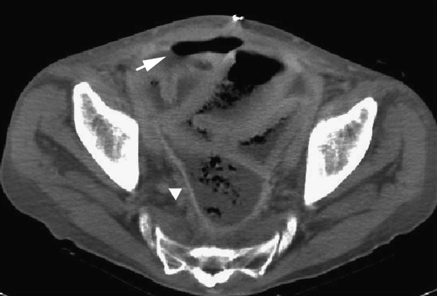
Large pelvic hematoma containing air bubbles (arrowhead). Pneumoperitoneum also is present (arrow).
Because of concerns about an anastomotic leak or breakdown with leakage of feculent material into the pelvis, the patient was brought to the operating room for re-exploration. He received a single dose of preoperative antibiotics (ampicillin-sulbactam and gentamicin) prior to creation of the skin incision. A gush of air was encountered on entering the abdomen. Contrary to expectations, careful inspection of the entire gastrointestinal tract by two surgeons did not find any signs of perforation. The anastomosis was grossly intact, and there was no contrast extravasation from the recent CT preparation. Instead, a large, foul-smelling pelvic hematoma was identified, evacuated, and sent for gram stain and cultures. The peritoneal cavity and pelvis were irrigated thoroughly with saline.
Gram stain of the hematoma showed many white blood cells (>25/low-power field). The aerobic cultures had no growth; the anaerobic cultures revealed 1–2 + anaerobic gram-negative bacilli (beta-lactamase negative), not Bacteroides fragilis. The patient was treated with a seven-day course of ampicillin-sulbactam and gentamicin and recovered uneventfully.
Discussion
The finding of pneumoperitoneum usually is associated with a perforated viscus and thus is a surgical emergency necessitating prompt diagnosis and intervention. More than 80% of patients with a ruptured viscus present with pneumoperitoneum [8]. In patients who recently underwent laparoromy, extraluminal gas usually disappears within 48 h, as judged by abdominal radiographs [9]. If free air is detected beyond this time, especially in the presence of fever, leukocytosis, and signs of peritonitis, disruption of the anastomosis or unrecognized enterotomy during surgery should be considered. Timely evaluation and urgent re-exploration may be prudent if clinical suspicion is high.
Absence of evidence of visceral perforation has been reported in 5% to 14% of all occurrences of pneumoperitoneum [8,10]. The sources have been described as intrathoracic, intra-abdominal, gynecologic, and miscellaneous in origin [1,2]. Rupture of intraperitoneal abscesses because of gas-forming organisms is an uncommon cause of pneumoperitoneum. The occurrence of pneumoperitoneum secondary to infection of a hematoma with gas-producing organisms is rare. The presence of air bubbles within a hematoma on CT scan strongly implies infection with gas-forming organisms. In patients who recently had surgery, it is especially difficult to attribute the cause of pneumoperitoneum to an infected hematoma, given that multiple other potential sources of free air such as residual air from recent laparotomy, unrecognized enterotomy during surgery, or disruption of the intestinal anastomosis with leakage of enteric contents into the peritoneal cavity, are possible.
Hitherto, numerous gas-forming organisms have been isolated as causes of emphysematous infection or pneumoperitoneum [11,12]. There was no aerobic growth from the hematoma in our patients, and only beta-lactamase negative gram-negative bacilli (not Bacteroides fragilis) were recovered. It is possible that the preoperative antibiotics or specimen transport from the operating room precluded further identification.
In patients without clinical signs of peritonitis, percutaneous drainage under radiographic guidance (such as ultrasound or CT scanning) together with antibiotic therapy is safe and effective management for an infected hematoma [13]. Broad-spectrum antibiotics covering both aerobic and anaerobic organisms should be started empirically and replaced with appropriate narrow-spectrum antibiotic(s) once the causative agent(s) has been isolated. If this is unsuccessful, surgical exploration with washout and drainage will be necessary. In selected patients, laparoscopic surgery has a unique advantage because there is less surgical trauma, decreased postoperative pain, and a shorter hospital stay.
In summary, infection of a hematoma with gas-forming organisms is a potential cause of pneumoperitoneum in patients who have had recent surgery. Careful clinical and radiographic evaluation may help to clarify the diagnosis and avoid unnecessary laparotomy.
Footnotes
Author Disclosure Statement
No conflicting financial interests exist.