
Abstract
Abstract
Background:
The presentation of primary abscess of the greater omentum is similar to other acute abdominal disorders. It is a rarely reported phenomenon, and correct diagnosis is typically made during abdominal exploration.
Methods:
Case report and review of pertinent English language literature.
Results:
In a patient who presented with symptoms consistent with an acute surgical abdomen, an unusual intraabdominal pathogen was found within a primary omental abscess upon abdominal exploration.
Conclusion:
Abdominal exploration and resection is a reasonable approach to primary abscess of the greater omentum.
Case Report
A 49-year-old Colombian male with no substantive medical problems or prior surgical history presented to the emergency department with an acute exacerbation of left-sided abdominal pain that he had been experiencing for the previous three months. He also reported new-onset fevers to 102°F and anorexia for the previous two days. He denied any abdominal trauma or recent travel outside the United States.
On physical examination, he was febrile (101.3°F), and his pulse was 109 beats/min. His abdominal examination was notable for a warm 5-cm palpable mass in his left midabdomen with focal signs of peritonitis. Laboratory evaluation was revealed for a white blood cell count of 11,900 mm3 with 76.1% neutrophils. He also had mildly high liver enzyme concentrations (alkaline phosphatase, aspartate aminotransferase, and alanine aminotransferase).
A computed tomography scan was performed and revealed a large area of omental fat stranding with a phlegmon containing central necrosis in the left upper quadrant to mid-abdomen (Fig. 1). The diagnosis of an intraabdominal abscess was made. The patient was started on intravenous ertapenem for broad-spectrum antimicrobial therapy, and the decision was made to operate.

Computed tomography scan demonstrating the omental phlegmon.
At laparotomy, the greater omentum was densely adhered to the anterior abdominal wall. This area was dissected free, revealing a large area of omental induration with foul-smelling purulent material extruding from its necrotic center. Manual and visual inspection of the stomach, small bowel, and colon failed to demonstrate a perforation and were normal. The greater omentum was resected (Fig. 2). For abdominal closure, a running 1-0 looped polydroxanone (PDS; Ethicon, Inc., Somerville, NJ) suture was used to close fascia. The skin and subcutaneous space was irrigated extensively with saline. Skin was then re-approximated loosely using skin staples, with wicks of 1″ strip gauze packed between the staples in hopes of decreasing the chance of surgical site infection.
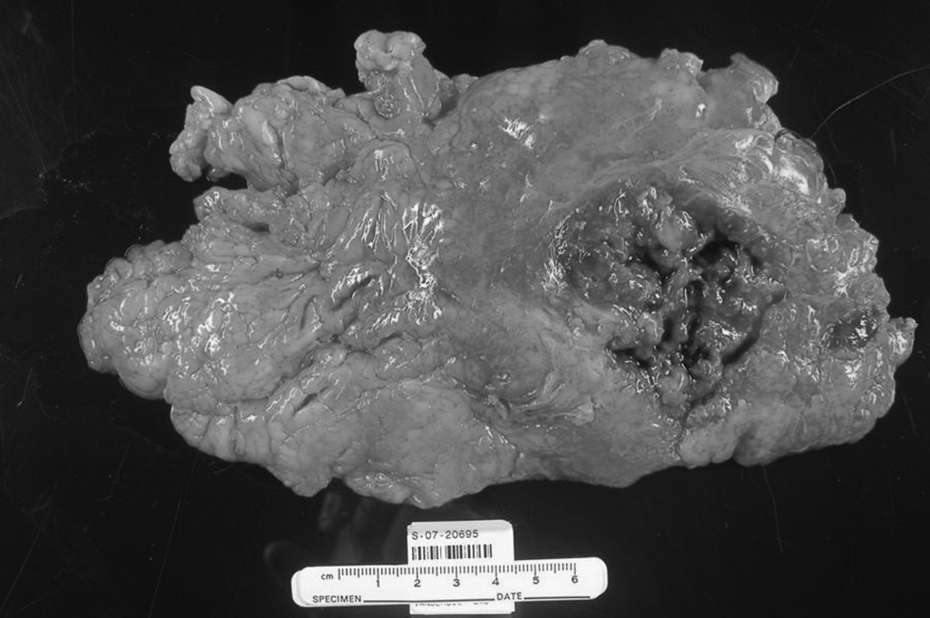
Gross pathologic specimen after resection.
Microbiologic analysis of the purulent material revealed a single organism: Pan-sensitive Streptococcus constellatus. Pathologic analysis of the omentum revealed a 5- x 5-cm area of necrosis terminating 1.5 cm from the area of colonic insertion without evidence of malignant tumor or foreign body, leading to a diagnosis of a primary omental abscess. The patient continued on intravenous ertapenem while hospitalized because of the gross purulence of the specimen and likely surgical-field contamination. The gauze wicks were removed on postoperative day two, and there were no signs of a surgical site infection. He defervesced and was discharged home on postoperative day five.
A deep incisional surgical site infection, which was diagnosed on postoperative day 10 when he complained of purulent drainage from the incision, complicated his postoperative course. This was treated with opening of the incision and wet-to-dry dressing changes to allow the wound to heal by secondary intention. Ten months after his initial operation and after complete closure of his midline wound, he returned complaining of a painful bulge in his midline incision. Physical examination confirmed a symptomatic incisional ventral hernia. He has since undergone incisional herniorrhaphy with primary closure of his fascia and an underlay of biologic mesh. He has done well postoperatively.
Discussion
A primary abscess of the omentum is rare, and in 2004, the last time this disease was described, only 13 cases existed in the combined English and Japanese literature, with the vast majority reported from Japan [1]. This is the first report in which S. constellatus was isolated from the abscess.
The cause of a primary abscess of the omentum is unknown, but other causes leading to the development of an abscess of the omentum have been described. Lupovitch et al. reported a case in which microscopic-sized foreign particles were located within the abscess cavity, raising the possibility of an antecedent clinically silent perforation of the bowel or appendix [2]. Omental infarction secondary to intraabdominal adhesions with subsequent abscess formation has also been described [3]. An omental abscess requiring operative resection in a patient who had recently undergone a cesarean section has been described [4]. An abscess of the omentum has also been reported in the postoperative setting of inguinal herniorrhaphy [5]. In this case, suture material was located amidst the abscess; thus, this was felt to be secondary to the primary operation. Hematogenous spread may also secondarily cause an omental abscess.
In our patient, the location of the abscess cavity was near the omental insertion to the transverse colon. This raised the possibility of a microscopic colonic perforation as the source, but the colon was grossly normal appearing without inflammatory changes at laparotomy, and the organism that was ultimately cultured is not typical of colonic flora.
Streptococcus constellatus is a member of the Streptococcus milleri group. The members of this group of bacteria have a striking tendency to cause abscesses [6,7]. In one report, 73% of clinical isolates of S. constellatus were associated with deep abscess formation. Furthermore, this particular organism is more likely to be associated with intraabdominal infections than the other members of the group [6].
Summary
Primary abscess of the greater omentum simulates other acute abdominal disorders such as acute appendicitis. The presentation includes fever, abdominal pain, and leukocytosis. It is a rarely reported phenomenon, and correct diagnosis is typically made during abdominal exploration. This report of an omental abscess with an unusual single intraabdominal pathogen adds to 13 previously reported cases. In these cases, one can speculate about the inciting process that led to abscess formation, but often no clues can be found. The natural clinical course of a primary omental abscess is not well known because operative intervention is usually undertaken. It can be assumed that abdominal exploration and resection is a reasonable approach for those who are appropriate surgical candidates.
Footnotes
Author Disclosure Statement
No conflicting financial interests exist.