
Abstract
Abstract
Background:
Infection is one of the most feared complications of surgery. New instrumentation is being developed to reduce deposition of bacteria.
Methods:
We investigated 45 major surgical procedures (21 radical nephrectomies [RN] and 24 radical retropubic prostatectomies [RRP]) in our urology department during 2007. In about one-half of the interventions, an ultraclean air flow mobile (UAF) unit was used. Bacterial sedimentation was evaluated by nitrocellulose membranes placed on the instrument tray and by settle plates positioned at four points in the operating room. In 27 operations, an additional membrane was located near the incision.
Results:
Bacterial counts on the nitrocellulose membranes during RN were 230 colony-forming units (cfu)/m2/h with the UAF unit and 2,254 cfu/m2/h without the unit (p = 0.001). During RRP, the values were 288 cfu/m2/h and 3,126 cfu/m2/h respectively (p = 0.001). The membrane placed near the incision during RN showed a microbial count of 1,235 cfu/m2/h with the UAF unit and 5,093 cfu/m2/h without the unit (p = 0.002); during RRP, the values were 1,845 cfu/m2/h and 3,790 cfu/m2/h, respectively (difference not significant). Bacterial contamination detected by settle plates during RN showed a mean value of 2,273 cfu/m2/h when the UAF unit was used and 2,054 cfu/m2/h without the unit; during RRP, the values were 2,332 cfu/m2/h and 2,629 cfu/m2/h with and without the UAF unit, respectively (NS). No statistically significant differences were detected in the clinical data registered in patients operated on under standard conditions and while the UAF unit was functioning.
Conclusions:
The UAF appears able to reduce microbial contamination at the operating table, reaching a bacterial number obtained in ultraclean operating theatres.
Several factors can influence the incidence of SSI: The type of operation, the surgical technique, the surgical preparation and antibiotic prophylaxis, the insertion of foreign material or implants, the immune state of the patient, and contamination of the environment [2,13–17]. For most SSIs, the source of pathogens is endogenous, the flora of the patient's skin, mucous membranes, or hollow viscera; however, exogenous sources, including surgical personnel, operating room environment, tools, instruments, and materials brought to the sterile field during the operation generally are accepted as the main factor causing SSIs after clean operations [2,14,17–21]. A correlation between the numbers of airborne bacteria and the incidence of SSI was demonstrated in hip and knee replacement surgery [19]; however, in other kinds of surgery as well, in particular when foreign material is inserted, the causes of infection are similar, and low levels of airborne contamination are needed [14]. When foreign material is inserted at the surgical site, because of the susceptibility of the tissue, the number of contaminating micro-organisms required to cause infection is low: It has been estimated that as few as 10 colony-forming units (cfu) are sufficient to cause deep space infection in a prosthetic replacement arthroplasty [18,19].
Appropriate staff dress and good theatre discipline to minimize the bacterial spread from operators, associated with a controlled ventilation system, are used to reduce microbial contamination of the air [2,15,21]. For operations carrying a high risk of airborne infections, in particular implant surgery, an ultraclean ventilation system is recommended to ensure low levels of air contamination. However, the installation of an ultraclean system is expensive and may require extensive rebuilding; hence, there have been proposals to use an independent mobile unidirectional air flow (UAF) unit, which can be added to conventional operating theatre ventilation. The unit has been evaluated during sham operations and during hernia operations [22,23] and coronary angiography [24]. The aim of the present study was to evaluate the efficiency of the UAF unit in reducing air contamination of the surgical area during standard urologic procedures performed in a conventionally ventilated operating theatre.
Methods
Operating theatre
The study was performed at the Urology Unit of University Hospital of Parma. The operating theatre, 46.31 m2 × 154 m3, is supplied with a turbulent ventilation system with 15 air changes per hour.
Mobile unidirectional air flow unit
The UAF unit (Fig. 1) consists of a box equipped with a fan and a high-efficiency particulate air (HEPA) filter, HP14, with an efficiency of 99.995% (EN 1822). The air is sucked into the box and then filtered through the HEPA filter. The unit (0.57 × 0.43 m) has a central zone of air flow at 0.6 m/sec and a peripheral zone with 0.4 m/sec. The high pressure in the center of the flow prevents mixture with the contaminated air. The additional UAF unit was located at the foot of the operating table with the air flow directed toward the surgical area.
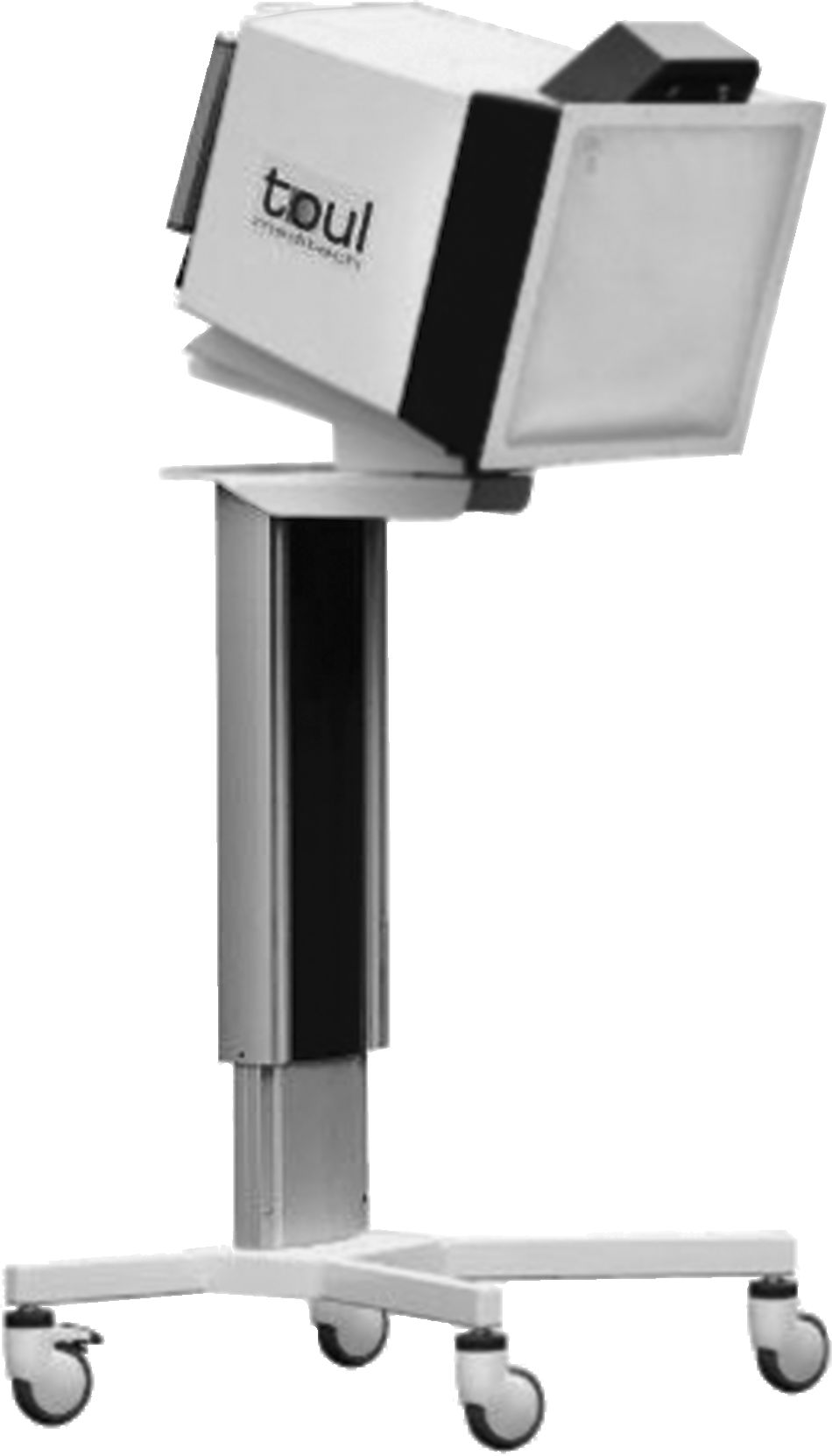
Unidirectional air flow apparatus.
Surgery
A total of 45 consecutive patients undergoing urologic procedures between January and December 2007 were evaluated; of these, 21 had a radical nephrectomy (RN) and 24 had a radical retropubic prostatectomy (RRP). Eleven RNs and 14 RRPs were performed under standard conditions; 10 RNs and 10 RRPs were performed while the UAF unit was functioning. The mean age of the patients was 64 years (range 58–80 years). All patients were admitted to the hospital approximately 16 h before the operation. No patients with infection underwent operation; positive urine culture led to specific antibiotic therapy until a negative result was achieved. All patients were shaved before the operation; nobody had a preoperative antiseptic shower. All patients received antibiotic prophylaxis half an hour before general anesthesia was started.
After skin preparation with antiseptic foam, all patients underwent incisional laparotomy. The prostatectomy was performed through a subumbilical abdominal laparotomy access (incision length ∼12 cm), and the mean duration of the operation was 165 minutes (range 90–280 min); whereas for the nephrectomy, a lumbotomy access was created (incision length ∼20 cm), and the mean duration of the operation was 105 minutes (range 60–315 min). After the RN, one drainage tube was inserted beside the surgical incision, which was removed on the third or fourth day, and the bladder catheter was removed on the second or third day. The RRP was completed by placing two drains at separate points with respect to the surgical incision; these were removed between the third and fifth days, and the bladder catheter was removed on the ninth day.
The first medication was delivered 24 h after the end of the operation, and all patients received intravenous or oral antibiotics until the bladder catheter was removed. The incision status, white blood cell count, and temperature were evaluated regularly until discharge. The patients were followed up for one month after surgery to detect any infections; the surveillance was performed in accordance with the U.S. Centers for Disease Control and Prevention (CDC) definitions [2].
Operating team
The operating team had six to eight persons (three or four surgeons, one scrub nurse, one circulating nurse, one or two anesthetists). The average number of people in the operating theatre was 7.2 (range 5–10) during operations performed in standard conditions and 7.3 (range 6–10) during operations performed while the UAF unit was functioning. The entire team wore conventional cotton gowns for all operations.
Monitoring
Settle plates and nitrocellulose membranes were used to evaluate aerobic bacterial sedimentation starting when the surgical incision was created. Petri dishes 9 cm in diameter containing plate count agar (PCA) were positioned 1 m from the floor and about 1 m from relevant obstacles and left open for 1 h at four points in the operating room (one in the patient area and three around the perimeter) in compliance with the Index of Microbial Air (IMA) contamination standard [25] (Fig. 1). At the same time, a 47-mm nitrocellulose membrane was placed on the instrument table, and after 1 h of exposure was transferred to a Petri dish containing PCA. During 13 RNs and 14 RRPs, an additional membrane was located near the incision (Fig. 2). The plates were incubated at 36°C for 48 h.

Schema of operating theatre showing position of unidirectional air flow unit and sampling points.
For easier comparison, the results obtained using both settle plates and nitrocellulose membranes were expressed in cfu/m2/h. The following threshold values were considered in the interpretation of the results: For an operating theatre with a conventional ventilation system: 3,930 cfu/m2/h; for operating theatres with an ultraclean system: 350 cfu/m2/h (ideal value) and 786 cfu/m2/h (alert value) [25–27].
Statistical analysis
The Statistical Package for Social Sciences version 11.5 was used for the analysis (SPSS, Inc., Chicago, IL). A non-parametric statistical test (Mann-Whitney) was used to assess significant differences between variables. A p value ≤ 0.05 was regarded as significant.
Results
Under standard ventilation conditions, the average bacterial sedimentation rate on the nitrocellulose membrane on the instrument table was 2,254 cfu/m2/h during RN and 3,126 cfu/m2/h during RRP, whereas when the UAF unit was used, sedimentation decreased significantly, reaching average values of 230 and 288 cfu/m2/h, respectively (p = 0.001) (Table 1). The reduction in the rate of deposition was 90% for RN and 90.8% for RRP. The nitrocellulose membrane placed near the incision also showed lower bacterial counts while the UAF unit was functioning. During RN, it decreased from 5,093 cfu/m2/h to 1,235 cfu/m2/h (p = 0.002), a 76% reduction. However, during RRP, the reduction, from 3,790 cfu/m2/h to 1,845 cfu/m2/h (52%), was not significant (p = 0.12) (Table 2).
Table 3 shows the average values of bacterial contamination detected by the settle plates placed in the patient area and the perimeter (average values obtained at the three points) of the operating theatre. The lowest mean value (1,620 cfu/m2/h) was obtained during RN performed while the UAF unit was functioning, the highest (2,927 cfu/m2/h) at the perimeter during RN performed while the UAF unit was functioning. No significant difference was found at the perimeter between samplings performed with and without the UAF unit. Under standard conditions, no significant difference was observed between the air sedimentation obtained on the settle plates in the patient area and on the nitrocellulose membranes at the instrument table during either RN (p = 0.53) or RRP (P = 0.46), whereas when the UAF unit was functioning, a significant difference was observed: RN (p = 0.001) and RRP (p = 0.001).
No SSIs were observed during the postoperative period. No statistically significant differences were found between clinical data (temperature and white cell count) registered in patients operated on under ordinary conditions and patients operated on while UAF unit was functioning. No significant correlation was found for either surgical procedure between bacterial contamination of the incision and instrument table site (with or without UAF), and the patients' clinical data.
Discussion
The risk of airborne postoperative infections increases the demand for operating theatre asepsis to prevent even small numbers of bacteria from reaching the incision. According to the British Health Technical Memorandum 03-01 (HTM), in an ultraclean operating theatre, on average, air sampled within 300 mm of the incision should not contain more than 10 cfu/m3; in conventionally ventilated theatres, a maximum value of 180 cfu/m3 is acceptable [28] (Table 4). Considering the correlation between active and passive sampling of the European Commission Good Manufacturing Practices (EC GMP) for Pharmaceutical Industries, the value 10 cfu/m3 corresponds to 5 cfu/9-cm plate/4 h, which means 1.25 cfu/plate/h, whereas a value of 180 cfu/m3 may be considered to correspond to about 25 cfu/settle plate/h (3,932 cfu/m2/h) (Table 1). According to the Index of Microbial Air Contamination (IMA) standards, IMA: 5 and IMA: 25 are considered the threshold values for ultraclean and conventional operating theatres, respectively [25]. Friberg et al. proposed a value of 350 cfu/m2/h for ultraclean operating theatres, which corresponds to 2.2 cfu/9-cm plate/h [27]. Considering the proposals regarding microbial air contamination in a controlled environment, the following values were considered thresholds: For an operating theatre with a conventional ventilation system <3,932 cfu/m2/h; for operating theatres with an ultraclean system ≤350 cfu/m2/h (ideal value) and 786 cfu/m2/h (alert value) [25–27] (Table 4). To evaluate bacterial contamination, we used passive instead of active air sampling, as it gives an estimate of the number of bacteria sedimenting from the air onto the surgical incision, which represents the critical surface exposed to the microbial hazard [25,27,29]. Settle plates were used at four points in the operating theatre, whereas sterile light nitrocellulose membranes were used directly at the critical points (surgical incision and instrument table). The results obtained on settle plates positioned at the perimeter are consistent with those of a conventional operating theatre; in standard conditions, similar results were obtained on the settle plate in the patient area and on the nitrocellulose membrane positioned on the instrument table. The results obtained while the UAF unit was functioning suggested that the unit is able to reduce microbial contamination of the air in the surgical area. The low number of organisms observed on the instrument table during both RN and RRP while the UAF unit was used was similar to that expected in an ultraclean operating theatre. In the incision area, the mean cfu values were higher than at the instrument table, and the reduction was lower than on the instrument table; a significant reduction was observed only during RNs, even though a reduction also was seen during RRP. This finding may be explained by the fact that the membrane was placed downstream of the incision area and was sometimes at a lower level than the incision itself, so that it was difficult for the flow to reach. In particular, in RRP, because of the operating position, the membrane was lower than in RNs, and this could explain the lesser reduction in the number of microorganisms obtained for RRP. It also should be considered that the air flow could not have been directed perfectly toward the incision and that the assistants' position could hinder the air flow during surgery. To ensure the correct position of the UAF unit and the proper air flow direction, the new model of the UAF unit has a sensor to direct the air flow correctly.
Colony-forming units (cfu) on settle plates 90 mm diameter after 4 h of exposure.
Colony-forming units on settle plates 90 mm in diameter after 1 h of exposure, calculated as one-quarter of the value indicated by EC GMP after 4 h of exposure.
Colony-forming units on settle plates 90 mm in diameter after 1 h of exposure.
Colony-forming units on settle plates 140 mm in diameter after 1 h of exposure.
Figures in parentheses are cfu/m2/h.
We did not find any significant difference in air bacterial contamination at any point in the operating theatre under standard conditions or while the UAF unit was functioning; this means that the air flow is limited to the surgical area and does not influence other sites in the operating theatre. No differences in white blood cell counts or body temperature were found for either operation between the patients operated on under ordinary conditions and those operated on while the UAF unit was functioning.
No conclusion can be drawn regarding the efficacy of the UAF unit in reducing incision infections, although this is the most important outcome indicator. A larger number of patients should be followed to evaluate this point. However, considering that Whyte et al. estimated that about 98% of the bacteria in the joint replacement incisions of patients operated on in a conventionally ventilated operating theatre derived from the air, either directly or indirectly transferred by vehicles (e.g., instruments) [17], we can postulate that a smaller number of bacteria found in the incision while the UAF unit was functioning should lead to fewer incisional infections. It should be considered also that the antibiotic prophylaxis and subsequent therapy may have influenced the results. This is an issue of a great importance, as our protocol did not comply with international recommendations [30].
The results of our study confirm the efficacy of the UAF unit in reducing bacterial contamination to the level reached by an ultraclean system and represent a further evaluation of this equipment. In case of operations associated with a high risk of airborne infection, the use of a UAF unit offers a particular advantage for operating rooms with conventional air flow, as it can be added without any renovation and can be utilized immediately. In urology, the UAF system could be used during surgical procedures at high risk of airborne infection where a sterile field is mandatory; e.g., penile prosthesis insertion or implantation of artificial urinary sphincters or synthetic incontinence devices. This UAF equipment can be seen as a valuable proposition for surgery under poor hygienic conditions such as in military hospitals and flying operating rooms (e.g., a SMILE project aircraft).
Footnotes
Author Disclosure Statement
No conflicting financial interests exist.