
Abstract
Abstract
Background:
Descending necrotizing mediastinitis is a serious condition with few cases reported in the literature. Surgical treatment is controversial and may include wound exploration, local drainage, and even mediastinal debridement approached by thoracotomy.
Methods:
Description of a case of descending mediastinitis caused by group A Streptococcus as a complication of thyroidectomy.
Results:
Aggressive debridement was required for source control and treatment of septic shock.
Conclusion:
Post-thyroidectomy descending necrotizing mediastinitis is a rare and dangerous infection. It should be treated aggressively with appropriate cervical and mediastinal drainage combined with optimum medical care.
Case Report
The patient was a 60-year-old female known for well-controlled diabetes and dyslipidemia. She underwent an uncomplicated standard left thyroid lobectomy for a suspect asymptomatic thyroid nodule at a community hospital. No antibiotic prophylaxis was given, and no drain was left in place. No break in sterile technique was reported.
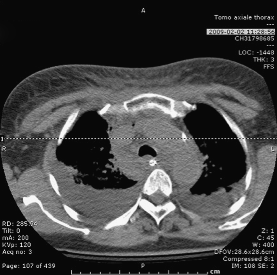
The first night after surgery, the patient complained of rigors and pain in her neck and upper chest. The following day, the patient deteriorated, complained of progressive neck and chest pain, developed fever, and was delirious. Physical examination revealed incision site erythema without crepitus. A chest computed tomography (CT) scan revealed fat infiltration in the incision and mediastinum (Fig. 1). The patient was transferred to a tertiary-care center.

Computed tomography (CT) scan of chest showing mediastinal air and soft tissue infiltration. This image is from the scan made in the referring center.
On arrival, she appeared toxic, with a temperature of 38.4°C and tachycardia. Laboratory investigations revealed acute kidney injury and coagulopathy. The patient was taken to the operating room for source control. The incision was opened, and brown purulent liquid was evacuated, but there was no fasciitis. The upper mediastinum was explored, but no signs of soft tissue infection were present. Penrose drains were placed, and the incision was left open. The patient was brought to the intensive care unit with the intention to re-explore in the event of deterioration. Gram stain of the fluid showed gram-positive cocci in chains, suggestive of streptococci.
The following 24 h were marked by labile blood pressure necessitating vasopressors and slow progression of the erythema around the incision. A CT scan showed progression of the air in the mediastinum (Fig. 2). The neck was re-explored and debrided (Fig. 3). Mediastinoscopy was then performed to assess the middle mediastinum and to provide drainage. The tissue anterior to and around the trachea appeared normal. Next, a right thoracotomy was undertaken, during which the mediastinal pleura was opened to establish further drainage of the middle and posterior mediastinum
Computed tomography scan of the chest showing residual mediastinal air, soft tissue infiltration, and bilateral pleural effusions. This image is from the scan done in our center the morning of the second operation.

Neck wound after debridement and thoracotomy. Cellulitis is visible beyond the margins of the incision.
Blood cultures drawn on admission and mediastinal cultures obtained during both operations showed GAS. Unfortunately, no strain typing was done to characterize the isolates. The patient received immunoglobulin, 7 days of clindamycin, and 42 days of penicillin. The plastic surgery department was consulted to assist in reconstruction and site closure. The patient eventually was discharged from the hospital, and she was doing well after one year of follow-up.
Discussion
Thyroidectomy is a clean procedure rarely complicated by infection. Consequently, post-thyroidectomy descending mediastinitis caused by GAS is extremely rare: Only 13 cases have been described previously in the medical literature. One team reported six cases [1], whereas another group described seven cases worldwide of which the majority was caused by the emm 1 strain. Three of these cases involved the same surgeon during a short period of time [2].
Because infection after thyroidectomy is rare, prophylactic antibiotics are not used routinely. A recent randomized controlled trial showed a post-operative infection rate of <1% and discouraged prophylactic antibiotics for standard thyroidectomy (with or without use of drains) in the absence of co-morbidities such as immunosuppression, advanced neoplastic disease, or hematologic disease. It also is reported that pre-operative antibiotics are not effective for preventing invasive GAS infection [3].
The diagnosis of descending mediastinitis is based chiefly on clinical presentation. The patient complains of pain and often has signs of severe sepsis or septic shock. The condition evolves rapidly, usually within 24 to 48 h. Chest CT can aid the diagnosis, but should never delay treatment.
The optimal treatment is not clearly defined. One group recommends cervical drainage only if CT scan shows no infection below the carina, and both cervical and tube thoracostomy drainage otherwise [4]. Others recommend open thoracotomy at presentation in view of their experience with descending mediastinitis, showing a reduction in the mortality rate from 40% to 20% with thoracotomy [5]. Finally, in one case, cervical drainage and thoracic drainage alone was successful without the need for debridement [6]. In the case we present, a conservative approach was adopted at initial surgery because no macroscopic evidence of necrotizing fasciitis and no gross contamination of the mediastinum were found. For the second operation, aggressive treatment was chosen because improvement had not been observed in response to the treatment already given.
It also is common practice to administer intravenous immune globulin (IVIg) in invasive GAS infections. The rationale for its use relies on the data implicating extracellular exotoxins as mediators of shock and organ failure. However, only two publications evaluated the efficacy of IVIg in the treatment of streptococcal toxic shock syndrome. One was underpowered [7], and the other showed a significant reduction in the mortality rate compared with historical controls [8]. Despite the use of IVIg in our patient, its utility remains unclear. Likewise, corticosteroid therapy has been used for severe GAS infections to unclear benefit [9].
The patient in this case received high-dose penicillin and clindamycin as antimicrobial treatment. Despite the fact that all strains of GAS remain sensitive to penicillin, the addition of clindamycin is recommended for severe streptococcal infections because penicillin-binding proteins are not expressed during stationary-phase growth of GAS, and thus, penicillin is ineffective in severe deep infections in which a large inoculum is present [10–12]. Clindamycin also suppresses exotoxin production better than does penicillin [13]. No controlled trials of medical therapy have been conducted to guide the duration of treatment in cases of mediastinitis. This duration is determined by multiple factors but usually is for no longer than two weeks.
A case of post-operative GAS infection, as defined by the U.S. Centers for Disease Control and Prevention in 2002 [14], is the isolation, during the hospital stay or the first seven days after discharge, of GAS from a sterile site or a surgical incision in a post-operative patient for whom the indication for surgery was not GAS. Given the potentially disastrous complications of these infections, even one case of post-operative GAS infection should prompt an epidemiologic investigation by the hospital's infection control personnel. This investigation involves enhanced surveillance and screening of healthcare workers present in the operating room during procedures or who change dressings on open wounds (sites to screen: Anus, skin lesions, throat, and vagina). Epidemiologically linked carriers of GAS should be treated in order to eradicate the carrier state.
In the case presented, it is not known where the patient acquired GAS, but it is generally accepted in the literature that early post-operative necrotizing infection arises from either self-contamination of the patient or from healthcare workers involved with the operation or immediate post-operative care [15]. Unfortunately, screening was not conducted in the referring center, so confirmation of the bacterium's origin cannot be obtained.
Conclusion
Post-thyroidectomy descending necrotizing mediastinitis is a rare and dangerous infection. Diagnosis should be made promptly on clinical grounds with or without radiologic evidence. Treatment must be initiated promptly. Various approaches are reported in the literature, but a general rule is that this life-threatening infection should be treated aggressively with appropriate cervical and mediastinal drainage combined with optimal medical care.
Footnotes
Acknowledgments and Author Disclosure Statement
YC reviewed the chart, reviewed the literature on necrotizing descending mediastinitis, and wrote the article. MS was the patient's surgeon and supervised and counseled during the writing of the article. AC acted as a consultant for the infection discussion in the article. JCLW contributed to the review of the manuscript.
No outside funding was supplied for this paper.
None of authors is associated with any organization that would bias the writing of this article.