
Abstract

To the Editor:
Chest computed tomography (CT) scanning revealed multiple areas of ground-glass opacity, alveolar infiltrates, and nodular lesions, located mainly in the posterior segments of both lower lobes (Fig. 1). Bronchoscopy with bronchoalveolar lavage and gram stain showed no microorganisms, but a Grocott–Gomori methenamine silver stain identified Pneumocystis jiroveci. Intravenous co-trimoxazole and methylprednisolone was initiated. The patient's condition improved after five days of non-invasive ventilation, making it possible to discontinue supportive ventilation. After 22 days in the ICU, the patient was discharged to the ward. A subsequent CT scan showed radiographic amelioration, together with disappearance of the alveolar infiltrates.
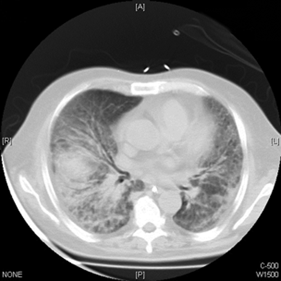
Computed tomography scan shows multiple areas of ground-glass opacity, alveolar infiltrates, and nodular lesions, mainly in the posterior segments of the lower lobes.
Despite the known important relation between P. jiroveci and pneumonia in immunocompromised patients, it is necessary to take into account the asymptomatic carrier and the population at risk [1,2]. Risk factors associated with the development of P. jiroveci pneumonia are smoking, chronic obstructive pulmonary disease [3,4], lung cancer, interstitial lung disease, corticosteroid therapy, and working directly in patient care [5]. Iatrogenic immunosuppression by corticosteroids has been linked to a higher risk of P. jiroveci colonization. A high index of suspicion and early diagnosis are essential to achieve a positive outcome. Any patient treated with corticosteroids for a cerebral space-occupying lesion should be considered at risk to develop P. jiroveci pneumonia.