




Abstract

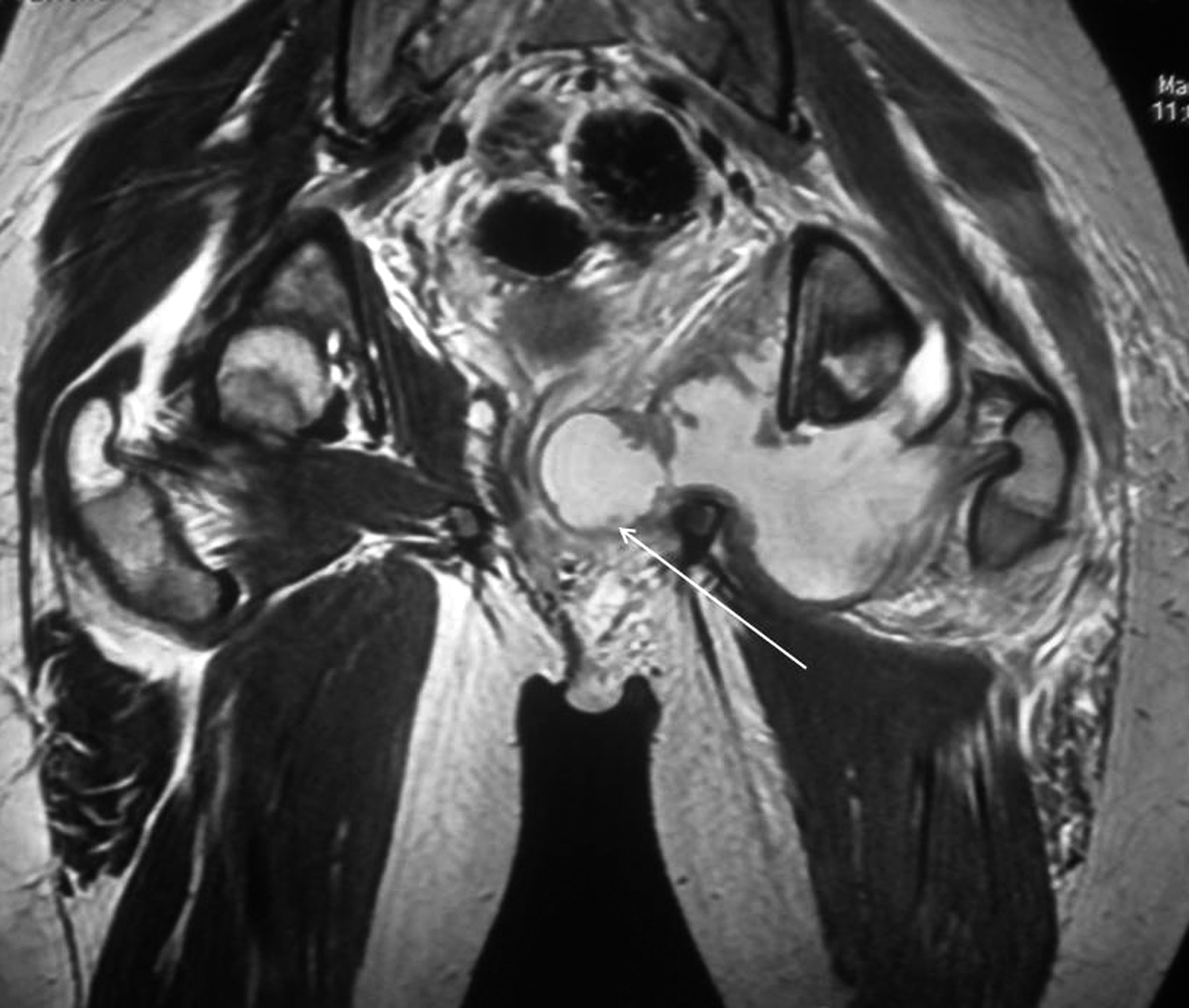
T2-weighted magnetic resonance image showing a multilobulated abscess measuring approximately 11x6 cm, involving the left obturator internus muscle and bulging into the left lateral vaginal wall (white arrow) and deviating it to the right.
Discussion
Primary pyomyositis of the obturator internus muscle is the result of colonization of traumatized ischemic muscle during an episode of transient bacteremia [1]. Imaging is the mainstay of diagnosis because the clinical features of the condition are easily confused with other common causes of hip pain, and laboratory findings in the condition are often nonspecific. Magnetic resonance imaging is the modality of choice for imaging the obturator internus muscle and to ascertain the diagnosis of pyomyositis [2]. In a developing country such as India, where MRI is restricted to a few centers, a thorough clinical examination at the patient's bedside is invaluable for the diagnosis of pyomyositis of the obturator internus. Because of the close proximity of the muscle to the rectum and female reproductive organs, a routine digital rectal examination or vaginal examination can facilitate the diagnosis. The characteristic finding of a tender, fluctuant bulge of rectal or vaginal mucous membrane confirms the presence of a pelvic abscess and the need for prompt drainage. Also suggesting an abscess of the obturator internus in female patients is edema of the ipsilateral labia majorum [3]. The choice between medical and surgical management of an obturator internus abscess is guided by the clinical course of the disease and demonstration of a well-defined pus pocket on imaging studies. The patients with localized symptoms and signs who show no features of systemic toxicity and have no fluid collection in the area the of obturator muscles on imaging can be managed with a 2–4-wk course of intravenous antibiotics [3]. The lacki of a response to medical management, detection of a pus-filled cavity around the obturator muscles, and development of complications mandate surgical intervention. This can consist of image-guided percutaneous drainage or open surgical drainage of the abscess. Selection of the most appropriate access route is guided by precise location of the abscess cavity on imaging. Although all previously reported patients with obdurator internus abscess benefited by either medical or surgical intervention, ours is the first reported case in which spontaneous transvaginal rupture of the abscess during the natural progression of the condition had a favorable outcome.