
Abstract
Abstract
Background:
Melioidosis, caused by Burkholderia pseudomallei, an important human pathogen in tropical regions, has protean multi-system clinical manifestations.
Methods:
Case report and review of pertinent English-language literature.
Results:
A 33-year-old male, who had been treated for pulmonary tuberculosis and multiple splenic abscesses four years previously, presented with a five-day history of acute-onset high-grade fever, abdominal pain and distension, and dyspnea. Laparotomy and drainage was done for paraumbilical abdominal wall abscess. Omentectomy was performed because the omentum was infarcted and studded with tubercles. Pus culture was positive for B. pseudomallei. The patient developed septic shock and succumbed on the fifth day after surgery.
Conclusion:
This report emphasizes an unusual presentation of melioidosis and the diagnostic challenge posed by its clinical similarity to tuberculosis.
Case Report
A 33-year-old male presented to the emergency department with high-grade fever, abdominal pain and distension, and dyspnea for five days. He had been treated for primary pulmonary tuberculosis four years previously. He developed splenic abscesses six months thereafter and responded well to multi-drug anti-tuberculosis therapy. Chest radiography on presentation showed diffuse bilateral opacities. Ultrasound scanning of the abdomen revealed a 4.3×1.2×6.6-cm septated collection in the left paraumbilical region, surrounded by inflamed mesentery. The spleen contained multiple hypoechoic areas with central calcification.
An emergency laparotomy was performed, and 100 mL of pus was drained from the mid-abdomen. Necrotic omentum that was studded with tubercles (Fig. 1) was resected. Serosa of small bowel and sigmoid colon also was studded with tubercles (Fig. 2), but visceral peritoneum of the liver and spleen were not. The patient was started on ceftriaxone and metronidazole but developed septic shock post-operatively. Culture revealed B. pseudomallei sensitive to ceftazidime and ciprofloxacin; therefore, ceftazidime was added. He deteriorated progressively and succumbed on post-operative day 5. The omental tubercles revealed abscesses; there was no evidence of tuberculosis.

Intraoperative photograph showing necrotic omentum studded with tubercules.
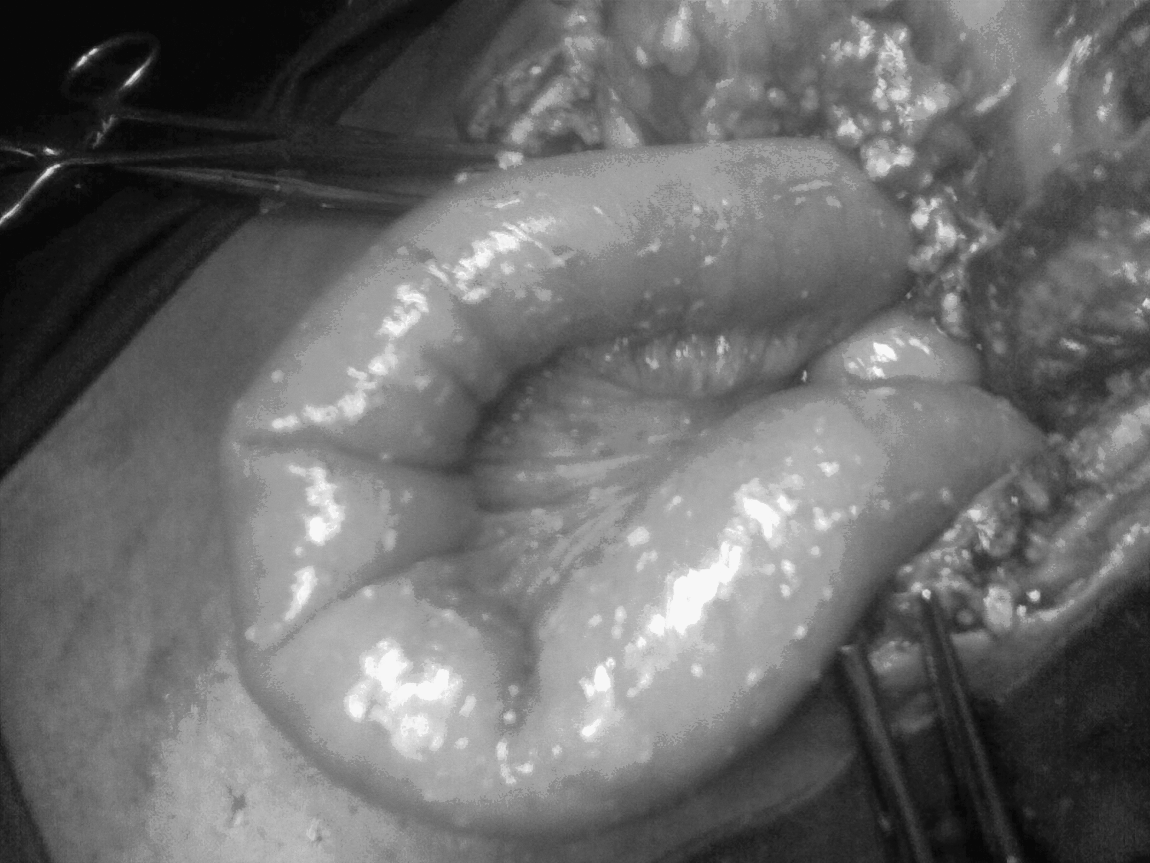
Intraoperative image of small bowel studded with tubercules.
Discussion
Tuberculosis of the gastrointestinal tract is the sixth most frequent presentation [3,4] and often mimics other diseases, both common and rare [5]. Clinicians must suspect tuberculosis, which is treatable. Peritoneal tuberculosis should be considered with thickened parietal peritoneum with or without tubercles, or when the omentum, liver, or spleen is studded with tubercles. The condition also may present as a fibroadhesive variant [4,6].
Melioidosis is caused by B. pseudomallei, an aerobic, gram-negative, bipolar-staining bacillus. The disease causes abscesses at unusual sites such as the central nervous system, parotid gland, neck, adrenal gland, and prostate [7,8], but there is no report heretofore of an intra-abdominal abscess. In non-endemic areas, the diagnosis of melioidosis presenting with leukocytosis alone is missed frequently [8]. Although the clinical spectrum of melioidosis ranges from chronic abscesses to fulminant sepsis [9], the latter is more common [1]. The acute clinical and radiologic features of melioidosis are non-discriminative from other pyogenic infections. Melioidosis should be suspected when a chronic relapsing infection occurs in immunocompromised patients. Diagnostic confusion with tuberculosis may exist, especially in endemic areas. On histopathologic examination, chronic melioidosis is expressed as granulomatous lesions confined to a single organ, and bacteria are demonstrable rarely in tissue sections [10].
The causative organism is sensitive to chloramphenicol, trimethoprim/sulfamethoxazole (co-trimoxazole), ceftazidime, and imipenem-cilastatin. Ceftazidime and imipenem-cilastatin are the first-line antimicrobial agents employed in bactericidal regimens for melioidosis [11,12]. A combination of ceftazidime with parenteral co-trimoxazole for three weeks in the acute stage, followed thereafter by oral co-trimoxazole for two months, is the minimum recommended [13]. The mortality rate of the acute disseminated form with sepsis is as high as 87% in patients not receiving prompt, appropriate treatment [14]. A study that compared a three-drug combination (chloramphenicol, doxycycline, and co-trimoxazole) with ceftazidime alone demonstrated a 50% reduction in the mortality rate, from 80% to 35%, with combination therapy [15]. In a subgroup with severe sepsis, meropenem was associated with a lower mortality rate than ceftazidime [16]. Appropriate antibiotic therapy is defined as delivery of antibiotics active against B. pseudomallei within 48 h.
In our patient, the diagnosis of melioidosis was not considered until a microbiologic diagnosis was made post-operatively. The diagnosis was missed initially because of the history of pulmonary tuberculosis and the presence of tubercles on the omentum and bowel. Hence, empiric broad-spectrum antibiotics (ceftriaxone and metronidazole) were started only post-operatively; however, melioidosis should be considered in the differential diagnosis of intra-abdominal abscesses in the appropriate setting. The institution of empiric ceftazidime or imipenem-cilastatin could have been a better choice and resulted in the salvage of the patient.