
Abstract

To the Editor:
A 37-year-old Turkish female presented with an abdominal mass that she had realized was present two weeks before. She had a history of 12 pregnancies (five abortions), gave birth to her last child four months earlier, and had never used an intrauterine device (IUD). Her serum Ca 12–5 concentration was three-fold higher than normal, and the C-reactive protein concentration was two-fold above normal. The other laboratory tests were normal. On physical examination, she had no signs of acute abdomen. Her appendix and cecum were found by colonoscopy to be severely inflammed, but nothing indicated a colonic neoplasm. Abdominal computed tomography scanning revealed terminal ileal wall thickness as well as a 70×50-mm mass originating from the retroperitoneal area invaded the cecum and the mesentery of the terminal ileum (Fig. 1). A gynecologic examination to investigate possible remnant fetal tissues, which can cause an ascending infection, showed no abnormal findings. Acquired immunodeficiency syndromes, such as an human immunodeficiency virus infection and anti-phospholipid syndrome, were eliminated after laboratory tests and physical examination.
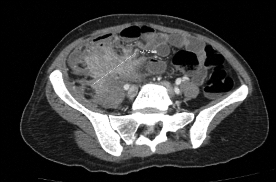
Computed tomographic view of retroperitoneal mass.
Exploration laparotomy showed a mass originating from the retroperitoneal area and invading the right iliac crest, cecum, and terminal ileum. Right hemicolectomy was performed, but the mass could not be resected totally from the iliac crest. Pathologic examination indicated a chronic abscess and basophilic, branched, and filamentous microorganisms that were gram-positive, periodic acid Schiff-positive, and Grocott-positive. Ehrlich-Ziehl-Neelson staining applied for the differential diagnosis of Nocardia and Actinomyces was negative. With these findings, the case was diagnosed as an Actinomyces infection.
Actinomycosis occurs mostly in the cervical or fascial region (60%), abdomen (20%), and thorax (15%) [2,3]. Pelvic actinomycosis is a rare (3%) complication associated with the use of IUDs [2,4], and can lead to pelvic inflammatory disease, abdominal wall and gluteal abscesses, vesico-uterine fistulas, hydroureter, hydronephrosis, ileopelvic fistulas, and pyometrium. In fact, invasive pelvic actinomycosis occurs nearly exclusively in women using IUDs [4]. It can mimic ovarian tumors and, rarely, colonic or retroperitoneal pathologies as well. Abdominal actinomycosis has been reported occasionally as a complication of perforated diverticular disease [5].
The patient described here had never had an IUD or gastrointestinal tract surgery despite the fact that these are both causes of pelvic actinomycosis according to the literature. Her last pregnancy ended after giving birth to a child four months earlier. Therefore, we suggest that this was a chronic ascending infection as our hypothesis.
Bowel obstruction caused by an actinomycosis-related mass orginating from the retroperitoneal area and the lack of an IUD are unusual presentations of this case. Actinomycosis, especially in a woman of reproductive age, IUD users, and post-partum patients, should be included in the differential diagnoses of all unusual bowel obstructions and intra-abdominal masses. Although it is a rare entity, it must be kept in mind, especially in patients with pelvic and retroperitoneal masses and predisposing factors.