
Abstract

To the Editor:
The patient's recovery from surgery was complicated by progressive respiratory failure requiring prolonged ventilatory support. On post-operative day five, suspicion of pulmonary infection and progression of radiographic opacities prompted bronchoscopy, which yielded Pseudomonas aerigunosa on non-quantitative culture of the bronchoalveolar lavage fluid. Although P. aeruginosa is a frequent cause of tracheal colonization in our ICU, the patient's antibiotic therapy, guided by in vitro sensitivity testing, was changed to meropenem. On day 18 of hospitalization, blood cultures yielded Klebsiella pneumoniae sensitive to meropenem and colistin. Multiple blood and urine cultures on standard media on post-operative day 30 were sterile.
On day 33 of the patient's stay in the ICU, following improvement of what may have been ventilator-associated pneumonia, he developed a fever of new onset. Findings on abdominal ultrasonography included sludge, gallbladder wall thickening (>8.0 mm), and the absence of calculi, which were suggestive of acute acalculous cholecystitis. On post-operative day 42 the patient underwent cholecystectomy, and his condition improved subsequently.
However, his clinical condition deteriorated 3 d after cholecystectomy, with a marked increase in his inotrope agent (norepinephrine) requirement (10 mcg/mL). His pulse rate was consistently above 120 beats/min, and his core temperature was 39°C. A reevaluation of his thoraco-abdominal computed tomography (CT) scan was negative. With the provisional diagnosis of septic shock, the patient's antibiotic regimen was broadened in view of his immunocompromised status. On post-operative day 46, when nurses turned and cleaned the patient, a swelling over the right parotid area with associated erythema was present that had not been noticed previously. Pus was expressed with palpation of the gland. The clinical impression was that of acute parotitis. A culture of the secretions from the right parotid duct grew P. aeruginosa with in vitro sensitivity to colistin only. A CT scan showed findings suggestive of a right parotid abscess (Fig. 1). Incision and drainage yielded a further 1 mL of pus. Despite this treatment, the patient's condition deteriorated within a week and he died.
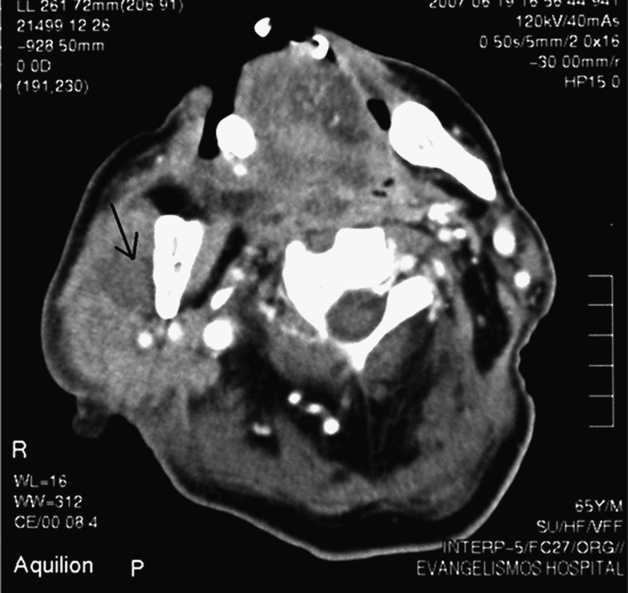
Computed tomographic scan enhanced with intravenous contrast, showing a large right parotid gland mainly in the superficial parenchymal lobe, with rim enhancement and intra-parotid lymph nodes.
The patient's condition represented a rare case of acute post-operative parotitis caused by P. aeruginosa and complicated by abscess formation in a setting of immune compromise. The crucial factors predisposing to this were the patient's recent surgery and his underlying condition [1]. He had undergone two major surgical procedures within 45 d. The interval between surgery and the onset of parotitis varies from a few hours to many weeks [1]. Besides his debility, the patient had prolonged hospitalization in an ICU environment, which may promote parotitis as a new type of health-care–associated infection. His upper respiratory tract was colonized with P. aerigunosa, which may ascend into the parotid gland. Although Staphylococcus aureus is the pathogen most commonly responsible for acute parotitis [1], gram-negative pathogens have also been reported [2], especially in children from areas with endemic melioidosis [3], and in an adult with a complex medical history [4]. Although the parotid gland is supposed to be a sterile site, it could serve as an anatomic harbor for occult pathogens capable of triggering septic shock. Instances of P. aeruginosa parotitis with or without abscess formation remain rare in the published literature, with only two cases reported to our knowledge.