
Abstract

A 30-year-old man presented with a two-day history of acute anal pain. The physical examination was unremarkable, but digital rectal examination was slightly painful on the right side. An anal ultrasound scan was indicated. The three-dimensional anal ultrasound revealed a fluid collection in the right ischiorectal fossa (Fig. 1). Surgical exploration confirmed an ischiorectal abscess, which was drained. The patient was discharged without any problems.
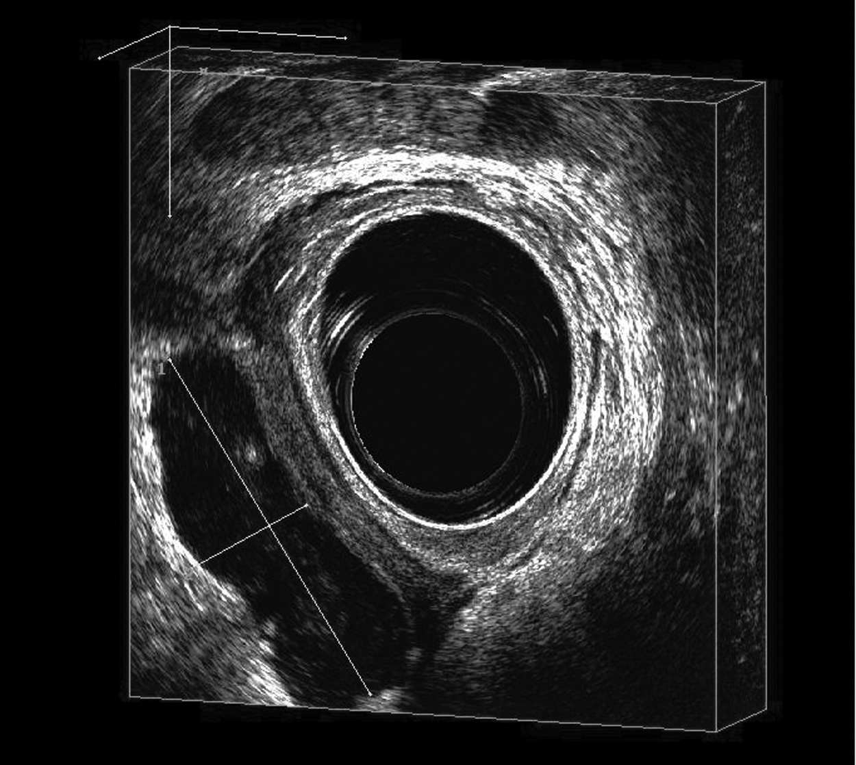
Three-dimensional anal ultrasound image showing fluid collection in right ischiorectal fossa, confirming a diagnosis of abscess.
Anorectal abscess generally is diagnosed by physical examination [1,2]. The principal symptom is pain, which may be associated with local symptoms accompanied in some cases by systemic signs such as fever and toxicity [1]. However, in other cases, there are no relevant physical signs. If an abscess is suspected but cannot be diagnosed, imaging or examination under anesthesia must be arranged [1].
Anorectal ultrasonography has been established as a relevant imaging tool for anorectal pathology assessment [3]. Recently introduced three-dimensional anal ultrasonography provides excellent imaging of the anal canal and sphincter complex [4]. This technique can provide crucial information regarding the presence of abscess and its exact location as well as extension when physical examination is insufficient. The appearance of the lesions is variable, and their echogenicity depends on their contents [4]. A highly inflammatory purulent collection, as in our case, is hypoechogenic and delimited, and in some cases, gas has been identified in the fluid collection [4]. Three-dimensional anorectal ultrasound scanning exhibits high performance for the evaluation of abscess close to the anorectal ring, but the yield declines for the evaluation of supralevator collections [4]. If the suspicion is of a pelvic or a supraelevator abscess, magnetic resonance imaging or a computed tomography scan may be considered [5].
Footnotes
Author Disclosure Statement
The authors have no conflicts of interest to disclose. No funding was received for this work.