
Abstract
Abstract
Background:
Primary pyomyositis is a rare bacterial infection that affects large muscle groups mainly in immunocompromised patients. Treatment options include antibiotic treatment with surgical or radiologic interventions.
Case:
A 22-year-old immunocompetent athlete was diagnosed with bilateral obturator muscle pyomyositis involving pelvic floor muscles and the urinary bladder after muscle injury during training. Intravenous antibiotic treatment was administered, resulting in eradication of the infection.
Conclusion:
This is the first case of bilateral obturator pyomyositis with coexisting involvement of pelvic floor muscles (levator ani) and viscera (urinary bladder) treated exclusively and with success by the administration of appropriate antibiotic therapy. A non-operative approach may could be attempted for the avoidance of postoperative morbidity and complications, especially when early clinical suspicion and diagnostic work-up lead to early diagnosis.
P
We present a case of an immunocompetent athlete, diagnosed with bilateral pyomyositis of the obturator muscles who was treated successfully with antibiotics. Involvement of the urinary bladder and the rectum were suspected by imaging studies.
Case Report
A 22-year-old male athlete with no notable past medical history was admitted to our clinic with rigors and fever up to 39.2°C, tenderness located in the lower abdomen, and pain in the adductor muscles bilaterally. Pain initiated four days prior to admission after intensive football training. Physical evaluation did not reveal specific signs and the pain was attributed to muscle injury, therefore, intramuscular diclofenac was administered with moderate improvement. The next day, pain increased and extended to the lower abdomen, mostly supravesically, and the patient had persistant with concomitant diarrhea. Three days later, he was admitted to our clinic.
Laboratory investigation on admission revealed leukocytosis [white blood cell count (WBC) 20×109 cells/L with 80% neutrophils], erythrocyte sedimentation rate (ESR) 99 sec, C-reactive protein (CRP) 19,8 mg/dL, and creatine ophosphokinase (CPK) 2,300 IU/L. Urinalysis revealed moderate pyuria and microscopic hematuria. Abdominal ultrasonography (US) was performed without findings and an abdominal Computed Tomography (CT) scan revealed opacity of the retroperitoneal fat adjacent to the left lateral wall of the urinary bladder with the presence of air bubbles, thickening of the bladder wall, as well as diffuse inflammation of the right obturator internus and externus and greater adductor magnus muscles. Additionally, the left portion of the levator ani muscle was thickened with marked opacity of the perirectal fat (see Figs. 1–4). Magnetic resonance imaging (MRI) was conducted subsequently, confirming the findings of the CI scan. Considering the diagnosis of pyomyositis, the patient was given empiric antibiotic treatment with daptomycin and piperacillin with tazobactam intravenously. Blood and urine were sampled and cultured for aerobic and anaerobic bacteria. Viral tests for hepatitis B, C, and human immunodeficiency virus (HIV) I and II were negative. Serum investigation was negative for Treponema, Brucella or Salmonella. Immunologic and hematologic evaluation were also negative for any findings implying immunodeficiency. Endoscopy of the lower gastrointestinal tract combined with biopsy was performed without pathological findings. Although no microorganisms were cultured in urine, two consecutive blood cultures were positive for methicillin-resistant Staphylococcus aureus (MRSA), thus confirming the diagnosis. The extent of affliction increased suspicion of the presence of a Panton-Valentine leukocidin (PVL), positive strain of community-acquired MRSA (CA-MRSA), which was not confirmed. Four days later, with fever that did not subdue, repeat MRI of the abdomen, pelvis, and upper third of the lower extremities depicted abscesses in all the muscles involved in the inflammation, as well as opacity of the perivesical fat and an abscess medial to the left obturator internus muscle.
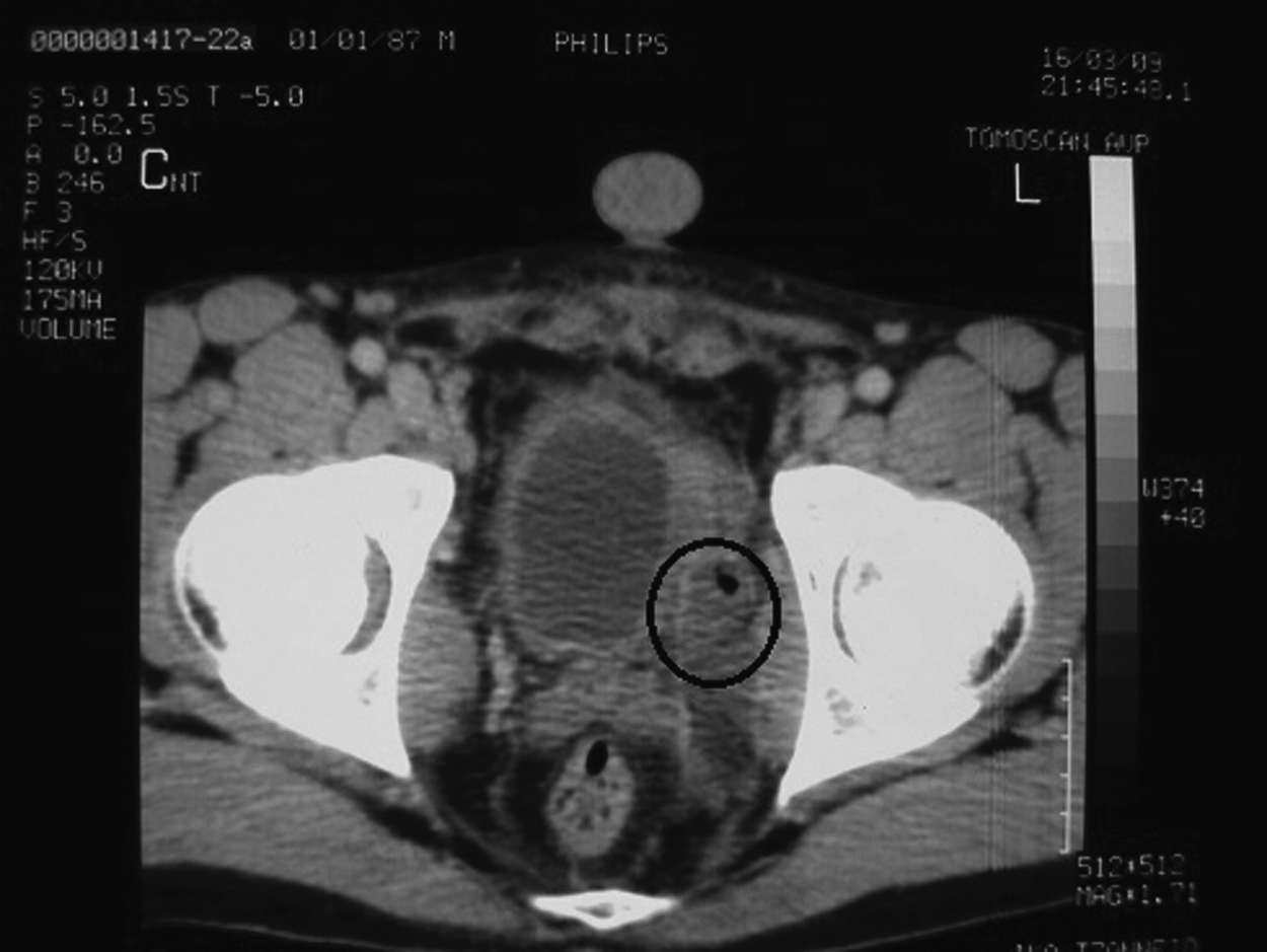
A fluid collection is present medial to the left obturator muscle. It abuts the left lateral wall of the urinary bladder, which appears thickened and ill-defined. A bubble of air is seen within the collection.
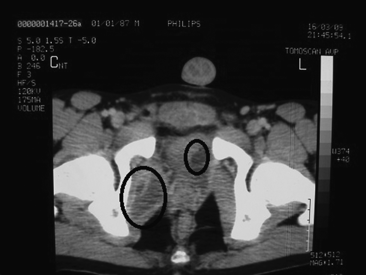
Slice 2 cm caudal to Fig. 1. A fluid collection is present within the right internal obturator muscle. The lowest part of the left-sided collection is also noted.

Fluid collections within the right external obturator muscle.

Fluid collections within the right adductor magnus muscle.
The patient gradually became responsive to the treatment and became afebrile six days later, while pain decreased substantially. Pelvic MRI depicted remission of the abscesses. After remaining completely afebrile for seven days, the patient was discharged with linezolid (600 mg bid), according to antibiotic sensitivity, for four weeks and with instructions for outpatient re-evaluation twice per week. The patient was healthy clinically, inflammation markers including WBC, ESR, CRP, and CPK were normal during follow-up. Four weeks after the discontinuation of linezolid, pelvic MRI confirmed complete remission of the abscesses. Six months later, the patient was healthy. Another MRI scan confirmed the complete remission of the lesions.
Discussion
Pyomyositis is a purulent infection of muscle, commonly afflicting large muscle groups. It was first described by Scriba [1] in 1885, despite the fact that both Osler and Virchow had already reported undetailed cases of the disease. The disease was associated initially with tropical climates, mainly African countries, hence the original description as myositis tropicalis. However, current literature reports an increasing incidence of the disease in temperate climates, to such an extent, that previous travel in a tropical country has been excluded from the diagnostic criteria for the disease [2].
The increased incidence in tropical climates is attributed to the low socioeconomic status that is associated with malnutrition, and increased incidences of parasitic infections and trauma, constituting up to 1 to 4% of hospital admissions [3]. The incidence in temperate climates has been estimated at 0.5 cases per 100,000 persons annually by Block et al. [4], and appears usually in immunocompromised patients suffering from HIV infection, diabetes mellitus, malignant disease, and patients undergoing chemotherapy or immunosuppressive treatment [2,4]. Males are afflicted more often than females and the disease is most prevalent in childhood and adolescence [5].
The pathophysiology of pyomyositis remains unclear. However, experimental data suggest the existence of underlying muscle injury in combination with transient bacteriemia as necessary conditions for the development of the disease, because normal muscle is not susceptible to bacterial colonization. Miyake [6] in 1904 showed in animal studies that following injection of sublethal doses of S. aureus myositis did not occur until the muscles were crushed, electrocuted, or rendered ischemic. It was proposed that fibronectin-binding receptors on myocytes serve as the path of bacterial entry, and that prior muscle trauma is necessary for the development of pyomyositis. However, prior local trauma has been documented in only 21% to 66% of reported pyomyositis cases [2,5,7].
Literature agrees in the recognition of three stages [2,7]. The disease initiates with non-specific alterations of the muscles such as edema and is characterized by pain attributed to bacterial invasion. In this stage, the disease presents with low-grade fever and tenderness. Abscesses are not obvious until 2–3 wks later, during which suppurative phase most of the cases are diagnosed because imaging techniques and biopsy demonstrate more specific findings. The aforementioned symptoms are aggravated, the characteristic “woody” texture of the affected area manifests and, depending on the location of the abscesses, symptoms and signs may include those of retrocecal appendicitis, arthritis, or polymyositis. The final and most ominous stage is that of sepsis, including bacteremia with rigors and high fever, hemodynamic instability, abscesses in remote organs and tissues, and multiple organ dysfunction syndrome.
Staphylococcus aureus is the most common pathogen involved in the pathophysiology of pyomyositis, being identified in 70% to 90% of cases, being followed by group A streptococci and less commonly gram-negative bacteria such as Escherichia coli, in patients with underlying hematologic malignant tumors [2,8].
Pyomyositis often affects the limbs, most commonly large muscles of the lower extremities and less often larger muscle groups of the torso [9]. Additionally, a single group is afflicted in most cases [2]. However, cases of multi-focal pyomyositis have been described [10,11], as have cases of unusual location such as the forearm, with concomitant compartment syndrome [12]. Obturator pyomyositis is rare. After the review of 22 cases by King et al. [5], there have been sporadic reports of pyomyositis of the obturator muscles [13–17], mostly in children and young adults. Bilateral obturator involvement is even more rare, with the first case described by Mukhtyar et al. [18]. However, ours is the first case of bilateral obturator muscle involvement with coexisting involvement of pelvic floor muscles (levator ani) and possibly viscera (urinary bladder).
The diagnosis of pyomyositis is based on clinical suspicion and laboratory evaluation, but key is the use of imaging modalities. In particular, MRI with gadolinium enhancement is believed to be the investigation of choice in acute pyomyositis, delineating pelvic muscle anatomy and locating abscesses [2,5,9,19]. However, CT may provide adequate imaging in certain cases [2].
The treatment of pyomyositis depends on the stage at which the diagnosis is made. Among patients diagnosed while still in the first stage, antibiotic treatment alone may be sufficient. Given that the most common isolated pathogen is S. aureus, empiric treatment should include anti-staphylococcal penicillins, or vancomycin in centers where MRSA infections have a high incidence. Antibiotics effective against gram-negative bacteria should also be considered in immunosuppressed patients. Additionally, an active agent should be administered in the presence of anaerobes [2,4,20].
The regimen should be modified accordingly, once cultures and susceptibility tests are available. Treatment should be administered for at least 3–4 wks, according to response [2,20]. Antibiotics should initially be administered intravenously to achieve high concentrations rapidly, and may be administered orally later, according to response. Once abscesses formation is confirmed, or when drainage of the abscesses should be considered, whether percutaneously with image guidance, or through an open surgical procedure along with the administration of antibiotics.
Current literature reports that approximately 50% to 55% of cases are treated adequately by the sole administration of antibiotic therapy [4,5,10–18]. On the other hand, cases presenting with either SIS and large abscesses (usually larger than 3 cm), patients at extremes of age—mostly children—are treated predominantly with abscess drainage. There is no evidence that any drainage modality is superior in terms of remission and survival [4,5] as long as source control is ensured. In our patient, percutaneous drainage was not attempted, because the abscesses were not accessible anatomically [21]. Moreover, the clinical condition and the clinical course of the patient did not render mandatory any type of source-control procedure. The patient was young, well-nourished, and lacking any other co-morbidity, and the peritoneal cavity was not affected [22]. Therefore, we preferred to avoid a drainage procedure with putative complications. The rationale for this approach was that the patient did not demonstrate signs of deterioration, along with the facts that the abscesses were multiple, small in size, and inaccessible via percutaneous route. Finally, the alternative of surgical drainage was deferred because an extensive operative procedure could be accompanied by complications that might affect in the quality of life and lifestyle of the patient. The presented case is, to the best of our knowledge, the first of bilateral obturator pyomyositis with such an extensive affliction of pelvic muscles that was treated exclusively by the administration of antibiotic therapy directed against CA-MRSA [23], and the effort for the least invasive strategy, resulting in the lowest morbidity rates possible. Unfortunately, the lack of large case series renders the suggestion of a specific treatment algorithm implausible. The management of such patients, unless amenable to the standard treatment protocol, should be tailored and considered case by case.
Footnotes
Author Disclosure Statement
No competing financial interests exist.