
Abstract
Abstract
Background:
Basidiobolomycosis is a rare fungal infection caused by the fungus Basidiobolus ranarum. Gastrointestinal basidiobolomycosis (GIB) is an unusual presentation of the fungal infection that is reported sparsely in the literature, but is an emerging infection in the southwestern United States. Lack of awareness of GIB has resulted in its delayed diagnosis and in extensive morbidity and mortality in patients with GIB.
Methods:
Case report and literature review.
Case Report:
We report the rare case of a young female with GIB that presented as perforated appendicitis with abscess formation.
Conclusion:
Although GIB is rare, immediate and aggressive therapy should be initiated when it is diagnosed. Both long-term medical and surgical treatment is required for its definitive management.
B
Case Report
A 34-year-old female with a body mass index (BMI) of 20.8 kg/m2 presented to the emergency department with a chief complaint of abdominal pain in the epigastric region that had persisted for 1 mo. The pain was sharp, intermittent, 6/10 in intensity, radiating to the groin, and worsening on food intake, and was associated with vomiting, constipation, and weight loss.
The patient's past medical history was notable for diabetes mellitus, chronic pain syndrome, and gastroesophageal reflux disease (GERD). She reported daily use of rofecoxib and ranitidine.
On physical examination, the patient was afebrile with normal vital signs. Her abdomen was soft and tender with normal bowel sounds. Laboratory tests showed an elevated white blood cell (WBC) count of 13×103 WBC/mm3, eosinophilia (absolute eosinophil count of 2.2×10³/mm3), and an elevated cardiac troponin concentration of 0.64 mcg/L. The patient's serum glucose concentration was 133 g/dL.
An electrocardiogram (ECG) revealed sinus tachycardia, a shortened PR interval, and inverted T waves. The patient was given sublingual nitroglycerin and admitted for cardiac catheterization, which revealed a normal ejection fraction and occlusion of a branch of the left circumflex coronary artery.
Despite treatment with nitroglycerin and cardiac catheterization, the patient's pain persisted and her WBC count continued to increase. A urine analysis was positive for red blood cells (RBC) at 24/high-power field (HPF) and WBC at 40/HPF, and treatment with antibiotics was begun on the suspicion of a urinary tract infection (UTI). Despite this, the patient's condition continued to deteriorate and a CT scan revealed perforated appendicitis with abscess formation in the right lower quadrant, and narrowing and thickening of the terminal ileum caused by inflammatory changes (Fig. 1). Computed tomography-guided drainage was done and a pigtail catheter was placed in the right lower quadrant. Culture of the abscess fluid was positive for coagulase-negative staphylococci, lactobacilli, mixed flora, filamentous fungi, and yeast.
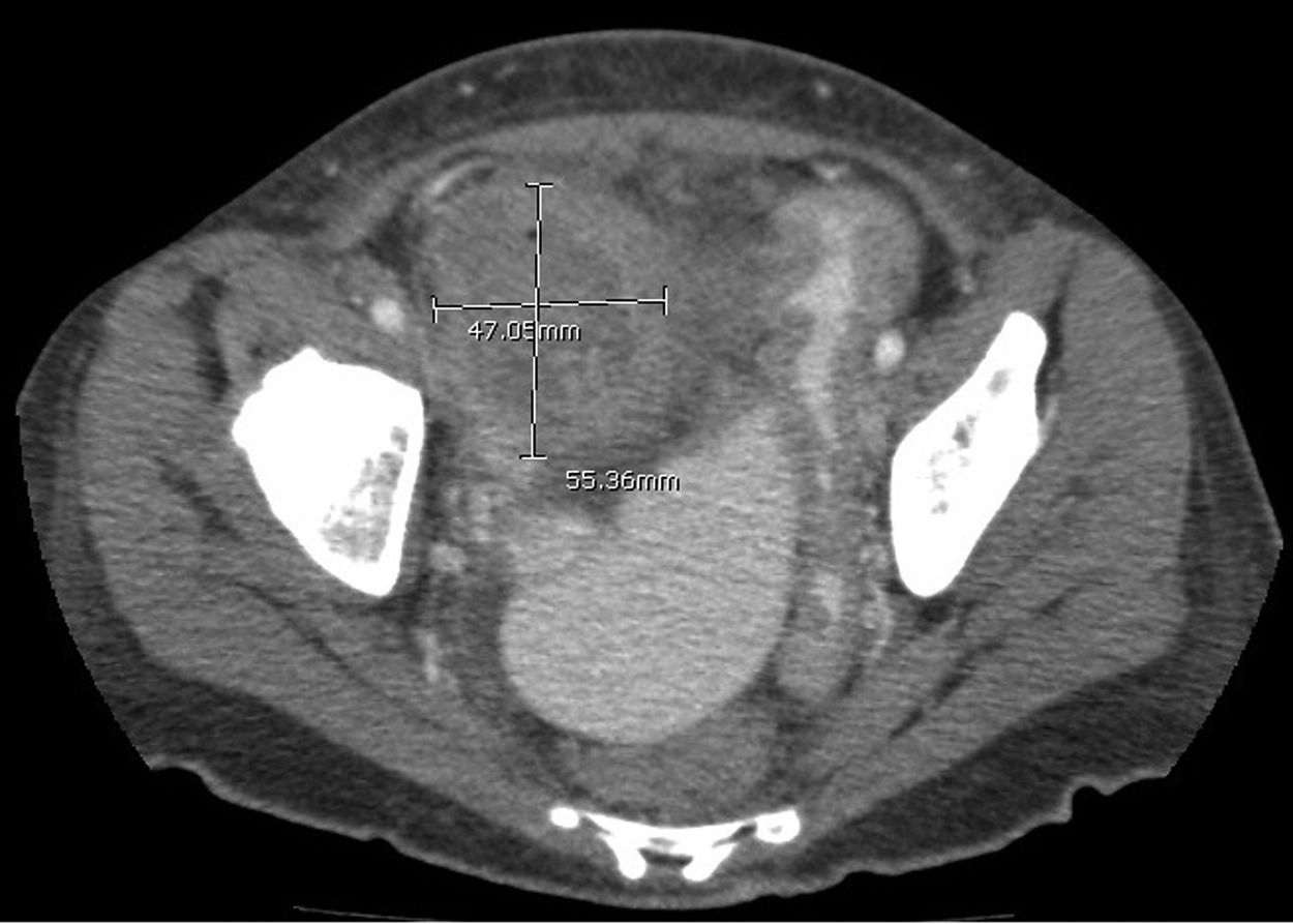
Computed tomographic image of patient's abdomen demonstrating perforated appendicitis with abscess formation.
The patient was given single doses each of 1 g IV of ceftriaxone and piperacillin/tazobactam 3.375 gm IV. The patient was also started on oral ciprofloxacin 250 mg BID over a 5-d period. The patient's condition improved with antibiotic therapy over the course of her hospital stay, and she was discharged home with the drain and a prescription for oral amoxicillin-clavulanic acid 500 mg BID for 15 d and amphotericin B lipid complex 130 mg daily for 2 wks. The patient was advised to come to the outpatient clinic for follow up at two weeks after her discharge, for removal of the drain and planning for an interval appendectomy.
However, the patient returned to the emergency department within one week after her discharge with a complaint of abdominal pain similar to that which had prompted her initial presentation. Fluoroscopy of her abscess cavity revealed that the cavity had a connection to the cecum, and a colocutaneous fistula was documented (Fig. 2). She was re-admitted to the hospital and taken to the operating room for an exploratory laparotomy. Through a midline incision, inflammation was noted in the terminal ileum and cecum due to perforated appendicitis. Exudate was adherent to the anterior abdominal wall and was taken down together with the adhesions. The inflamed portion of the terminal ileum and cecum was resected with a primary side-to-side anastomosis. A drain was placed, after which the abdomen was closed and the resected specimen was sent for pathology evaluation.
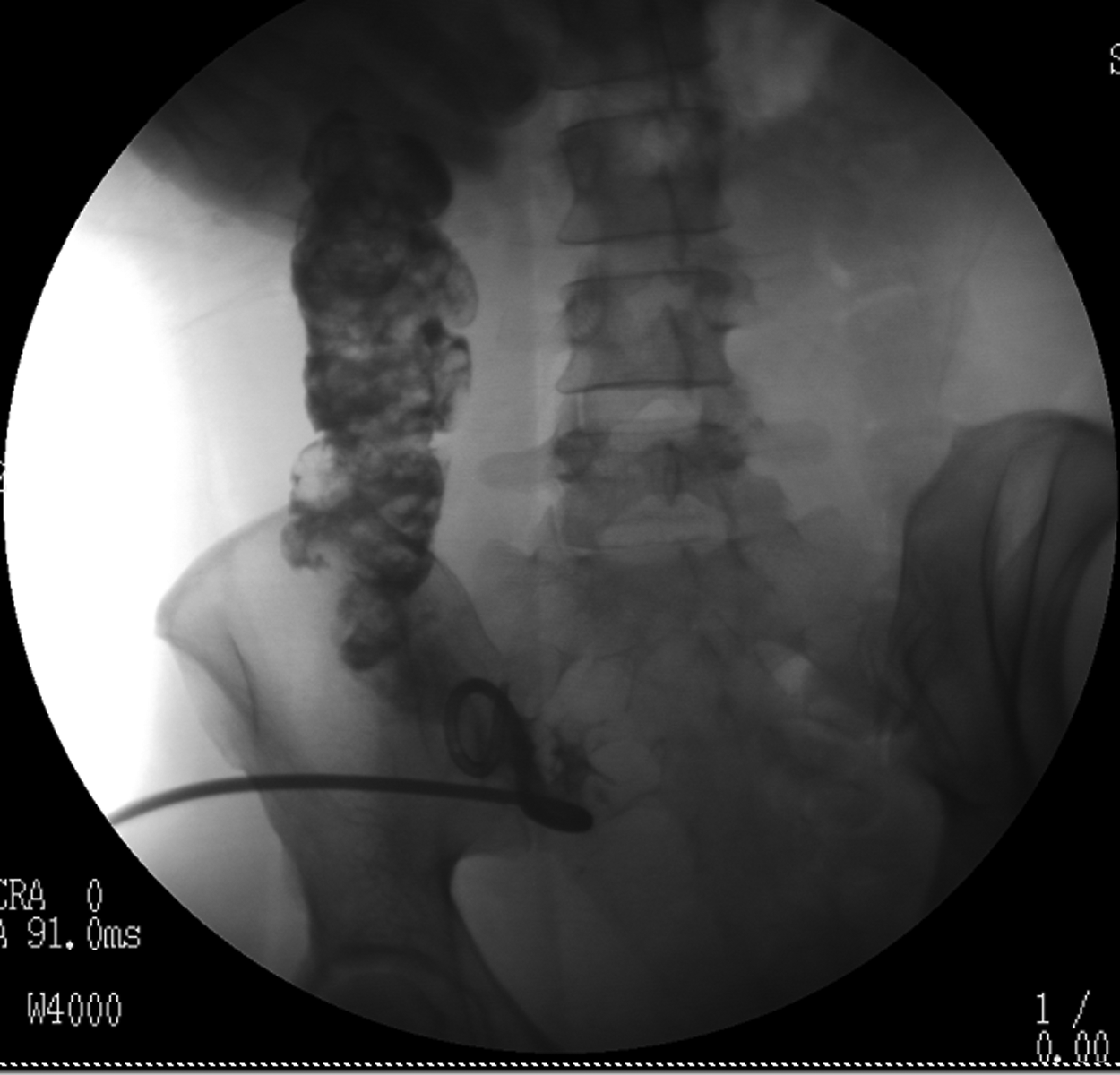
Fluoroscopic image of patient's abscess cavity demonstrating a colocutaneous fistula.
Surgical pathology revealed extensive necrotizing granulomatous inflammation of the ileocecal specimen and invasion by fungal hyphae corresponding to those of Basidiobolus fungi. Microbiologic testing of the abscess fluid demonstrated filamentous fungi, and Basidiobolus fungi were identified.
Post-operatively the patient was given piperacillin-tazobactam 3.375 g IV 96 h, oral itraconazole 200 mg BID for 5 d, and amphotericin B lipid complex 200 mg IV for 2 wks. The patient improved gradually and she was discharged home in stable condition with a peripherally inserted central catheter (PICC) line and a regimen of oral itraconazole 200 mg BID and amphotericin B lipid complex 130 mg IV daily for 6 wks.
Nevertheless, the patient had a complicated post-operative course and returned 3 wks after her discharge with recurrent right lower quadrant pain. She was found to have a recurrent abscess, which was treated through interventional radiology (IR), and the patient was re-treated with anti-fungal medications that consisted of a second round of oral itraconazole 200 mg twice daily and amphotericin B lipid complex 130 mg IV daily and had a complete recovery.
Discussion
Gastrointestinal basidiobolomycosis is a rare presentation of infection by the fungus B. ranarum, which is acquired through ingestion of contaminated soil or vegetables and high-risk activities associated with environmental exposure to this fungus, such as gardening and landscaping [7]. Patients with GIB present with non-specific signs and symptoms that delay a definitive diagnosis and treatment of the disease. Marshall et al. reported that patients with GIB present commonly with a range of sub-acute symptoms, with abdominal pain being the most important [6]. Fever, vomiting, and weight loss are also common presenting symptoms and mimic those of other common gastrointestinal diseases, resulting in a delay of appropriate treatment. Adults with GIB usually present with complaints of constipation, whereas children with GIB often present with diarrhea [8,9]. Gastroesophageal reflux disease and prolonged habitation in tropical climates are considered potential risk factors for development of GIB [6].
In patients with GIB, CT scanning demonstrates commonly a gastrointestinal mass consistent with inflammatory changes or malignant disease [6]. On the basis of its presenting symptoms and findings on CT scanning, GIB is often mistaken for inflammatory disease (Crohn disease or ulcerative colitis), especially in elderly populations. Hematology, serum immunology, and blood culture are used routinely for the diagnosis of GIB. Wardlaw et al. reported that patients with GIB have an elevated WBC count and eosinophil count commonly [10]. On blood culture, white or grey colonies with radial folds are diagnostic for the growth of B. ranarum [1]. Blood culture has a higher sensitivity for the diagnosis of GIB than does testing for specific serum antibodies to B. ranarum [9,11].
Basidiobolus ranarum infects the colon and the rectum commonly, followed by the small bowel, liver, and stomach. The lack of clarity in the diagnosis of GIB delays its definitive treatment, and the optimal management of patients with the disease therefore rests on a combination of medical and surgical techniques. Medical management includes the use of oral or intravenous antifungal drugs. Itraconazole is considered the drug of choice for treating GIB, whereas amphotericin B is considered ineffective for its treatment [15,16]. Patients with both GIB and subcutaneous manifestations of the disease must also be treated with potassium iodide [12–14]. Evidence suggests that antifungal agents belonging to the new generation of these drugs, consisting of derivatives of fluconazole, voriconazole, and posaconazole, are effective adjuncts for the treatment of GIB [2,9]. Antifungal treatment of our patient was begun with itraconazole during her first hospitalization, but her symptoms did not improve and she required a more invasive approach. Although other authors report poor results with amphotericin B in treating GIB, our patient responded to this therapy.
Surgical excision with histopathologic assessment of the resected specimen is the standard course of management for patients with GIB who do not respond to medical therapy. Holenarasipur et al. reported that most patients with GIB in their series underwent surgical excision for its treatment [2]. They also documented delays in its surgical management owing to the lack of definitive diagnosis of the disease.
Patients with GIB often have a prolonged and complicated hospital course. Most require surgical intervention with long-term antifungal therapy. High mortality rates have been reported for patients who undergo surgical resection without antifungal treatment [2]. The patient in our case received antifungal medications in addition to the surgery. Furthermore, the post-operative course of our patient was complicated, with recurrence of her abscess, which eventually resolved after drainage. Hence, we recommend early and aggressive medical therapy and surgical intervention for patients with a confirmed diagnosis of GIB.
Conclusions
There is an urgent need to spread awareness of GIB among health care professionals, especially in states where it is highly prevalent, such as Arizona. Although GIB is rare, immediate and aggressive therapy should be initiated when it is diagnosed. Both long-term medical and surgical treatment is required for its definitive management.
Author Disclosure Statement
No competing financial interests exist.