
Abstract

A
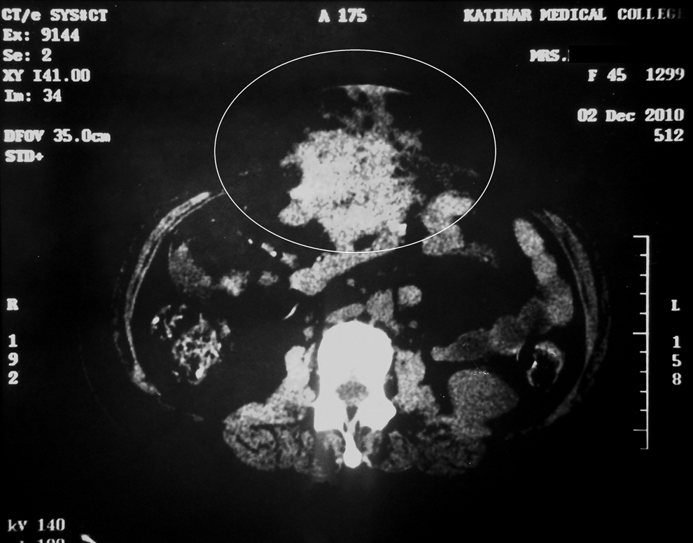
Computed tomography scan showing abdominal wall mass.
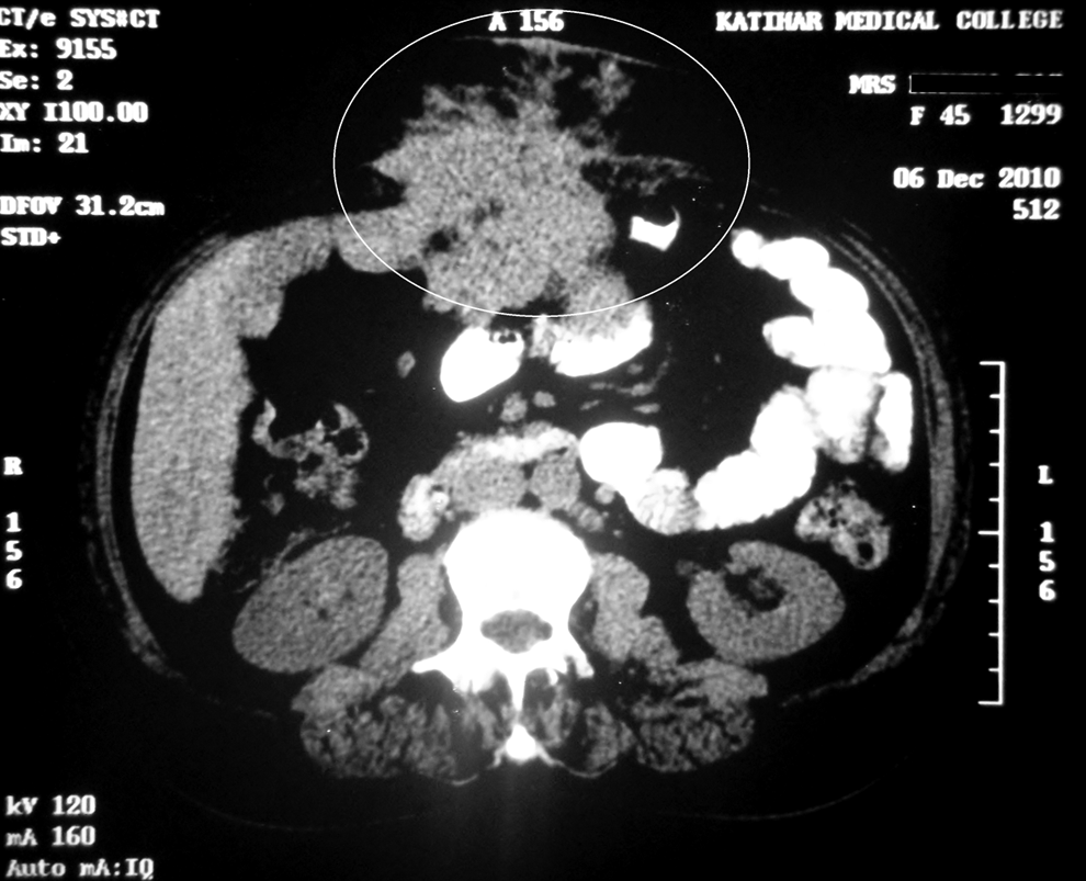
Contrast computed tomography scan shows enhanced solid contents in inhomogeneous mass.
Considering the size of the mass and an earlier poor response to empiric antibiotics, surgical intervention was decided upon. Pre-operatively, the patient was administered penicillin—500,000 IU/6 h and Dapsone—50 mg OD for four weeks. Laparotomy was done and three separate masses were excised. The final histopathology specimen showed “sulfur granules” consistent with Actinomyces (Fig. 3). Post-operatively, the patient was administered ceftriaxone-tazobactam for one week followed by oral amoxicillin for two weeks. The patient had an uneventful recovery.

Histopathologic specimen showing target-like sulfur granule surrounded by inflammatory cells, suggesting actinomycosis.
Abdominal actinomycosis presents as an indolent chronic suppurative process with atypical symptoms that are often misinterpreted as malignant disease; the correct diagnosis is usually achieved only at operation [1]. Isolated abdominal wall actinomycosis is rare; by 2008, only 22 cases have been reported in the literature [2]. Whereas some cases have been reported after insertion of an intra-uterine device (IUD), this is the first reported case of isolated abdominal wall actinomycosis following hysterectomy. The pathogenesis of abdominal actinomycosis is not well understood, but it seems to require injury to the normal mucosa to penetrate and cause disease [3]. Once the bacteria invade tissue, a granulomatous inflammatory response ensues, leading eventually to necrosis and reactive fibrosis. Histologically, “sulfur granules” are characteristic [4]. Actinomycosis should be included in the differential diagnosis when a CT scan shows an infiltrative mass with unusual aggressiveness and dense inhomogeneous contrast enhancement, especially in patients with leukocytosis, fever, long-term use of an IUD [5], or hysterectomy.
Footnotes
Acknowledgment
The authors acknowledge the help of Dr. Rajesh Kumar, Department of Pathology, Katihar Medical College in providing the histopathological image.