
Abstract
Abstract
Background:
Non-thermal dielectric-barrier discharge plasma (non-thermal plasma) is being investigated for use in wound healing. Alginate gel, already in clinical use, is non-toxic but has no meaningful antimicrobial property. This study reports that a non-thermal-plasma-treated alginate wound dressing has strong antimicrobial properties.
Methods:
Alginate gel was treated with non-thermal plasma in room air and inoculated with bacterial pathogens. At 15 min after this, bacterial cell viability was determined by colony assay or 2,3-bis-(2-methoxy-4- nitro-5-sulfophenyl)-2H-tetrazolium-5-carboxanilide (XTT) assay. The anti-biofilm efficacy of the non-thermal-plasma-treated alginate gel was investigated and the treated gel was tested against vascular endothelial cells for a cytotoxic effect. The proliferation and migration of bacterial cells before and after exposure to the treated gel were investigated with an in vitro wound testing assay. Scanning electron microscopy was used to observe changes in the gel surface associated with exposure to bacterial pathogens. The treated gel was tested against Acinetobacter baumannii, Escherichia coli, Staphylococcus aureus, S. epidermidis, Candida albicans, and C. glabrata as representative pathogens (at 106–109 colony-forming units [CFU]/mL), and the thickness of a plasma-treated gel dressing and distance between a glass dielectric-barrier discharge plasma probe and the gel surface were kept constant.
Results:
Non-thermal-plasma-treated alginate gel exhibited a strong biocidal property and inactivated all of the pathogens included in the study at counts of 108 CFU/mL and within 15 sec of treatment. The treated gel inactivated 109 CFU/mL of the organisms within 1 min, and 3 min of exposure to the treated gel inactivated pathogens embedded in biofilms. The plasma-treated gel showed no significant cytotoxicity, and endothelial cells exposed to the treated gel proliferated and migrated well across a wound area over a period of time. Dressings made with the treated gel retained their biocidal effects for about a month. Scanning electron microscopy showed no damage to the surfaces of treated gels, but damage to the bacterial pathogens on plasma exposure.
Conclusion:
A non-thermal-plasma-treated alginate gel dressing has the clinical potential to decontaminate wounds, prevent surgical site infection, and promote wound healing.
M
Research has shown the therapeutic effects of alginate dressings. One recent study examined full-thickness wounds on the dorsum of rats and demonstrated more rapid healing with alginate dressings in treated animals than in untreated controls. In the same study, alginate was found to bind elastase, decrease the expression of pro-inflammatory cytokines, and increase the expression of collagen I [7]. In 1989, a comparative trial of calcium alginate dressings and paraffin gauze dressings on 130 split-thickness skin graft donor sites found that the alginate dressings decreased healing time from 10 d to 7 d and significantly improved patient comfort [8]. Researchers have also demonstrated that calcium alginate dressings have hemostatic [9] and some bacteriostatic [10] properties. The anti-microbial effect of wound dressings is particularly important because it is believed that even sub-clinical bacterial colonization impedes wound-repair mechanisms [11]. Many alginate wound dressings have been modified with bactericidal materials, such as chlorhexidine or silver-ion-releasing compounds, to improve their efficacy of protection against wound infection. We explored the advantages of a new technology conferring a bactericidal effect on calcium alginate gels.
Previous research in our laboratory demonstrated that non-thermal dielectric-barrier discharge plasma (thermally cold), generated at room air temperature, has a potent anti-microbial effect through its generation of free radicals, reactive oxygen species (ROS), and reactive nitrogen species, such as hydrogen peroxide (H2O2), superoxide, singlet oxygen, nitric oxide, and ammonia [1,12–15]. The non-thermal plasma used in the present study is typically micropulsed and generated by a plasma generator from cold plasma pulsed at high voltage between a gap with the glass or quartz-surface-covered electrode at one end and the surface of a biologic sample or fluid (which serves as a second electrode) at the other end. The high-voltage electrode is covered with a dielectric barrier that makes it safe for treating heat-sensitive materials and fluids. The plasma used in the present study was tested previously for sterilizing capability by the direct application of a plasma-generating probe to the pig skin and other biologic materials cultures of gram-positive and gram-negative bacteria (direct plasma application). Both the direct application (wherein a biologic material of interest is directly exposed to a non-thermal-plasma discharge) and indirect application (wherein a vehicle such as a fluid is exposed to a non-thermal plasma discharge) methods are effective in killing bacterial cells, and generate reactive oxygen and nitrogen species [12,14]. The reactive species thus generated, or their products, are responsible predominantly for the inactivation or killing of bacterial cells. Our laboratory has demonstrated that fluids treated with a non-thermal plasma of the type used in the present study become capable of transferring the anti-microbial effect of such treatment to a biomaterial of interest, which can then be placed in contact with microbes to inactivate them (Joshi Laboratory, personal communication). This technique creates a potent anti-microbial effect that, according to preliminary results, is non-toxic to eukaryotic cells. In the present study we used a non-thermal dielectric barrier discharge plasma and confer anti-microbial properties of treated alginate dressings with potentially less tissue toxicity than might be produced by the anti-microbial agents suspended in currently used alginate dressings, but still inactivating a range of wound-contaminating pathogens.
Materials and Methods
Wound dressings
For the preparation of non-thermal-plasma-treated alginate wound dressings, sodium alginate (M:G ratio 1.6; Sigma-Aldrich, St. Louis, MO) was suspended in de-ionized H2O, filter sterilized, poured into a mold, and cross-linked with calcium to form circular hydrogels (26 mm in diameter). The hydrogels were then treated for 3 min with a non-thermal plasma generated by a micropulsed discharge from a dielectric barrier [12,14,16] (1.2 W/cm2, 31 kV, 1500 Hz). The non-thermal plasma was generated with an electrode constructed with a 38×64-mm copper plate insulated in silicone and a 1-mm-thick glass sheet. The discharge gap between the bottom of the glass-barrier and the treated surface of each alginate hydrogel was fixed at 1 mm (Figure S1). The energies calculated as being deposited in the hydrogels (J/cm2) were 0 J/cm2 at 0 s of exposure, 4.65 J/cm2 at 15 s, 9.3 J/cm2 at 30 s, 13.95 J/cm2 at 45 s, 18.6 J/cm2 at 60 s, 27.9 J/cm2 at 90 s, 37.2 J/cm2 at 120 s, and 55.8 J/cm2 at 180 s. Silver-containing test dressings (Acticoat; Smith & Nephew, London, United Kingdom), made from alginate fibers coated with silver nanoparticles, and chlorhexidine-containing dressings (Tegaderm CHG; 3M, St. Paul, MN), made from sodium alginate with chlorhexidine in suspension, were used for comparison with the plasma-treated and untreated alginate test dressings investigated in the study. A sterile, non-woven absorbent material (Sontara; DuPont, Wilmington, DE) was used as a control. Discs (26 mm) made of each dressing and of the two comparison control dressings were prepared in an aseptic manner and kept in sterile vials for use.
Test organisms
Acinetobacter baumannii ATCC 19606, Escherichia coli ATCC 23922, Staphylococcus aureus ATCC 23923, and Staphylococcus epidermidis ATCC 12228 were purchased from American Type Culture Collection (www.atcc.org; ATCC, Manassas, VA), and Candida albicans and Candida glabrata were a generous gift from Dr. Thomas Edlind (Department of Microbiology and Immunology, Drexel University College of Medicine, Philadelphia, PA). Cultures of the test organisms were revived and grown overnight essentially in accord with the directions on tryptic soy broth or agar for the bacterial pathogens and yeast extract–peptone dextrose broth or agar for the fungal pathogens included in the study.
Colony count assay
Untreated alginate hydrogel and Sontara were used as control dressings. A single colony of each test organism, isolated from an overnight culture plate, was inoculated in 5 mL of tryptic soy broth and incubated for 4–6 h at 37°C to reach an optical density (measured at 600 nm) of 0.2 (∼0.5 McFarland turbidity). A total of 50 microliters of the test bacterial or fungal suspension was transferred to a non-thermal-plasma-treated or untreated dressing in a sterile Petri dish (1-in diameter). After 15 min of incubation at room temperature, the dressings containing the bacterial or fungal pathogens were washed for 5 min with 10 mL of sterile phosphate-buffered saline (PBS), with gentle agitation used to dislodge and harvest the pathogens in solution. The solution from each dressing was appropriately diluted and 50 microliters of the diluted sample was spread on a culture plate with a sterile disposable spreader, with the plate then incubated at 37°C for 24 h through 72 h (plate read every 24 h). The numbers of surviving bacteria were obtained through a standard plate-count procedure, and the logarithms of the reduction in numbers of bacteria were calculated as the difference between the logarithms of the numbers of bacteria surviving on the control dressings and of the numbers surviving on the test dressings. The plates made from the different non-thermal-plasma-treated alginate gel dressings and the control dressings remained under incubation for 72 h to assess for delayed microbial reactivation or to investigate microbial dormancy. For Candida species, yeast extract–peptone dextrose broth plates or agar plates were used. All experiments were done at least three times and in duplicate.
Antibiofilm activity assay
Biofilms were developed for E. coli and S. aureus essentially as described previously [12]. An overnight culture of each organism was diluted to 1:100 with sterile tryptic soy broth, and 200 microliters of the resulting suspension was applied to 96-well microplates (Costar; Corning, Inc., Corning, NY) in triplicate and incubated for 24 h at 37°C without shaking. On the following day, fluid from the biofilm-containing 96-well plates was gently aspirated and washed three times with sterile PBS to remove any planktonic or loosely attached bacterial cells before appropriate treatment with the supernatant from the non-thermal plasma-treated alginate dressings or silver-coated alginate or chlorhexidine–alginate control dressings. Metabolic markers were then detected using the 2,3-bis-[2-methoxy-4- nitro-5-sulfophenyl]-2H-tetrazolium-5-carboxanilide (XTT) assay. The gel supernatant for the anti-biofilm assay was prepared by incubating either non-thermal-plasma-treated alginate gel (treated on both sides for 1 min each) or untreated alginate gel with 2 mL of sterile PBS with gentle agitation (50 rpm) over periods ranging from 0 h through 24 h, and was collected by centrifugation at 10,000 rpm for 2 min. The biofilms were then either left untreated or were treated with non-thermal-plasma-treated alginate gel supernatant and incubated at 37°C for 15 min. Afterwards, the biofilms were briefly washed three times with sterile PBS, and the XTT assay was performed as described [12].
Viable cell counts by assay with XTT
The XTT assay measures a cell's respiration rate (by reading the orange-colored product generated during metabolism of XTT) and thus detects the numbers of viable cells in a specimen of cells. For each XTT assay, fresh XTT reagent solutions were prepared as described previously [12,13,17]. Aliquots of the reagent solutions containing 0.5 mg XTT (Molecular Probes; Invitrogen, Carlsbad, CA) and 1 μmol/L menadione (Sigma) were used to prepare working solutions in 1×PBS. After appropriate treatment or no treatment with the supernatant from the non-thermal-plasma-treated alginate dressings or silver-coated alginate or chlorhexidine–alginate control dressings, incubated respectively with the biofilm of E. coli or S. aureus organisms, 200 microliters of XTT reagent was added gently to appropriate wells of 96-well microtiter plates and the plates were incubated at 37°C for 2 h. After incubation, the absorption of the supernatant (100 microliters) from each plate used in the assay, containing an orange-colored XTT metabolic product, and was measured photometrically at 492 nm with a microtiter-plate reader (Synergy Mx; BioTek, Winooski, VT). The readings were normalized, and the percentages of surviving cells in each biofilm were calculated against the percentages in cultures of E. coli and S. aureus that were not exposed to the non-thermal-plasma-treated alginate or control dressings.
Endothelial-cell viability assay
The gels investigated in the study, either untreated or treated for 1 min on each side through exposure to non-thermal plasma, were pre-incubated (submerged) for periods of 0 h through 24 h in serum-free ATCC formulated DMEM cell-culture medium with agitation at 50 rpm. Multiple samples of the medium were taken over a 24-h period and cultures of endothelial cells were exposed to the supernatant from the culture medium. For this purpose, immortalized human umbilical vein endothelial cells of the EA.hy926 line were cultured according to the recommendations of the cell supplier (ATCC), with or without serum as indicated. The cells were seeded into the wells of 96-well plates at densities of ∼1×10⁴ cell/200 microliters per well and allowed to attach to the surfaces of the seeded wells through continuing incubation at 37°C in humidified standard cell-culture incubators with a 5% CO2 atmosphere. When the cells were near confluence, they were either left untreated or were treated with plasma-treated gel supernatant, and were assayed for cell viability over time. This was done with the XTT assay and a high-throughput 96-well microplate reader. The photometric densities of the reduction products of the XTT reagent were read in a microplate reader at 492 nm as described above. The assay was done on untreated cells or cells subjected to all three of the treatment conditions used in the study (cells treated with the supernatants of non-thermal-plasma-treated sodium alginate gels or untreated alginate gels, and of chlorhexidine-containing gels). Untreated gels and chlorhexidine-containing alginate gels were included as controls.
In vitro scratch wound assay
Cell scratch assays done with human fibroblast cells and the Oris Cell Migration Assay kit (Platypus Technologies, Madison, WI) were used to measure cell proliferation and migration across a wound site, according to the kit manufacturer's directions, with a slight modification. The plate used in the assay is commercially available in a pre-tricoated form (containing all three standard coatings of tissue culture, collagen I, and fibronectin) that generates highly reproducible, uniform slit wounds, and the assay is an ideal in vitro assay of wound healing (without the need for commercial trans-well membrane assembly). A total of 20,000 human vein endothelial cells were seeded into wells in the plate and allowed to adhere. The stoppers on the wells were then removed. The 200 microliters of cell culture medium from cells that were exposed either to untreated or non-thermal-plasma-treated alginate gel supernatant, collected over the period from 0 h–24 h, was added to the wells. The plate was then incubated for 2 h and the cell culture medium was aspirated and replaced with serum-containing medium, and the cells were observed over a period of 48 h. The cells were washed twice with 1×sterile PBS and labeled with Hoechst 33342 stain to compare the migration patterns of untreated cells and cells treated under the three different experimental conditions, using an Olympus BX60 Fluorescence Microscope (Olympus, Shinjuku, Tokyo, Japan). The images were saved as TIFF files and analyzed and edited with Adobe Photoshop CS (Adobe Systems, San Jose, CA).
Scanning electron microscopy
The morphologies of alginate hydrogel surfaces were observed before and after exposure of the hydrogel surfaces to non-thermal plasma or to pathogens inoculated on the surfaces of the hydrogels. The observations were made with a Zeiss Supra 50VP Scanning Electron Microscope (Zeiss, Oberkochen, Germany), according to the manufacturer's protocols. Samples were fixed, dehydrated, and sputter-coated with a platinum–palladium alloy before SEM.
Statistical analysis
For group comparison, significance was analyzed with a two-tailed Student t-test, and a value of p<0.05 was considered significant. All data were analyzed with Prism software (GraphPad, San Diego, CA). Bar indicates a standard deviation (SD). All experiments and experimental conditions were repeated at least three times and in triplicate (n=3), unless otherwise stated.
Results
Exposure of alginate gels to as few as 15 s of non-thermal plasma treatment resulted in complete inactivation of 7 log E. coli cells. The bactericidal effect was very rapid and the energy deposited during plasma treatments of the gels was sufficient to completely inactivate bacteria. Figure 1 shows a representative data set from a bacterial colony assay, with bacterial (E. coli) survival plotted against the duration of exposure in seconds.

Complete inhibition of E. coli at 107 CFU/mL by sodium alginate gels exposed for 15 sec to non-thermal plasma (bar=SD).
To determine the bactericidal capability of the non-thermal-plasma-treated alginate gel dressings, increasing concentrations of bacteria were exposed to gels treated with plasma for 15s–60 s. As illustrated in Figure 2, gels given 60 s of plasma exposure completely inactivated 109 colony-forming units (CFU)/mL of E. coli, and gels given 15 s of plasma treatment completely inactivated about 1×10⁸ CFU/mL of E. coli. The inactivation of E. coli was therefore proportionate to the amount of non-thermal plasma energy deposited on alginate gel dressings, and was cell-density dependent.
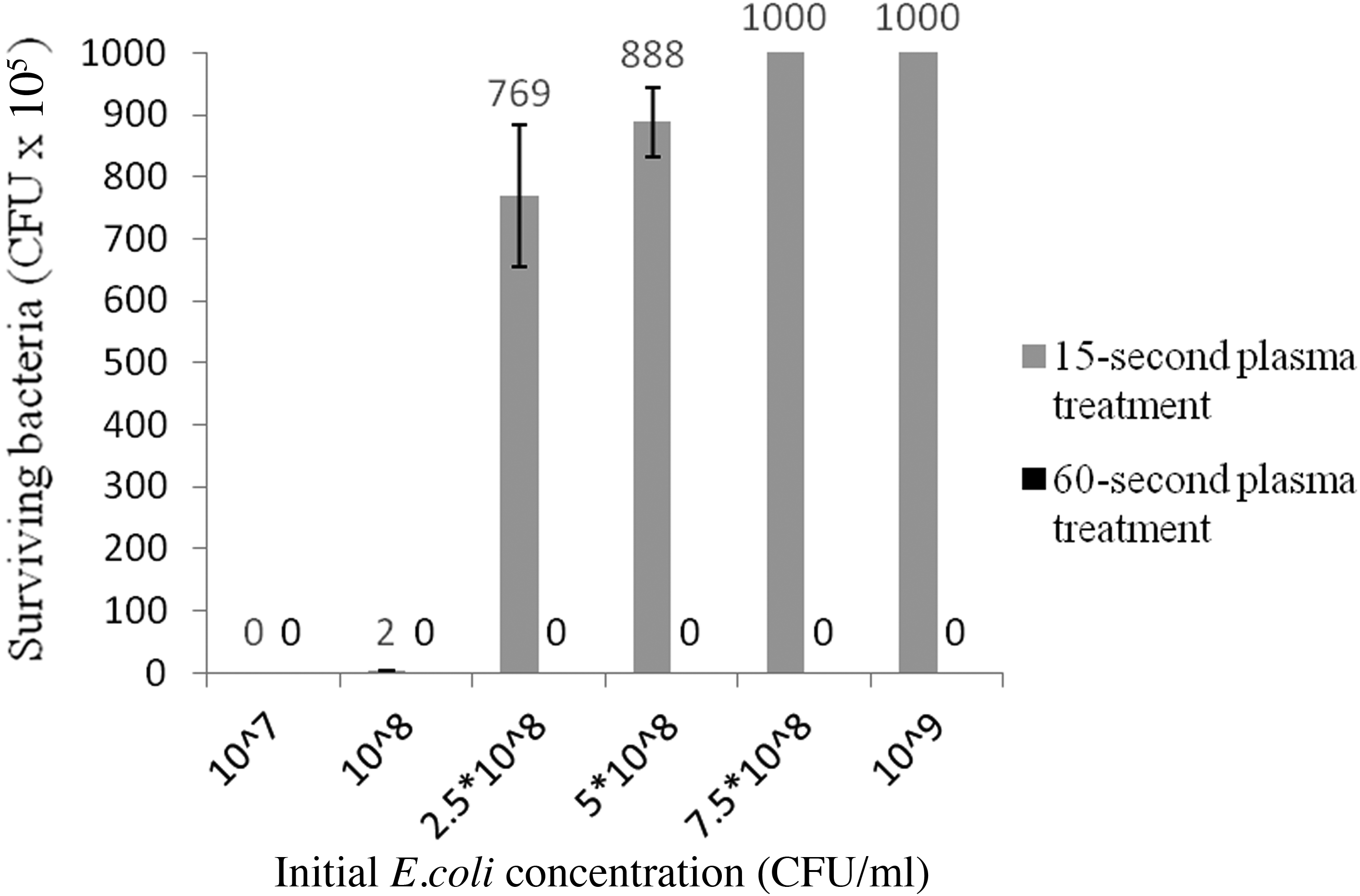
Complete inhibition of E. coli at 109 CFU/mL by alginate gels exposed for 60 s to non-thermal plasma (bar=SD).
Non-thermal-plasma-treated alginate gel dressings with a range of anti-microbial activities were also tested against a representative variety of gram-positive, gram-negative, and fungal pathogens commonly isolated from wounds (Fig. 3). As shown in Figure 3, gels treated with plasma for 15 sec or more completely inactivated almost all of the pathogens. Although there was some initial inactivation at zero exposure time of the pathogens during handling, exposure to plasma-treated gel produced statistically significant inactivation (p<0.05) of all of the representative pathogens tested (Fig. 3). As little as 15 s of plasma treatment was sufficient to induce inactivation of all pathogens except C. glabrata, which was completely inactivated by gels having less than 30 s of plasma treatment.
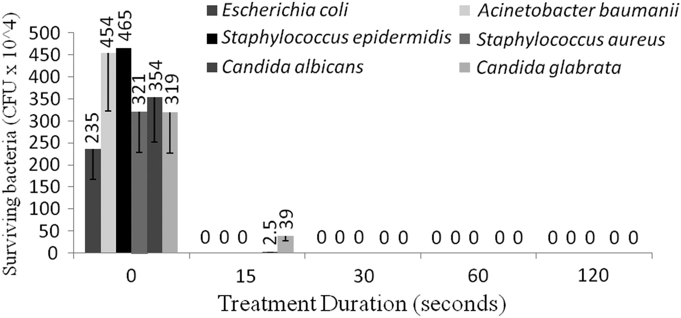
Alginate gels exposed for 15 sec to non-thermal plasma completely inhibited 107 CFU/mL of all bacterial pathogens included in the study. A value of p<0.05 was found for 15 sec of treatment of fungal pathogens with plasma-treated alginate gels vs. untreated gels under corresponding conditions (zero time). Complete inhibition was found with longer treatment times (bar, SD).
As in the case of non-thermal-plasma-treatment time, we also conducted experiments with different contact times to determine the optimal duration of contact required between a non-thermal-plasma-treated alginate gel and a pathogen. This assay also provides another important measure of antimicrobial efficacy, the rapidity of killing of pathogens. The plasma-treated gel dressings were compared with the commercially available chlorhexidine- and silver-containing dressings. The chlorhexidine-containing dressing produced the most rapid inactivation of pathogens, with a 1.5-log reduction in the numbers of pathogens at 1 min and complete inactivation of pathogens at 5 min (Figure 4). Non-thermal-plasma-treated gels completely inactivated pathogens in less than 10 min. The performances of the silver-containing dressing and the untreated alginate dressing were roughly equivalent over a period of 15 min.

Alginate gels exposed for 3 min to non-thermal plasma completely inhibited 107 CFU/mL E. coli after 10 min of contact. A value of p<0.05 was found for the effect of non-thermal plasma treated alginate gel on E. coli at 107 CFU/mL at 5 min (*) vs. an untreated dressing or silver-containing dressing with the same contact period. (CHG=chlorhexidine-containing dressing; bar=SD).
After treatment, the gels were stored either in a non-airtight container (in which the treated surfaces of the gels did not come into contact with anything but air) which was kept in a refrigerator at constant temperature and humidity, or in a vacuum-sealed plastic container (in which the treated surfaces of the gels came into contact with the container but not with air). Regardless of the storage method used, the anti-microbial effect of the plasma-treated gel dressing was maintained for 21 d but was diminished by 28 d (Fig. 5). Both the non-thermal plasma-treated and untreated alginate gels, inoculated with E. coli or uninoculated, were examined with SEM for surface-associated changes. Comparison of SEM images suggested that exposure to non-thermal plasma did not alter the hydrogel surface (Fig. 6). However, scanning electron micrographs of bacteria on the surfaces of treated hydrogels showed extensive damage to and leakage of the organisms' cell membranes as compared with those of organisms on untreated hydrogels.
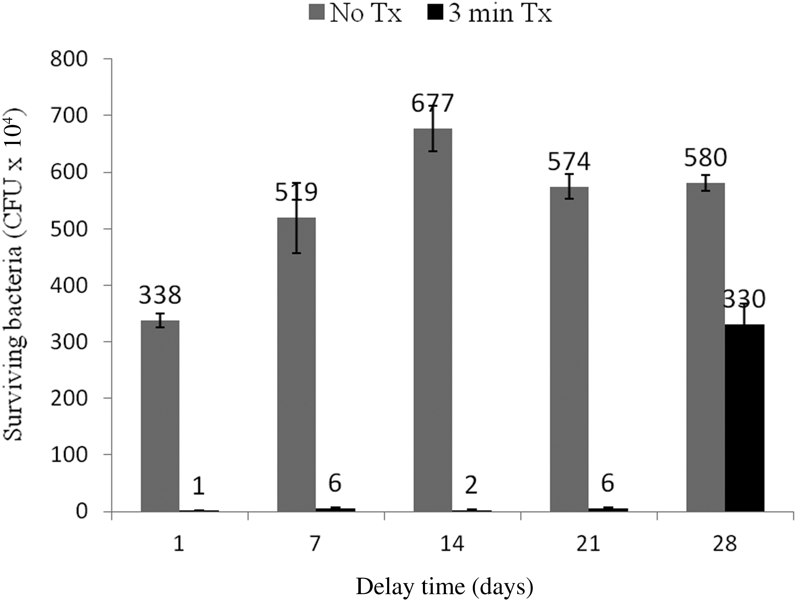
The bactericidal effect of gels exposed for 3 min to non-thermal plasma lasts for about 3 wks. A value of p<0.05 was found for the bactericidal effect of all alginate gels treated for 3 min vs. untreated gels. (Tx=plasma treatment; No Tx=no plasma treatment; min=minutes; bar=SD).

Images made with scanning electron microscopy of an untreated hydrogel without E. coli inoculation
Biofilms of early (24 h) duration, which typically correlate with the early colonization of wounds, were developed, and the inactivation response of pathogens embedded in the biofilms was observed over time. Assays showed that supernatants from non-thermal-plasma-treated gels had powerful anti-biofilm activity. Figure 7 shows a representative set of data for the response of an S. aureus biofilm to a plasma-treated alginate gel as a proof-of-concept experiment. Statistically significant inhibition of the biofilm (p<0.05) can be noted as compared with the supernatant from a gel not exposed to non-thermal plasma. A chlorhexidine-containing dressing was used as an internal control, and the data were normalized to biofilms without exposure to gels of any kind, which were taken as representing 100% survival of embedded pathogens.
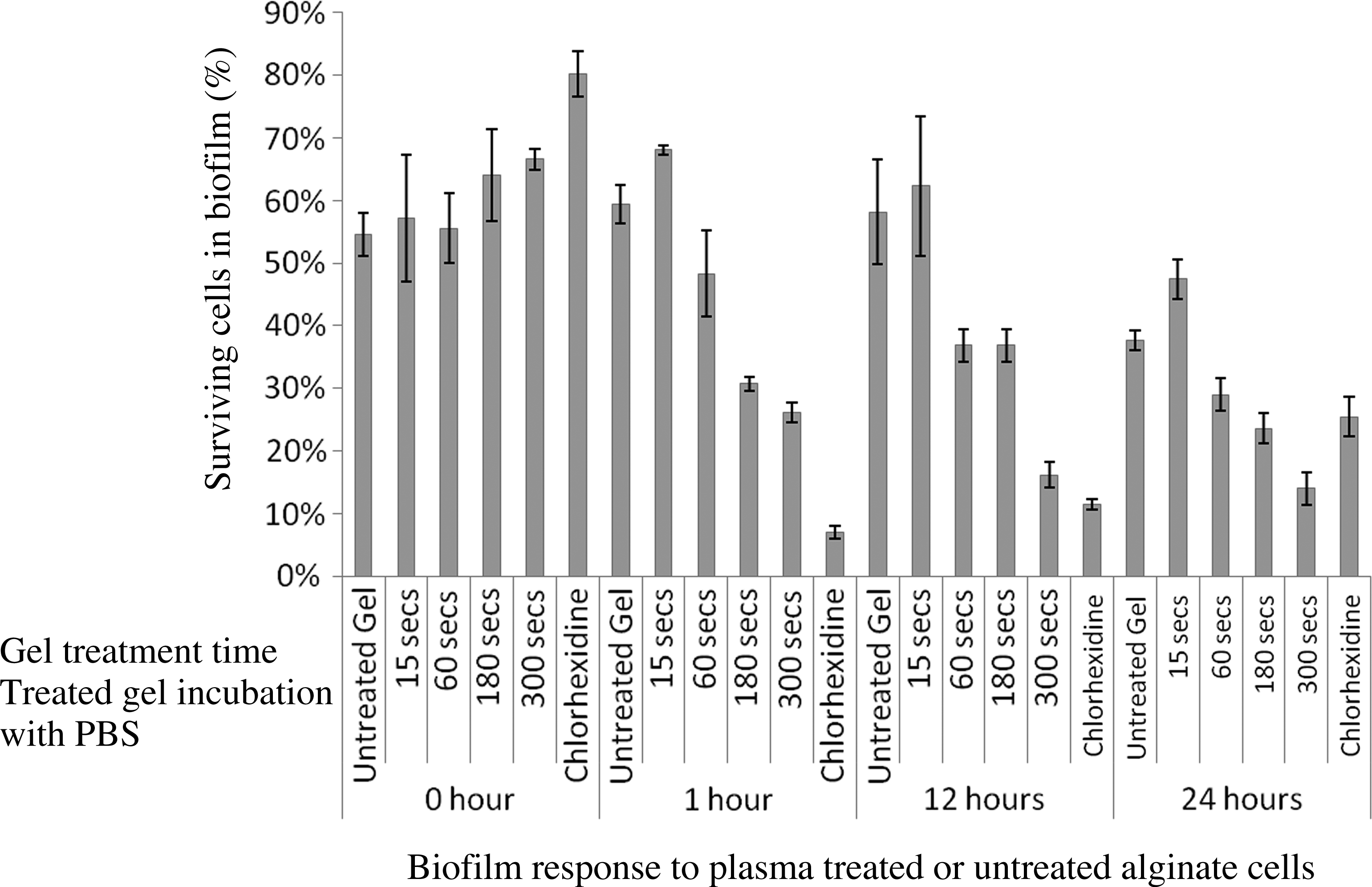
Three minutes of non-thermal plasma treatment of alginate gel had a significant inhibitory effect on Staphylococcus aureus in its biofilm form after 15 min of contact with the treated gel. The figure shows a histogram representative of the response of S. aureus biofilms grown for 23 h before gel exposure, demonstrating inactivation of biofilm-embedded S. aureus. The data are normalized against untreated biofilms that were not exposed to either non-thermal-plasma-treated or untreated gel. The treated or untreated gels were incubated for given period with biofilms in phosphate-buffered saline to test their anti-biofilm activity (see Materials and Methods). A value of p<0.05 was found for treated gels vs. untreated gels (synonymous with zero time) gel. (Bar=SD).
Assays of the viability of human umbilical vein endothelial cells exposed to plasma-treated gels, and of the gels' toxicity to these cells, showed that the gels caused no toxicity, but that on the contrary, the cells proliferated after exhibiting transiently retarded growth (up to 24 h). Non-thermal-plasma-treated and untreated alginate gels behaved almost similarly (Fig. 8), without causing any cytotoxicity, whereas the chlorhexidine-containing alginate dressing had significant toxicity (p<0.05 vs. both the plasma treated and untreated gels) under corresponding conditions. Comparing cells exposed for both 24 h and 48 h to plasma-treated alginate gels with cells exposed to untreated gels under corresponding conditions of exposure showed statistically significant cell proliferation at 48 h of exposure to the plasma-treated gels (p<0.05).

A graphical presentation of the responses of alginate gels exposed to non-thermal plasma treatment that shows cellular proliferation of human endothelial cells. The cells were allowed to recover after gel exposures for 24 h or 48 h at cell cultures incubator as described under methods. The cytotoxicity is significantly lower (p value<0.05) than chlorhexidine-containing dressing that is in clinical use, when the plasma treated gels are compared with the corresponding chlorhexidine gels (bar=SD).
In vitro scratch wound testing showed that endothelial cells exposed to non-thermal-plasma-treated gels not only proliferated, but migrated across the wound site much more rapidly than cells exposed to untreated control gels, suggesting that the treated gels met a criterion required for wound healing. Figure 9 shows that exposure of endothelial cells to plasma-treated alginate gels produced greater proliferative and migration effects and occluded simulated wounds more rapidly than with exposure of the cells to untreated gels. This effect became more apparent from 36 h onward. A commercially available chlorhexidine-containing gel had significantly greater cytotoxicity than did plasma-treated gels (data not shown as Fig. 8 demonstrates a parallel influence).
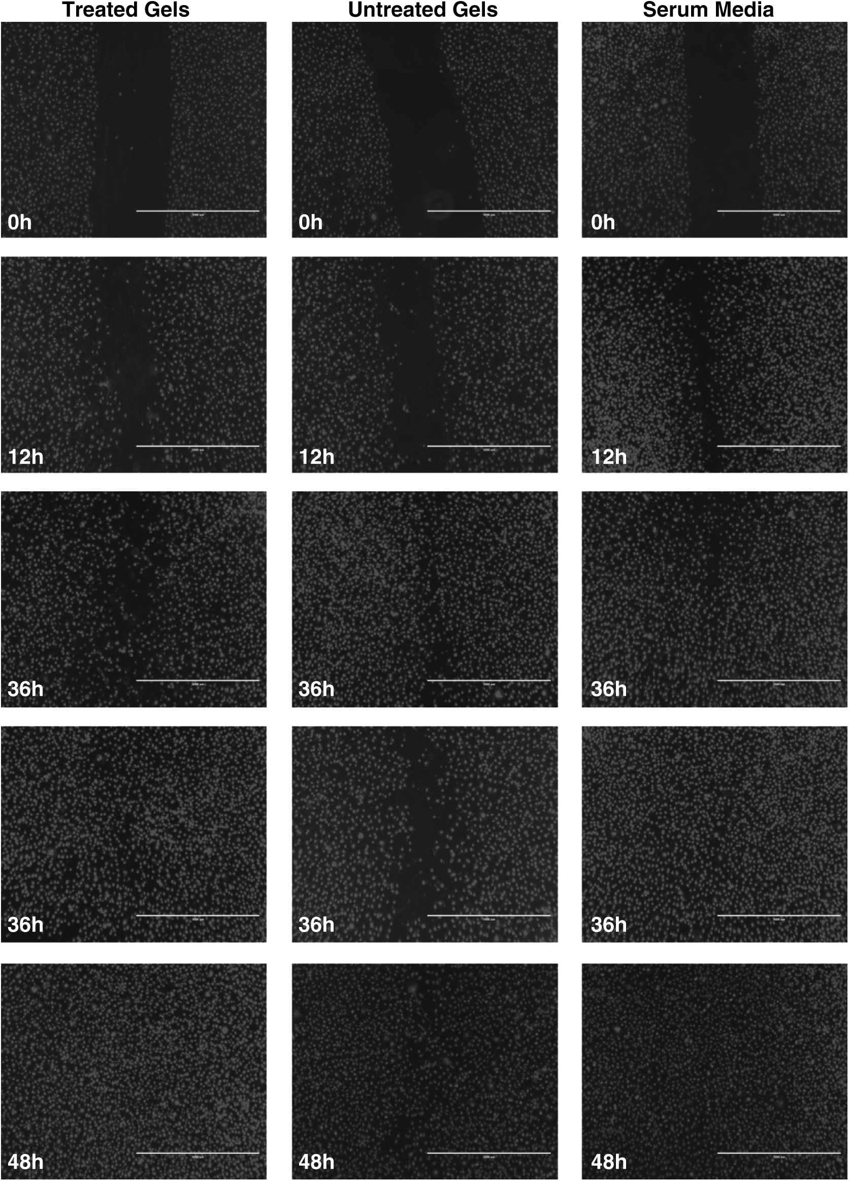
Non-thermal plasma treatment of alginate gels promoted cell proliferation and migration across wound sites as compared with cells not exposed to these gels in in vitro studies. The changes became more obvious at 36 h and beyond. (Bar=1,000 microns).
Discussion
Alginate wound dressings are biodegradable and have been shown to promote wound healing in vitro, in vivo, and in clinical trials [8,18,19]. These dressings play an important role in the management of acute and chronic, and as recently shown, of diabetic wounds [20–22]. However, the anti-microbial efficacies of these dressings alone, as compared with those of chlorhexidine- or silver-containing dressings, are negligible. Wounds are contaminated or colonized by a vast array of microorganisms whose presence and metabolic byproducts are detrimental to and delay wound healing [23–25]. In recent years, researchers have tried successfully to impregnate or trap various antimicrobial agents in alginate dressings to improve these dressings' antimicrobial coverage. Many of these antimicrobial agents are either cytotoxic or slow-acting, stain wound tissues and epithelial cells, or interfere physically in the healing process. Clinicians need a better antimicrobial dressing that has less cytotoxic effects on host tissues yet kills contaminant(s) rapidly and efficiently. We sought to design a novel antimicrobial alginate dressing that would control bacterial pathogens that commonly contaminate wounds, but would do so without compromising the proliferation and migration of prototype wound tissues and cells. Two such dressings in clinical use were included in the present study as positive controls against which to compare the efficacies of the non-thermal-plasma-treated alginate gel dressings that we designed and tested in the study.
The microbial organisms that reside in wounds exist in both planktonic form and in biofilms [12, 23], and their eradication is often difficult, partly because of the presence of wound fluids and debris and partly because the organisms have developed enhanced tolerance to antimicrobial agents. Considering the pathophysiology and microbiology of wound healing, antimicrobial wound dressings have significant effects, and an increasing number of broad-spectrum antimicrobial dressings (including chlorhexidine- and silver-containing dressings) are used in wound-management clinics. In the present study, non-thermal plasma treatment of calcium alginate gels conferred significant antimicrobial properties on the gels, and these properties were proportional to the amount of plasma energy deposited in joules per square centimeter and to the plasma treatment time (Figs. 1 and 3). Antimicrobial efficacy was retained for about 3–4 wks (Fig. 5). Thus, plasma-treated alginate dressings are potentially clinically useful wound dressings. Further optimization is required to prolong their effects, and is being studied.
The non-thermal-plasma-treated alginate dressings investigated in the present study compared favorably with silver-containing dressings in inactivating more rapidly pathogens than the latter (Fig. 4). A 9-log bacterial load was inactivated efficiently in 10 min of contact time with alginate gel treated with non-thermal plasma for just 60 sec (Fig. 2). This rapidity and efficiency in killing contaminants is important because bacteria may develop resistance to slower-acting or inefficiently acting agents [26]. With regard to the silver impregnation of gel dressings, the fastest rate of killing quoted for these dressings in the literature is 30 min and the slowest is 24 h [27]. Many bacterial pathogens reproduce about every 20 to 28 min, which correlates with a population size of ∼1021 colonies over a period of 24 h. An environment containing a constant level of antimicrobial agent has the potential to select for mutations that confer resistance in each new generation of organisms [28]. The chlorhexidine-containing dressing used for comparison in the present study acts far more rapidly than a silver-containing dressing, and chlorhexidine in solution has been shown to reduce the number of E. coli by 3.5 log after only 30 s of exposure [29]. Although the plasma-treated gels investigated in the present study do not act this quickly, a 10-min contact time with an alginate gel treated for 3-min with plasma (Fig. 4) inactivated pathogens relatively more rapidly than the average period for bacterial reproduction, probably impeding the development of resistance. The anti-biofilm efficacy of plasma-treated gels was highly evident (Fig. 7); was superior to that of a silver-alginate dressing in rapidly inactivating biofilm-embedded pathogens (data not shown).
Chlorhexidine is a potent bactericidal agent but also has relatively more cytotoxicity and animal toxicity [30]. Silver-containing wound-dressing preparations may include agents that release silver rapidly or slowly, depending on the polymer used for containing it, and resistance to these preparations is increasingly reported [11]. Thus, a non-thermal-plasma-treated anti-microbial alginate dressing is a novel concept, and the present study is proposed as a proof of this concept. Treatment with non-thermal plasma generates various reactive chemical and physical species that interact with an alginate gel matrix, producing chemical changes that render the gel biocidal. The chemical changes produced in the gel (either in the form of stabilized chemical species or the product(s) thereof, as is now being studied in detail) are relatively stable, and the anti-microbial properties of such a gel are therefore fairly consistent, lasting for close to one month, which is encouraging. Favorable findings were made in the present study with the plasma-treated gels in cytotoxicity assays and in an in vitro wound healing model in which the gels promoted cell proliferation and migration across wound sites. The treated gels inactivated rapidly all of the gram-negative, gram-positive, and fungal pathogens included in the present study. In earlier studies, we found that plasma-treated gels generated reactive oxygen species (ROS) inside bacterial cells, leading to their rapid death [14]. The cell wall thickness may not play a significant role. In addition to inactivating planktonic forms of the bacteria in the present study, the plasma-treated gels also inactivated bacteria in biofilms (which are usually more resistant), suggesting these gels have strong, broad-spectrum anti-bacterial activity (Fig. 7). In the case of many other antimicrobial agents, doses of 10- to 100-fold their minimum inhibitory concentrations (MICs) for planktonic pathogens are required to kill the same pathogens embedded in biofilms. Non-thermal-plasma-treated alginate gels may therefore be a better choice for dressing infected wounds.
The non-thermal-plasma-treated and untreated alginate gel dressings prepared in the present study were observed for surface changes associated with exposure to bacterial pathogens. Images made with SEM did not reveal any abnormalities in the surfaces of treated as compared with untreated gels, and damaged bacterial cells were visible on dressings inoculated with bacteria (Fig. 6). We therefore proceeded to cytotoxicity experiments to demonstrate the influence of plasma-treated anti-microbial gels on human umbilical vein endothelial cells. As expected, the non-thermal-plasma-treated gels initially slowed host-cell proliferation, and in an in vitro wound model enhanced both host-cell proliferation (Fig. 8) and the proliferation and migration of host cells across wound sites (Fig. 9).
In conclusion, non-thermal-plasma-treated alginate gels have better in vitro wound decontamination and wound healing capabilities, as well as broad-spectrum anti-bacterial activity and negligible cytotoxicity. Investigation in animal models of wound decontamination with non-thermal-plasma-treated gels is the next step in studying their therapeutic efficacy in contaminated and infected wounds.
Footnotes
Acknowledgment
The research reported in this paper was supported by funding from the Department of Surgery of the Drexel University College of Medicine. Dr. Alexander Poor is a resident research fellow and Utku Ercan is a PhD candidate at Drexel University who has a fellowship sponsored by the government of Turkey. The authors thank Ms. Diana Winters, Academic Publishing Services for critical reading of the manuscript.
Author Disclosure Statement
The authors declare that they have no commercial associations that would create a conflict of interest in connection with the work described in this paper.
References
Supplementary Material
Please find the following supplemental material available below.
For Open Access articles published under a Creative Commons License, all supplemental material carries the same license as the article it is associated with.
For non-Open Access articles published, all supplemental material carries a non-exclusive license, and permission requests for re-use of supplemental material or any part of supplemental material shall be sent directly to the copyright owner as specified in the copyright notice associated with the article.