
Abstract
Abstract
Background:
We report a 52-year-old male presenting with a graft–appendiceal fistula four years after placement of an aorto-bifemoral Dacron prosthesis.
Case Report:
After appendectomy and total graft removal, the patient was treated with an in-situ repair using a rifampicin–silver graft. This kind of repair has only been reported in an animal study.
Results:
This infected aortic graft was treated successfully with a rifampicin-silver graft with a follow-up of 15 months without complications.
Conclusion:
In-situ repair of a graft–appendiceal fistula with a rifampicin–silver graft seems a promising strategy. The long-term outcome needs to be studied.
A
Aorto-enteric fistulas themselves are rare, and involvement of the appendix has been reported only occasionally. The first case of a graft–appendiceal fistula was described in 1969 [4]. Since then, only ten more cases have been reported [5–14], with only four cases being reported over the last 20 years (Table 1) [6,7,10,12].
Treatment of aortoenteric fistulas follows the principles of the treatment of aortic graft infections: Surgical debridement, including graft removal; partial resection of the affected bowel; and revascularization of the lower limbs either by extra-anatomic reconstruction or by in-situ replacement. Established tools for in-situ reconstructions include venous grafts, allografts, rifampicin-soaked grafts, and silver grafts. The effects of a graft combining rifampicin and silver on the treatment of infected aortic grafts have been studied only in animals. This report presents a case of a patient with a graft–appendiceal fistula after an aorto-bifemoral Dacron reconstruction that was treated successfully by in-situ repair using a rifampicin-soaked silver-coated Dacron graft.
Case Report
A 52-year-old male with no notable medical history received a conventional aorto-bifemoral Dacron graft in another hospital because of bilateral critical limb ischemia. Proximal anastomosis was at the level of the inferior mesenteric artery. This procedure was uncomplicated.
Four years after the procedure, he was referred to another hospital because of fever and malaise. Physical examination did not reveal any abnormalities, and the patient was admitted for further study. On admission, laboratory testing showed elevated infectious parameters (C-reactive protein 67 mg/L, white blood cell count 14.3×10⁹/L, erythrocyte sedimentation rate 110 mm/h), and a slightly elevated serum creatinine concentration (1.22 mg/dL). Ankle–brachial indices proved normal (left 1.1, right 1.2).
Diagnostic computed tomography (CT) angiography demonstrated a fluid collection surrounding both sides of the distal prosthesis that was unsuitable anatomically for puncture aspiration. The appendix was in a close relation to the right limb of the graft (Fig. 1). Both ureters were obstructed, resulting in bilateral hydronephrosis and almost complete destruction of the right kidney.

Computed tomography (CT) angiography demonstrates fluid collection surrounding distal prosthesis. Appendix is closely related to right limb of graft (arrow).
Because of a high suspicion of aortic graft infection, the patient underwent positron emission tomography (PET) scanning using 18F-fluorodeoxyglucose (FDG) as a tracer. A PET-CT fusion image revealed focally increased uptake of FDG in the fluid collection and soft tissues surrounding the prosthesis as well as in the graft itself, confirming the suspicion of aortic graft infection (Fig. 2).
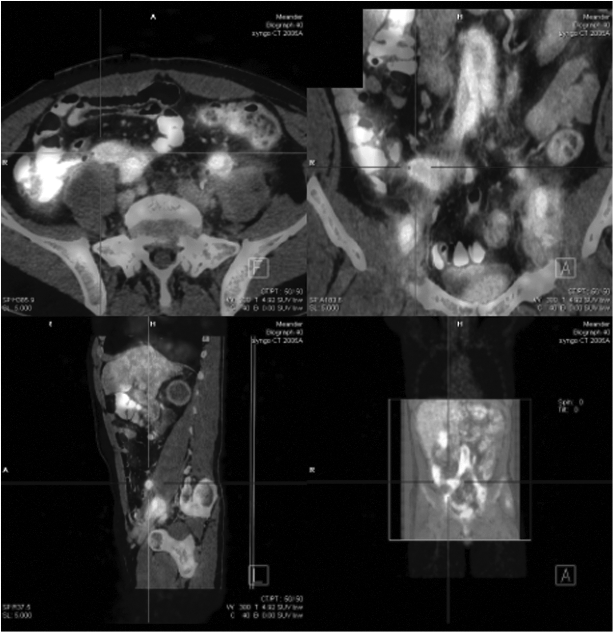
Positron emission tomography (PET) fused with computed tomography (CT) image reveals focally increased uptake of tracer in fluid collection and soft tissues surrounding prosthesis, as well as in graft itself.
Three months later, the patient was referred to our hospital for further treatment. The imaging studies were complemented with magnetic resonance angiography (MRA) to obtain a full view of the vascular tree and to plan a surgical reconstruction. Apart from the infected graft, no further stenoses were seen in the aorto-iliac–popliteal and crural regions. After a multidisciplinary discussion, the patient was scheduled for graft excision and in-situ repair. Preoperatively, double-J stents were inserted into the ureters to guarantee urine outflow and to assist with imaging during the procedure.
At laparotomy, the appendiceal region was found to be adherent to the distal end of the right limb of the prosthesis; the appendix itself was difficult to identify. Appendectomy and total removal of the graft were performed, combined with thorough debridement of the vascular bed. This procedure was complicated by a left ureteral injury, which was repaired primarily over the double-J stent. Lower-limb perfusion was restored with an in-situ aorto-bifemoral reconstruction using an InterGard silver-coated rifampicin-bonded graft (InterVascular, La Ciotat, France). The remainder of the procedure was uncomplicated.
Graft cultures showed infiltration with Staphylococcus lugdunensis and Enterococcus faecium. Until discharge, antibiotic treatment was given intravenously, consisting of amoxicillin-clavulanic acid 1200 mg three times a day combined with supplemental teicoplanin 200 mg once a day. The postoperative course was uneventful, and the patient was discharged after 11 days with ongoing antibiotic treatment (oral amoxicillin-clavulanic acid 625 mg tid and intravenous teicoplanin 200 mg qd).
At 15 months' followup, the patient had recovered completely, showing no signs of reinfection. Infectious measures had normalized, and the follow-up PET-CT scan showed normal FDG uptake. Antibiotic treatment was stopped.
Discussion
Most secondary aorto-enteric fistulas occur between part of the aortic graft and the transverse duodenum, representing 70% of all aortoenteric fistulas [15]. In only 0.6%–5% of aorto- or iliaco-prosthetic fistulas does involvement of the appendix occur [8,15]. Various theories have been proposed to explain the fistulization. This process is likely to be promoted by local inflammation, bowel injury, or a combination thereof. Local inflammation, which can be part of the normal healing process or a sign of low-grade graft infection, tightens the fibrous relation between graft and bowel, making the bowel more prone to pulsatile erosive damage. Bowel injury may be caused by the initial dissection or by erosion as a result of pseudoaneurysm formation, sutures, prolonged pressure on the bowel by a graft, or repetitive pulsating trauma [7,15,16]. The low incidence of graft–appendiceal fistulas may, at least partially, be explained by the fact that closure of the retroperitoneum after aortic surgery does not replace the appendix, thereby minimizing dissectional or frictional trauma [8].
Without prompt treatment, secondary aorto-enteric fistulas have a mortality rate of nearly 100% [12]. Even after accurate surgical intervention, the mortality rate for these fistulas is estimated to be 57% [15]. Although treatment of a graft–appendiceal fistula is not different from that of any other infected aortic graft, the clinical condition of the patient dictates which of the available treatment options is most appropriate. Most patients with a graft–appendiceal fistula are critically ill and often have serious co-morbidities that may limit the available therapeutic options. In a conservative approach, disconnection of the fistula and placing disease-free tissue, most commonly omentum, between the inflammatory tissues is performed, thereby avoiding resection of the infected graft [12]. Because of the high reinfection rates, however, the results often are poor [17].
The conventional surgical approach to aorto-enteric fistulas consists of three elements. First, extensive debridement is used to clear the abdomen of all infected material, including (part of) the graft. In the case of a graft–appendiceal fistula, this includes appendectomy. Second, the affected part of the bowel must be treated. Most often, this comes down to a small resection of the bowel and primary anastomosis or, in the case of a graft–appendiceal fistula, appendectomy. Third, lower-limb perfusion must be restored using an in-situ replacement with an autogenous femoral vein graft, cryopreserved allograft, rifampicin-bonded prosthetic graft, or extra-anatomic bypass [18]. The extra-anatomic reconstruction is a two-stage procedure and has worse results than in-situ grafts in terms of patency and reinfection rates [17]. The in-situ replacement as a one-stage procedure is the most up-to date therapeutic option. Although venous grafts are an excellent option for reducing reinfection, antibiotic-bonded or silver-coated grafts offer the great advantage of being “off-the-shelf.” Prebinding of grafts with a combination of an antibiotic and silver enhances their antibacterial activity [19]. In a recent porcine model of early prosthetic vascular graft infection, promising results were shown with an in-situ replacement with a rifampicin-soaked silver-coated polyester graft [19]. After implantation of aortic grafts inoculated with S. aureus and development of prosthetic vascular graft infection in 60 pigs, 52 pigs were randomized to undergo in-situ replacement with expanded polytetrafluoroethylene or rifampicin-soaked silver-coated polyester grafts [19]. In this model, S. aureus remained in only 4% of rifampicin-soaked silver-coated polyester grafts compared with 56% of expanded polytetrafluoroethylene grafts [19].
This paper reports the clinical treatment of an infected aortic graft with a rifampicin-soaked silver-coated polyester graft. Although we report only a short-term follow-up, our results confirm the positive in vitro results. As silver grafts do not affect antibiotic resistance, they offer a potentially powerful tool in the treatment of aortic graft infections. Although more studies are required, the combination of silver and rifampicin in one graft may well prove to be the next step in one-stage, off-the-shelf treatment of aortic graft infections.
Conclusion
A graft–appendiceal fistula is a rare and serious complication of surgical aortic repair. In-situ repair with a rifampicin–silver graft seems a promising strategy for the treatment of aortoenteric fistulas in the medium term. The long-term outcome needs to be studied.
Footnotes
Author Disclosure Statement
The authors declare no competing interests.