
Abstract
Abstract
Background:
The intercellular adhesion gene (ica) of Staphylococcus epidermidis is a key factor for bacterial aggregation. This study explored the effect of ica on the formation of bacterial biofilm on polyvinyl chloride (PVC) surfaces.
Methods:
Genes related to bacterial biofilm formation, including 16S rRNA, autolysin (atlE), fibrinogen binding protein gene (fbe), and ica were identified and sequenced from 112 clinical isolates of iatrogenic S. epidermidis by polymerase chain reaction (PCR) and gene sequencing. Based on the sequencing result, ica operon-positive (icaADB+/atlE+/fbe+) and ica operon-negative (icaADB−/atlE+/fbe+) strains were separated and co-cultivated with PVC material. After 6, 12, 18, 24, and 30 h of co-culture, the thickness of the bacterial biofilm and quantity of bacterial colony on the PVC surface were measured under the confocal laser scanning microscope and scanning electron microscope.
Results:
The positive rate of S. epidermidis-specific 16SrRNA in 112 iatrogenic strains was 100% (112/112). The genotype of ica-positive (icaADB+/atlE+/fbe+) strains accounted for 57.1% (64/112), and genotype of ica-negative (icaADB−/atlE+/fbe+) strains accounted for 37.5% (42/112). During 30 h of co-culture, no obvious bacterial biofilm formed on the surface of PVC in the ica-positive group, however, mature bacterial biofilm structure formed after 24 h. For all time points, thickness of bacterial biofilm and quantity of bacterial colony on PVC surfaces in the ica operon-positive group were significantly higher than those in ica operon-negative group (p<0.01).
Conclusions:
Iatrogenic S. epidermidis can be categorized into ica operon-negative and ica operon-positive strains. The ica operon plays an important role in bacterial biofilm formation and bacterial multiplication on PVC material.
S
Patients and Methods
Main materials, reagents, and instruments
Polyvinyl chloride material (Dongguan Kewei Medical Apparatus Co., Ltd. Guangdong Province, China) was cut into 1 cm×1 cm pieces, then fumigated and sterilized by ethylene oxide for preservation. Puregene Yeast/Bact Kit (Qiagen Inc., Almeda, CA); Live/Dead® Baclight™ Bacterial Viability Kit (Invitrogen Inc., Carlsbad, CA); API test strips (bioMérieux, Marcy d'Etoile, France) were used. MRC-1024ES confocal laser microscope (Bio-Rad Inc., Hercules, CA) and XL30ESEM scanning electron microscope (Philips Inc., Best, The Netherlands) were used.
Experiment strains
Standard strains of S. epidermidis RP62A (control strains of atlE, fbe, ica genetic testing-positive) and ATCC 12228 (control strains of ica genetic testing negative) [5] were all purchased from Institute of Microbiology, Chinese Academy of Sciences, Beijing, China. Clinical specimens were obtained from First Affiliated Hospital, Second Affiliated Hospital, Third Affiliated Hospital of Kunming Medical University and Kunming General Hospital of Chengdu Military Region from January 2005 to September 2009. One hundred twelve clinical isolates of iatrogenic S. epidermidis were isolated and cultivated from infections associated with biologic material implantation. All isolates were identified as S. epidermidis by the API kit. They were stored and preseved in 40% fetal calf serum glycerol at −80°C before the experiment. Among them, eight strains were isolated from infective endocarditis, 12 from implanted foreign body cultivation, 16 from blood samples of patients infected by biologic material implantation, 16 from chest drainage tubes, 30 from central venous catheters, 18 from endotracheal tubes, and 12 from other sources.
Target gene amplification and detection
Specific primers of icaADB, atlE, fbe, and 16S rRNA were designed according to the protocol by Arciola et al. [6], and were synthesized by a commercial company (TaKaRa Biotechnology (Dalian) Co., Ltd., Liaoning Province, China). The sequence of primers is shown in Table 1. Bacterial genomic DNA of iatrogenic S. epidermidis and standard strains RP62A, ATCC12228 were extracted using the Puregene Yeast/Bact Kit. With the genomic DNA as a template, atlE, fbe, and icaADB genes were amplified. To ensure that all iatrogenic clinical isolates were S. epidermidis, species-specific 16SrRNA was used as internal control. Standard strain RP62A, ATCC 12228 were selected as positive and negative control in the genetic testing. Polymerase chain reaction (PCR) conditions were as follows: 4 min pre-denaturation at 94°C, 30 sec denaturation at 94°C,40 sec renaturation at specific temperature, 1 min extension at 72°C, 30 cycles of amplification, and finally 7 min extension at 72°C. The PCR reaction renaturation temperature is shown in Table 1. The PCR results were confirmed through the gel imaging analysis system.
Sequencing of Target Genes
Based on the result of PCR amplification, three positive PCR products, namely 16S rRNA, atlE, fbe, and icaADB, were selected for gene sequencing. Purification and sequencing of PCR products were all conducted by TaKaRa Biotechnology (Dalian) Co., Ltd. Sequencing results were then compared with the corresponding gene sequences in GenBank using Blast software (U.S. National Laboratory of Medicine, Bethesda, MD).
Experiment of Bacterial Biofilm Formation on PVC Surface Preparation of Bacterial Suspension
A small amount of iatrogenic S. epidermidis was removed and inoculated into the sterile broth medium. After 16 h of cultivation at 37°C, it was then inoculated in LB plates. Bacterial colonies were obtained after 24 h cultivation at 37°C. A single colony was chosen and inoculated into the broth medium. After 12–16 h cultivation at 37°C, purified bacterial suspension was used for the study. We used the pour-plate method, the concentration of bacterial suspension was adjusted at 1×10⁵ colony-forming units per milliliter (CFU/mL) for the experiment.
Experimental Groups
Based on the results of gene sequencing, 106 strains of the obtained iatrogenic S. epidermidis were divided into two groups: ica operon-positive group (icaADB+/atlE+/fbe+; 64 strains) and ica operon-negative group (icaADB−/atlE+/fbe+; 42 strains) for bacterial biofilm formation test. Another 6 strains with the genotype of icaADB−/atlE−/fbe− or icaADB+/atlE−/fbe− were not included in the study. Five microliters of prepared bacterial suspension was added into a sterile bottle, together with 20 mL of broth medium and 10 pieces of PVC material. They were placed in 37°C water bath for cultivation. Two pieces of PVC material were removed from the culture bottle after 6, 12, 18, 24, and 30 h of cultivation, with one piece observed under the confocal laser microscopy and the other piece observed under the scanning electron microscopy.
Detection Index
Observation under the confocal laser microscope. Fluorescent dye was prepared in accordance with Live/Dead Baclight Bacterial Viability Kit manual. PVC pieces were washed three times with distilled water, and then stained with bacteria fluorescent dye for 20 min at room temperature before being put under the confocal laser microscope (argon laser [514/488 nm]) for observation. For each PVC plate one visual field was randomly selected to observe the number of bacterial colony. Then one bacterial colony was selected randomly to scan layer by layer from inside out and measure their thickness (bacterial biofilm thickness).
Observation under the scanning electron microscope
The PVC pieces were washed three times with sulfuric acid hydroxyethyl piperazine buffer solution (pH=7), then fixed on the loading platform of the scanning electron microscope. After CO2 supercritical drying, the fixed coating of ion sputtering surface gave the PVC plate golden-brown color. The surface structure of the bacterial biofilm was observed under the microscope.
Statistical Method
The SPSS 17.0 statistical software package (IBM, Inc., Armonk, NY) was used for analysis. Data are shown as mean±standard deviation. Analysis of variance for repeated-measurement was used for comparison between two groups, and p<0.05 was set as the significant value.
Results
Target gene test of clinical isolate strains of iatrogenic S. epidermidis
Results of agarose gel electrophoresis showed that positive rate of S. epidermidis-specific16S rRNA was 100% (112/112) in 112 clinical isolates, indicating that all strains were S. epidermidis. Existence of atlE and fbe gene was widespread, with a positive rate of 94.6% (106/112), among which genotype of icaADB+/atlE+/fbe+ accounted for 57.1% (64/112) and genotype of icaADB−/atlE+/fbe+accounted for 37.5%(42/112).
Target gene sequencing of clinical isolate strains of iatrogenic S. epidermidis
Sequencing results showed that sequences of target genes16S rRNA, atlE, fbe, and icaADB were consistent with gene sequences in GenBank.
Observation under the confocal laser microscope
The observation under the confocal laser microscopy showed that the number of bacterial colony and the bacterial biofilm thickness per unit area of the PVC surface in ica operon-positive group were significantly higher than those of ica operon-negative group. The differences were statistically significant (p<0.01; Table 2 and Table 3).
In this study, 112 genes of Staphylococcus epidermidis were detected. A total of 106 were found, including the iatrogenic S. epidermidis of positive icaADB, atlE, fbe genotype (64 strains, ica operon-positive group) and icaADB negative, atlE and fbe positive (42 strains, ica operon-negative group). Another six that included icaADB negative, atlE and fbe negative and icaADB positive, atlE and fbe negative were not included in the statistical analysis.
In this study, 112 genes of Staphylococcus epidermidis were detected. A total of 106 were found, including the iatrogenic S. epidermidis of positive icaADB, atlE, fbe genotype (64 strains, ica operon-positive group) and icaADB negative, atlE, and fbe positive (42 strains, ica operon-negative group). Another six that included icaADB negative, atlE and fbe negative, and icaADB positive, atlE and fbe negative were not included in the statistical analysis.
Observation under the scanning electron microscopy
Observation under the scanning electron microscopy indicated that during 30 h of cultivation there was no bacterial biofilm formation on PVC surface in the ica operon-negative group, nor did any mature bacterial biofilm structure appear. In the ica operon-positive group, the number of bacterial colony gradually increased with time on the PVC surface. Bacterial clusters were gradually wrapped by the mucus-like substance, and the volume of bacterial biofilm was also enlarged. The mature form of bacterial biofilm structure at 24 h appeared as compact, highly organized multi-cellular groups. After 30 h, the volume of bacterial biofilm tended to stabilize; some bacterial biofilm began pyknosis and collapsed (Fig. 1).
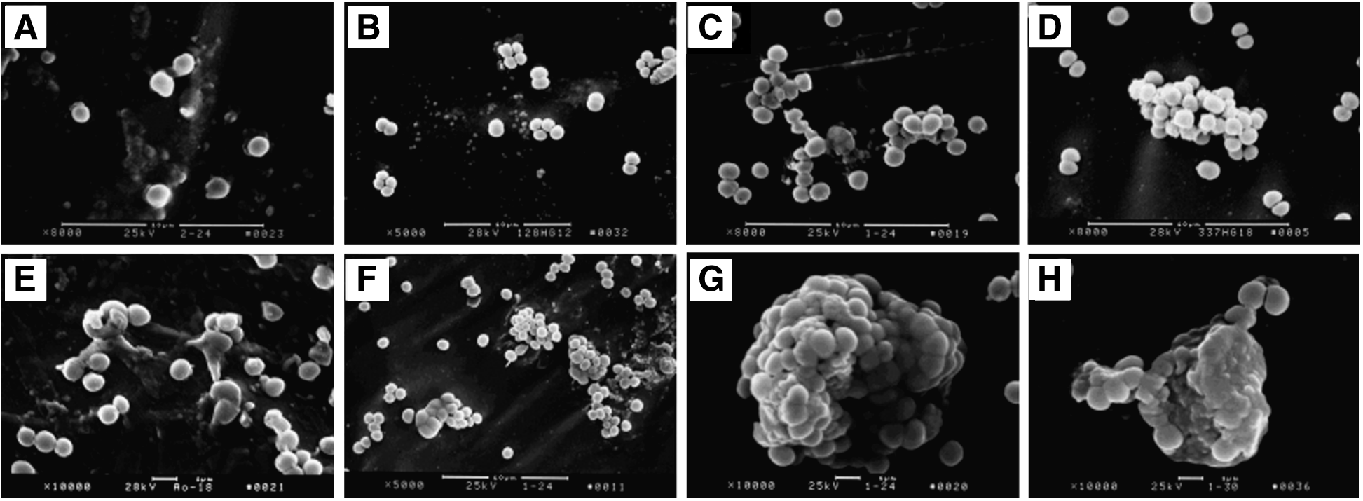
The image of the two group strains by scanning electron microscope examination at different times.
Discussion
In recent years, with the extensive application of biologic materials, S. epidermidis has become the major opportunistic pathogen in reparative and reconstructive surgery. Studies have shown that 40%–50% of endocarditis in patients after artificial heart valve surgery was caused by S. epidermidis. In patients who underwent peritoneal dialysis, S. epidermidis accounted for 40% of catheter contamination-related infections [1,6–8]. Staphylococcus epidermidis strains from different sources exhibit genetic differences. It is reported that S. epidermidis isolated from infections associated with clinical catheters differ from those isolated from normal human skin [6–9]. The positive rate of bacterial biofilm-related genes, such as fbe, atlE, and icaADB, varies with different sources as well. The results of our study were consistent with the above observations. The fbe and atlE genes existed in 112 clinical isolates of S. epidermidis in our study, accounting for 94.6% (106/112). The positive rate of icaADB gene was 57.1% (64/112). Staphylococcus epidermidis with different genetic background often showed different biologic characteristics, and whether S. epidermidis can form biofilm is the major criterion to distinguish between symbiotic and invasive S. epidermidis [3,9].
Studies on the biofilm formation of S. epidermidis showed that the enzyme responsible for biofilm formation is encoded by ica operon, which include icaR (regulatory gene), icaA, icaD, icaB, and icaC genes existing in series connection. The icaA gene alone shows low N-acetyl glucosamine transferase activity, but co-expression of icaA and icaD can increase the enzyme activity greatly, synthesizing the largest chain with up to 20 residues of oligomers. Four genes of icaADBC together can complete the synthesis of polysaccharide intercellular adhesion (PIA) [4,6], the necessary material to mediate the aggregation of S. epidermidis biofilm formation [2,6,8,9]. Studies on the biofilm formation of S. epidermidis based on planktonic conditions indicated that S. epidermidis lacking the ica operons has difficulty forming the mature bacterial biofilm, and S. epidermidis strains that can form bacterial biofilm, were shown to be mostly ica operon, positive, suggesting that biofilm formation by S. epidermidisis is related to the existence of ica operons. However, for the infections associated with biologic material implantation, the relationship between the ica operon of iatrogenic S. epidermidis and the bacterial biofilm formation on the surface of biologic material needs to be addressed further. Few relevant studies have been reported.
Our preliminary results showed that the biofilm formation of S. epidermidis is a dynamic process, with the initial adhesion to the biologic materials by the hydrophobic proteins or extracellular polysaccharides on the bacterial surface. Then the bacterium, with PIA as the main medium, aggregates with each other to form a bacterial biofilm. The number of bacterial colonies and the thickness of biofilm on biologic material surfaces are the indicators that reflect the phenotype of bacterial biofilms on the biologic material surface. The highly organized multi-cellular structure of bacterial biofilm on biologic material surface could be the source of clinical implantation-associated infections that are difficult to control. Thus, observation of bacterial biofilm structure in living conditions constitutes an important part of bacterial biofilm research [2,3].
In this study, PVC material, which is used commonly in reparative and reconstructive surgery, was used as the carrier of bacterial adhesion, and the role of ica operon of iatrogenic S. epidermidis on the bacterial biofilm formation on PVC surface was studied through the laser scanning technique. The results indicated that the number of bacterial colonies and the bacterial biofilm thickness of iatrogenic S. epidermidis with ica operon-positive on PVC surfaces were significantly higher than those with the ica operon-negative group. After further observation by scanning electron microscopy, it was found that after 24 h of cultivation, the ica operon-positive group can form the compact, highly organized bacterial biofilm structure on PVC surfaces, whereas the ica operon-negative group showed only part of the bacterial biofilm adhering to the PVC surface and no formation of any mature bacterial biofilm structure. One explanation could be that the latter contains the initial adhesion-related genes (atlE, fbe), which showed the initial adhesion function, but lack of ica operons results in failure of bacterial biofilm aggregation, therefore, they could not form a mature bacterial biofilm. This indicates that the ica operon of iatrogenic S. epidermidis plays an important role in the formation of bacterial biofilm on PVC surface.
There are many other factors that can influence the biofilm formation of S. epidermidis, such as pH value, and concentrations of glucose and NaCl in the culture medium [10–19]. Hypertonic saline solution (NaCl), ethanol, glucose, and the environmental pH value can change the expression of ica operon, regulating the biofilm formation of S. epidermidis [10–15]. Moreover, different factors regulate the biofilm formation of S. epidermidis via different pathways. Ethanol can directly or indirectly inhibit the repressor gene icaR transcription, leading to the increased icaADBC transcription, therefore promoting bacterial biofilm formation. Sodium chloride, on the other hand, works together with RsbU protein to activate SigB, the latter then promotes icaADBC transcription and bacterial biofilm formation [16–19]. However, current studies relevant to ica operon and the bacterial biofilm of S. epidermidis are based mostly on the planktonic growth pattern, and limited by the laboratory mutant bacterial strains. Also, for infections associated with biologic material implantation, the process of biofilm formation and organizational structure of the biofilm are different than those caused by planktonic infections [20–21]. Therefore, to characterize the genetic feature and biologic behavior of S. epidermidis on PVC surfaces would provide useful information for clinical treatment. By suppressing the expression of iatrogenic S. epidermidis ica operon, thus to effectively inhibit the bacterial biofilm formation on the implant surface, chances of implant-associated infections would be reduced. Our study therefore provides some insight for targeted therapy.
In summary, the presence of ica operon of iatrogenic S. epidermidis can increase the number of bacterial colonies and the bacterial biofilm thickness on the PVC surface. It plays a role in the bacterial biofilm formation for medical implant-associated infection, and may serve as the genetic basis to distinguish between symbiotic and invasive S. epidermidis. Studies in the related area could benefit clinical work by examining S. epidermidis phenotype (e.g., icaADB, atlE, fbe genotype) of biomaterial implant infection to determine the initial bacterial biofilm-forming ability.
Footnotes
Acknowledgments
We are indebted to Shang Bing, First Affiliated Hospital of Kunming Medical University and Lin Mighui, Kunming General Hospital of Chengdu Military Region, for their help. Thanks also to the teams of Key Laboratory of Medical Molecular Virology, Shanghai Medical College, Fudan University, for their support.
This experiment was supported financially by grants from the National Natural Science Foundation of China (No. 30872555, 81000672, 81260228) and Yunnan Provincia high-level personnel training fund (No. 2012HB032, D-201222), which were not involved in the study design, analysis of the results, or preparation of the manuscript.
Author Disclosure Statement
No competing financial interests exist.