




Abstract
Abstract
Background:
Silver-containing negative-pressure wound therapy (NPWT) foam dressings should reduce the microbial load of infected surgical sites and thereby promote healing. The effects of silver and an experimental copper coating of NPWT dressings on the growth kinetics of methicillin-resistant Staphylococcus aureus (MRSA) were investigated with a focus on the importance of the initial bacterial load and the incubation time.
Methods:
Commercially available foam samples with and without silver coating were inoculated in vitro with six MRSA suspensions differing in bacterial concentration (1.85×10³ to 1.85×10⁸ colony-forming units per milliliter [CFU/mL]). In a second series, uncoated, silver-containing and experimental copper-coated foam samples were inoculated with one MRSA suspension (1.85×10⁶ CFU/mL). The MRSA viable counts in the entrapped fluid were evaluated statistically after 1, 3, 7, and 14 d incubation.
Results:
Silver foam samples reduced MRSA counts by two decimal powers compared with the corresponding inocula. With respect to the uncoated samples, silver coating reduced MRSA concentrations by up to 7 logs, which was significant (p≤0.045) for all groups except the one with the highest MRSA concentration. The antibacterial effect of copper became apparent only after 7 d, but thereafter was far more pronounced than the effects of silver (p<0.01 after 14 d).
Conclusions:
Antimicrobial-coated foam dressings showed significant in vitro antibacterial properties and thus could be advantageous in the treatment of MRSA-infected incisions.
S
For reduction of the bacterial load of surgical site infections, and therefore, potentially faster, less complicated healing, NPWT can be combined with instillation of antiseptics [8] or with silver-containing foam dressings [9]. The ionized forms of silver, copper, and other metal ions are known for antibacterial and cytotoxic effects [10,11] depending on their concentration and chemical bonding. Although the molecular mechanisms of action are not understood, fully these effects have resulted in a broad spectrum of clinical applications [12,14–16]. For example, with respect to wound management, silver-impregnated dressings are used for care of burns and chronically infected wounds [17,18]. Such wounds are colonized frequently by a mixture of gram-positive cocci and gram-negative bacilli. Among these, methicillin-resistant Staphylococcus aureus (MRSA) are often involved increasingly as causative agents and pose an additional risk in the case of a complicating systemic infection [19,20].
Although the combination of physical suction and silver dressings is an established procedure in surgical site infection management, neither approach has been proved to be clearly superior to conventional dressings regarding the reduction of bacterial load and duration of incision healing [3,21–23]. Mouës et al. [24] quantified the bacterial load by taking repeated surgical site infection biopsies during treatment with either NPWT (n=29) or conventional moist gauze (n=25). In both patient groups the total bacterial load (colony forming units [CFU] per gram tissue) remained stable over 3-wk periods. Notably, NPWT reduced the numbers of non-fermentative gram-negative bacilli whereas the numbers of S. aureus, anaerobes, and Enterobacteriaceae increased or remained unchanged, respectively [24]. Furthermore, a recent Cochrane analysis did not provide sufficient evidence for usage of silver in different types of surgical site infections to prevent infection or promote healing compared with conventional dressings [23].
Negative pressure wound therapy foam dressings, especially when remaining in wounds too long or being clogged by debris, are even suspected of enhancing bacterial growth [25]. Thus, the question arises whether a commercially available silver coating could specifically affect the growth of MRSA on NPWT foam dressings, irrespective of the initial bacterial load and the in situ time of the dressing. Thus far, this is an unresolved issue. Also, a potential role of copper as an alternative to silver in such applications has not been investigated.
Therefore, one objective of the present in vitro study was to investigate the influence of uncoated and silver-coated NPWT foam dressings on the growth kinetics of MRSA with special focus on the initial bacterial load and time of incubation. Another objective was the comparison of uncoated, silver-coated, and copper-coated foam samples regarding their effects on MRSA growth or survival on such materials.
Patients and Methods
Test setup
In a first series of measurements, the untreated standard NPWT dressing and silver-coated foam samples were placed into sterile 24-well plates (Greiner Bio-One GmbH, Frickenhausen, Germany) and inoculated with MRSA suspended in Brain Heart Infusion (BHI) broth (Oxoid, Wesel, Germany) to defined concentrations between 103 and 108 CFU/mL. The closed containers with the inoculated foam dressing samples were incubated at 37°C under a 20% O2/5% CO2, water-saturated atmosphere (static conditions). Incubation periods were 1, 3, 7, and 14 d. At the end of the predetermined incubation period, MRSA numbers in the foam samples were evaluated for each type of foam dressing pad and concomitantly, each initial inoculum. In a second series of measurements, standard, silver- and copper-coated foam samples were inoculated with one MRSA suspension of 106 CFU/mL.
Preparation of the test suspensions
A MRSA isolate (Southern German variant, Robert Koch Institute, Wernigerode, Germany) was used to conduct the in vitro study. Pre-cultures were prepared as follows: material from a pure MRSA colony was transferred from a Columbia blood agar plate (5% sheep blood, Becton Dickinson, Heidelberg, Germany) into BHI bouillon and cultured under static conditions at 37°C in a 20% O2/5% CO2 atmosphere. After 16 h of incubation, the preculture was diluted serially with fresh BHI bouillon resulting in six inocula suspensions containing 1.85×10³ to 1.85×10⁸ CFU/mL in 10-fold dilution steps.
In parallel to each test series, the bacterial concentrations of the inocula were determined as follows: An aliquot of the corresponding pre-culture was diluted serially in phosphate-buffered saline (pH 7.4) and plated in 100 mcL aliquots onto BHI agar. Colony-forming units were counted after 24 h of incubation at the above-mentioned conditions.
Six MRSA suspensions containing 1.85×10³ to 1.85×10⁸ CFU/mL were used for the first test series comparing bacterial growth kinetics on uncoated and silver-coated foam dressing samples. For the second test series (comparison of uncoated, silver- and experimental copper-coated specimen) all types of foam dressing specimens were inoculated with one suspension containing 1.85×10⁶ CFU/mL MRSA.
Preparation of the foam samples and analysis of bacterial growth
All working steps were conducted under laminar flow conditions (Hera Safe, Thermo Fisher Scientific Inc., Waltham, MA) using sterile instruments. As a reference, standardized foam cubes with an edge length of 1.0 cm were cut out of commercially available NPWT dressings (V.A.C. GranuFoam™ Dressing, KCI Inc., San Antonio, TX) using a template. The second group consisted of the V.A.C. GranuFoam Silver® Dressing (KCI) with homogeneously distributed microbonded ionic silver [26]. The experimental foam samples consisted of the V.A.C. GranuFoam Dressing with a copper coating that was applied by vacuum sputtering (DOT GmbH, Rostock, Germany) using a pure copper cathode.
For the inoculation of the foam samples the wells of sterile 24-well plates (Greiner Bio-One GmbH, Frickenhausen, Germany) were each filled with 1 mL of the different MRSA suspensions. The foam dressing cubes were then pushed into the MRSA suspension with a sterile forceps to aspirate the bacterial suspension.
Subsequently, the specimens were transferred to new 24-well plates, lids were closed, and the plates were incubated at 37°C in a 20% O2/5% CO2, water-saturated atmosphere (static conditions). When the pre-determined end of the incubation period was reached, the bacteria-containing liquid was retrieved from the foam cubes by squeezing with a syringe. Viable counts were conducted as described above and protocolled as log CFU/mL.
Each experiment was performed in duplicate for each foam type and bacterial suspension (technical replicates) and repeated on at least three independent occasions (biologic replicates). For subsequent analysis, mean values from technical replicates were calculated for each experimental setup. With these values, the geometric mean ± one standard deviation (1 SD) CFU/mL was assessed for each biologic replicate.
Statistical analysis
Comparisons of the bacterial load between subgroups within each test series groups were made using a t-test for independent samples after testing the homogeneity of variances (Levene test). All p values resulted from two-sided statistical tests. Values of p<0.05 were considered to be statistically significant. Data were stored and analyzed using the SPSS statistical package 15.0 (IBM Inc., Armonk, NY).
Results
In the first test series 231 samples were analyzed with respect to dilution levels of the inoculum (six groups), incubation time (1, 3, 7, and 14 d), and foam type (standard versus silver coating). In the second series 59 samples were evaluated regarding incubation time and type of foam (standard versus silver versus copper).
Test series 1: Uncoated versus silver-coated foam specimen
Each foam type was inoculated with six different MRSA concentrations. In the uncoated foam samples, irrespective of the initial inoculum the MRSA load increased to approximately 109 CFU/mL (range, 7.13×10⁸ to 2.30×10⁹ CFU/mL) in the period between 1 d and 7 d incubation (Fig. 1). At the end of the second week, bacterial numbers decreased again by two logarithms independently from the initial inoculum numbers.
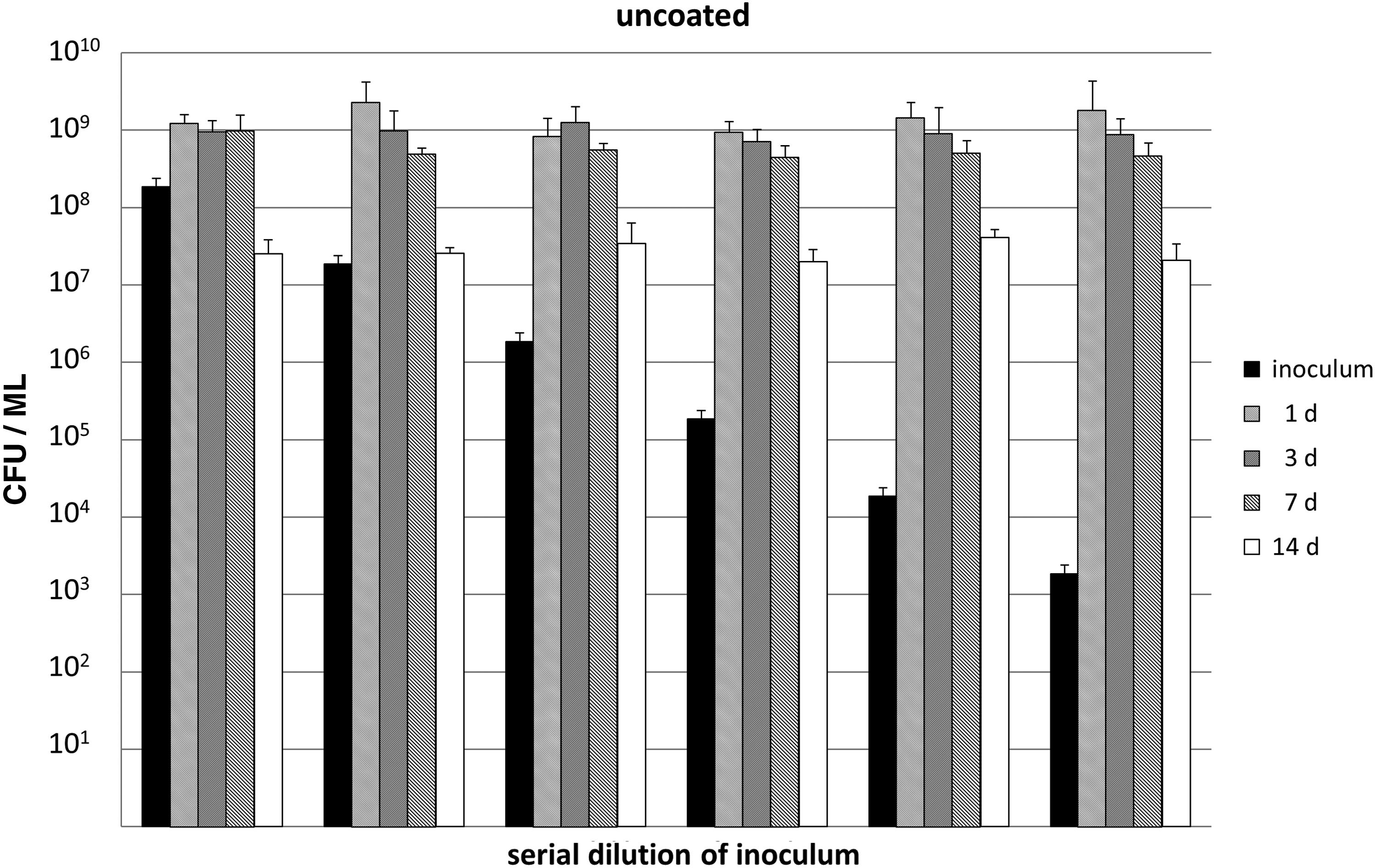
Methicillin-resistant Staphylococcus aureus (MRSA) concentrations after 1, 3, 7, and 14 d incubation with the uncoated foam dressing cubes. Tests were performed with six different initial inocula.
In the silver-coated foam samples the MRSA count in all groups was reduced by 2 logs after 24 h incubation compared with the corresponding initial inoculum size. Thereafter, further reductions depended on the individual initial inoculum size. Of note, in the group with the lowest initial inoculum, no culturable bacteria were found (3, 7, and 14 d) after incubation (Fig. 2).
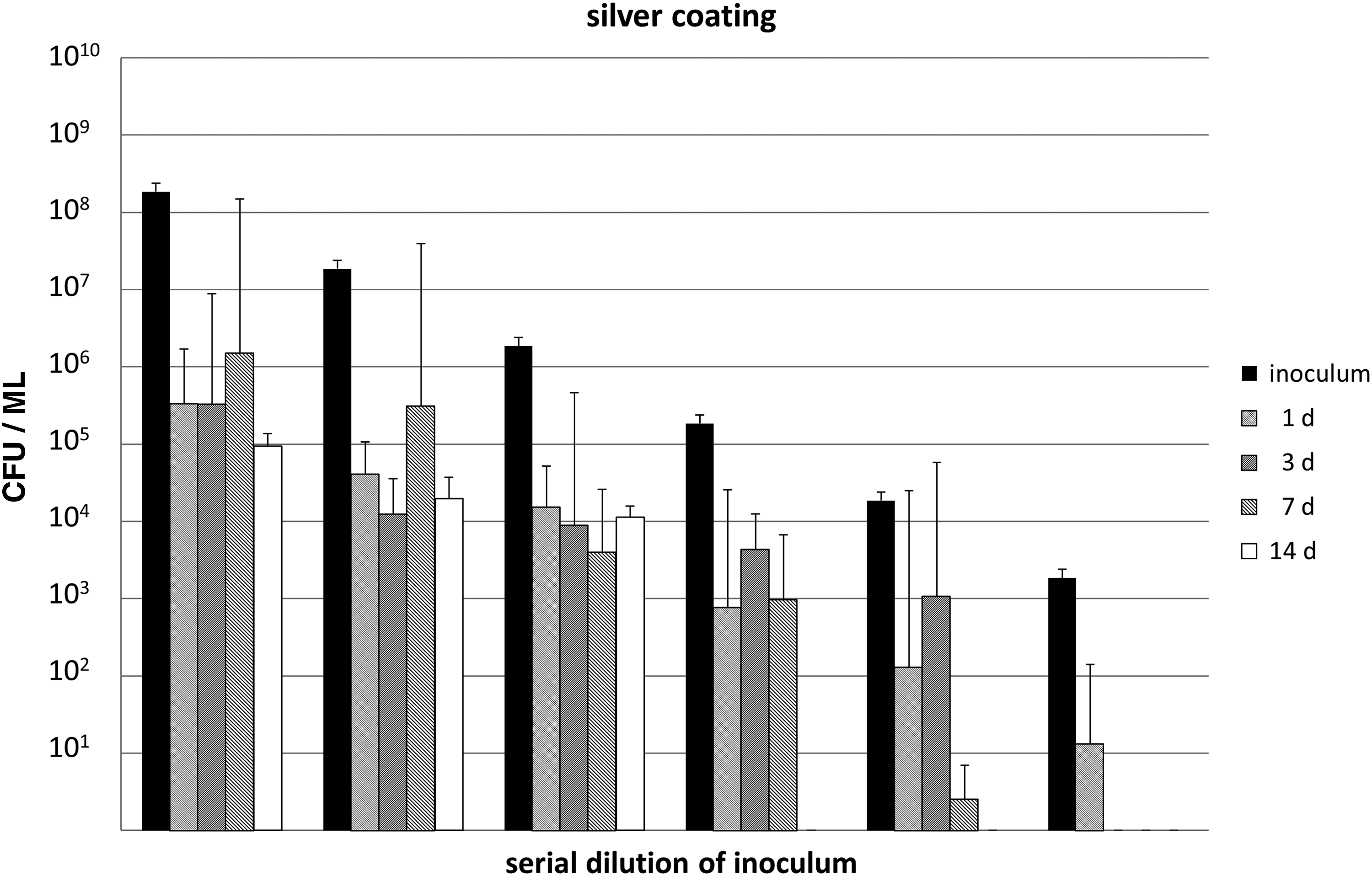
Methicillin-resistant Staphylococcus aureus (MRSA) concentrations after 1, 3, 7 and 14 d incubation with the silver-coated foam dressing cubes. Tests were performed with six different initial inocula.
Compared with the uncoated foam pads, the reduction rates ranged between three to five orders of magnitude throughout the complete observation period for the four groups with the higher initial bacterial load (1.85×10⁸ to 1.85×10⁵ CFU/mL). The reduction for the two groups with the least initial inoculum sizes were even higher.
Except for two incubation time points with the highest initial inoculum, the differences between silver-coated and uncoated pads were significant in the statistical analysis (Table 1). Within each inoculum subgroup in the silver dressing assays, viable count differences over the incubation periods 1 to 14 d were insignificant for the four higher inocula and significant for the two lower ones (data not shown).
Test series 2: Uncoated versus silver-coated versus copper-coated foam specimen
As before, the bacterial numbers on the uncoated dressing specimen increased significantly over the complete incubation period compared with the initial inoculum (1.85×10⁶ CFU/mL, Fig. 3 and Table 2). The silver-coated foam samples reduced significantly the viable counts by up to 5 logs compared with the initial inoculum (p≤0.005) and the uncoated specimen at identical incubation periods (p=0.027).
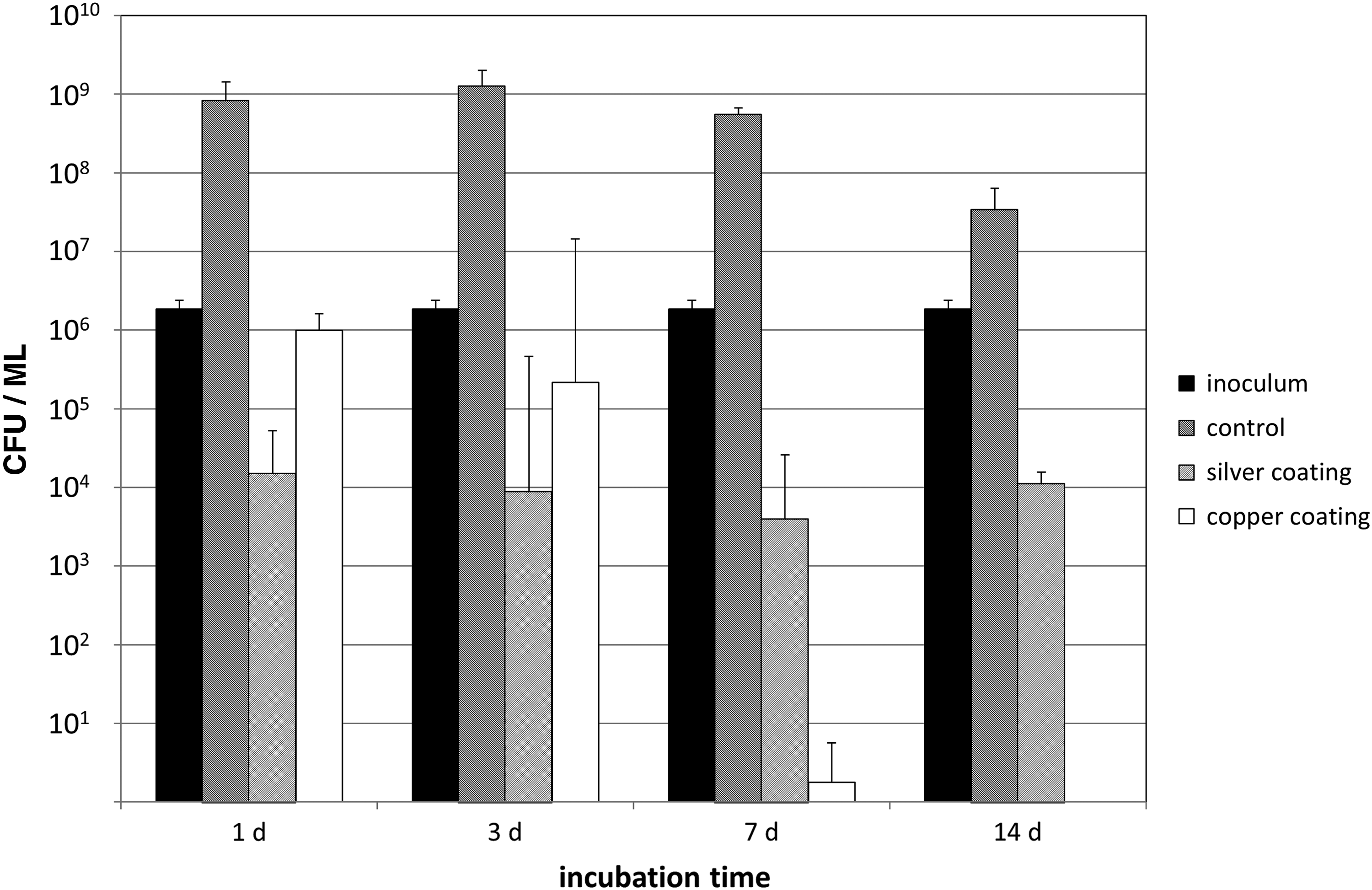
Methicillin-resistant Staphylococcus aureus (MRSA) concentrations after 1, 3, 7, and 14 d incubation in the uncoated, silver-, and copper-coated foam dressing cubes. One defined initial inoculum concentration (1.85×10⁶ colony-forming units per milliliter [CFU/mL]).
p values of the statistical comparison of the methicillin-resistant Staphylococcus aureus (MRSA) concentrations in different foam dressing samples with respect to the incubation time. For bacterial concentration values, see Figure 3.
Copper coating inhibited MRSA growth significantly compared with the uncoated dressing at any incubation time point (p=0.006). However, compared with the corresponding inoculum size, the reduction became significant only at 7 d and 14 d of incubation (p=0.001 and 0.003, respectively). Compared with silver coating, copper coating achieved a less pronounced reduction of bacterial loads in the first 3 d of incubation (1–2 orders of magnitude). However, at 7 d and 14 d of incubation copper coating reduced bacterial numbers by 3 to 4 logs better than silver coating (Fig. 3). Differences were statistically significant in favor of silver on incubation day 1 and in favor of copper on incubation day 14 (Table 2).
Discussion
The major objective of this study was to assess in vitro how specific metal coatings influence MRSA growth or survival in NPWT foam dressings by analyzing the bacterial numbers over an observation period of 14 d. An uncoated standard dressing was obviously colonized unrestrained, whereas silver-containing foam reduced MRSA numbers significantly compared with its own inoculum size as well as the uncoated samples. Of note, an experimental copper coating also revealed significant reduction effects on MRSA counts, which compared with silver-coating evolved slower but finally to a greater degree.
The present study has two limitations. First, the data were obtained exclusively with MRSA. Therefore, the findings may not be transferable to other bacteria involved in surgical site infections. However, S. aureus is the most frequent single pathogen causing wound and surgical site infections [27] and appears to be of importance specifically in VAC-treated wounds [24]. With respect to complicated skin and soft tissue infections, MRSA is an increasingly important subgroup within this species and any preventive measure decreasing the risk of MRSA infections could contribute to therapeutic success [19,20]. Second, the MRSA load was assessed exclusively in NPWT dressing samples, but not in an in vivo approach in adjacent animate tissue. Yet, the chosen in vitro approach allowed the investigation of the influence of antibacterial coatings under standardized conditions. Data from such studies on the effects of microbonded silver ions on NPWT dressings are still rare, and to the best of our knowledge, do not exist for the effects of other metal ions.
Several basic research studies have demonstrated the general antibacterial effects of silver and copper ions. In vitro tests with silver-containing coatings on titanium surfaces have been shown to be effective against S. aureus and S. epidermidis [28,29]. Some authors tested other metal ions concerning their biocompatibility and antimicrobial activity in such settings and found that copper showed a high antibacterial effectiveness in relation to its cytotoxicity [11,14,30,31].
The potentially favorable properties of copper and silver ions have resulted in clinical applications. Yet, the usefulness of silver-containing dressings in promoting wound healing remains questionable [23,32,33]. In contrast, copper appeared to combine antibacterial effect with the capacity to promote wound healing [34].
A potential association between bacterial colonization of NPWT foams and surrounding tissue has been addressed by Lehner et al. [25]. Tissue and foam dressing samples of 15 patients with a periprosthetic infection were acquired during their course of treatment with an instillation NPWT system. Of the initial foam samples, 88% showed bacterial contamination, most notably with staphylococci, streptococci, and enterococci. At the end of treatment, the foam samples of only one patient were contaminated persistently with MRSA. No quantitative analysis and evaluation of the in situ time of the foams were reported. However, at each time point the bacteriologic findings of the foam dressing samples and of simultaneously acquired tissue samples were identical.
In addition to antibacterial coating the negative pressure during NPWT could contribute to reduction of bacterial numbers. Assadian et al. [21] investigated the MRSA load of tissue and uncoated foam dressing samples in an in vitro model using NPWT and porcine skeletal muscle. Irrespective of the applied suction, no significant differences in bacterial load between tissue samples and uncoated polyurethane sponges were found after a 72-h observation period. After 36 h the MRSA counts increased to >108 CFU/g tissue or sponge.
Of note, within a similar period the MRSA count in both tissue samples and sponges reached numbers almost identical to the numbers counted for uncoated specimens in the present study. The authors concluded that in the absence of a functional host defense, physical suction alone does not reduce the bacterial load of wounds or NPWT foams [21]. These results are therefore consistent with our finding of unrestrained MRSA growth on pure polyurethane foams. Because an almost identical maximum bacterial load was reached irrespective of the initial inoculum size, nutrient availability appears to be the only limiting factor under such in vitro conditions (Fig. 1, Table 1).
Two studies [9,35] systematically investigated the antibacterial effects of silver combined with NPWT. Stinner et al. [9] compared plain NPWT and NPWT combined with silver-impregnated gauze for modifying effects on S. aureus and Pseudomonas aeruginosa growth in a goat open-fracture model. In both groups, NPWT was supplemented with a periodic surgical wound debridement. Compared with the initial bacterial load, the addition of the silver dressing halved the number of the residual bacteria after 6 d. However, after 2 d and 4 d, the S. aureus wound load before debridement in both silver and standard NPWT groups was higher than the initial load. Thus, the results demonstrate the difficulty of eliminating S. aureus in wounds by NPWT in addition to the bacterial load reducing general effect of surgical debridement. Compared with our in vitro study the S. aureus reduction rate in the silver group was much less marked, which could be because the in vivo design of that study and the different types of dressing.
Within an in vitro study, Payne et al. [35] placed uncoated and silver foam samples into solutions containing approximately 105 CFU/mL of either S. aureus or P. aeruginosa. A 4-log reduction for both organisms was registered after a remarkably brief (i.e., 0.5 h) exposure period. Even after 72 h simulated V.A.C. therapy the silver foam retained its antimicrobial activity.
By extending the findings of the published studies we believe that both silver and copper coatings are suitable for reducing MRSA numbers in NPWT foam dressings, although both metals display different kinetics of their antibacterial activities. Although there were clear inoculum size effects, we also demonstrated that the antibacterial effects of coated foam dressings affect bacterial concentrations that definitely exceed those present in surgical site infections.
In conclusion, our test results showed that commercially available silver foam dressings provide a significant antimicrobial effect on MRSA after 1, 3, 7, and 14 d incubation time compared with uncoated NPWT dressing samples. An experimental copper coating revealed delayed but ultimately a greater reduction of MRSA viable counts.
The literature indicates that the bacterial wound load correlates with the bacterial colonization of NPWT foams and that physical suction alone has no antibacterial effect. Our results indicate that silver-coated foam dressings have significant antibacterial properties beginning at the time of dressing insertion and lasting for the recommended treatment period. Therefore, silver coating could be advantageous, especially in the treatment of complex and deep MRSA-infected wounds where NPWT foam dressings may clog in some areas.
Compared with silver, copper coating may combine superior antimicrobial effects with the promotion of incision healing. However, a clinical application of copper-coated NPWT dressings would require essentially an earlier onset of the antimicrobial effect. The investigation and improvement of the copper ion release should be one aim of further studies.
Footnotes
Acknowledgments
The authors thank Susanne Finze, MA, and Prof. Dr.-Ing. Günther Kundt for their assistance in statistical analysis. Furthermore, we thank Prof. Dr. Hans-Georg Neumann and Dr. Cornelia Prinz from DOT GmbH, Rostock, Germany for the copper coating of the NPWT foam dressing samples.
Author Disclosure Statement
No competing financial interests exist.