
Abstract
Abstract
Background:
Necrotizing fasciitis (NF) can appear after various penetrating or non-penetrating skin lesions. This is the first reported case in which NF occurred after a central venous line placement. Because of intubation and sedation of the critically ill patient, only local conditions can indicate NF although other decisive symptoms, such as pain out of proportion to physical findings, are not evaluable.
Methods:
Case report and review of the literature.
Results:
A 71-y-old male patient was admitted to the intensive care unit after spine surgery due to post-operative delirium. Because of respiratory failure he had to be intubated and sedated. Ten days after central venous line placement in the right subclavian vein a reddening and swelling of the insertion site was observed but considered as extravasation. When aggravation of the local symptoms occurred, the suspected diagnosis of NF was made and a radical debridement was performed immediately. After a second-look operation, defect closure with a free-flap transfer and split-thickness skin grafting could be achieved.
Conclusions:
The first report on NF in a critically ill patient due to a subclavian central intravenous line aims to encourage checking for iatrogenic soft tissue condition in sedated intensive care patients. These patients may have a greater risk of developing NF, because they often have predisposing factors such as diabetes, end-stage renal failure, and immune suppression.
A 71-
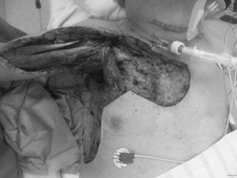
Defect after second-look operation.

Defect closure with free myocutaneous lateral vastus lateralis flap and split-thickness skin graft.
Discussion
Necrotizing fasciitis as catheter-associated infection
Central venous catheters (CVC) cause more than 90% of all catheter-associated infections. Typically, subclavian access is considered as the preferable approach for a CVC from an infectious point of view. However, severe complications such as catheter-related blood stream infections or cellulitis are possible. Therefore, strict criteria for CVC placement must be met and daily monitoring of the insertion site must be performed. Necrotizing fasciitis is a rare but severe soft tissue infection. The incidence is reported to be 0.4 cases per 100,000 population [1]. Three types are distinguished: Type I is caused by a polymicrobial infection consisting with at least one anaerobic or facultative anaerobic bacterium. Type II is monomicrobial, mainly triggered by β-hemolytic streptococci or staphylococci. Among those, a large variety of different bacteria causing NF is published in literature. Type III is reported to be induced by Vibrio species after minor injuries in salt water [2]. Necrotizing fasciitis can occur after different skin lesions and even in intact skin by hematologous spreading or by blunt trauma [3]. Necrotizing fasciitis after CVC placement has not yet been described. Another characteristic of this case is that the patient was intubated and sedated when NF took place. Thus, pain out of proportion, which is the major diagnostic criterion, could not be assessed [4]. This circumstance led to delayed diagnosis of NF of approximately 24 h, because NF was misdiagnosed initially as extravasation.
A laboratory scoring system was created for NF, namely, the laboratory risk indicator for necrotizing fasciitis (LRINEC) [5]. C-reactive protein, WBC count, and hemoglobin, sodium, creatinine, and glucose concentrations, are integrated into the score. On the day of NF diagnosis, the patient had a LRINEC score of 10, which is believed to be high risk with a probability of ≥75% for NF. However, the score is challenged by many authors because parameters are influenced by sepsis and critical illness in general [1]. Publications show that early diagnosis, within 24 h, is missed in 85%–100% of all cases. Late determination of NF is combined with increasing mortality. If the suspected diagnosis is made, surgical intervention in terms of radical and adequate debridement is required immediately. The operation is not only the therapy of choice, it is also the most sensitive and specific diagnostic approach to clarify the suspected disease. In this particular case the patient went into septic shock and hence potential entry points for an infection were re-evaluated. Otherwise, the local symptoms of the right shoulder could have been overlooked. As a result, special awareness regarding NF is needed in ventilated and sedated patients because of impaired clinical signs that may appear less impressive than expected and they often bring along predisposing factors, such as diabetes mellitus, end-stage renal failure, and immune suppression. We decided to perform a planned second-look operation 2 d after initial debridement, which is controversial because some authors recommend operating twice only when an improvement of a patient's condition fails to appear [1]. We tend to prefer the planned second look because infectious progress can progress without noticeable clinical symptoms in the immunocompromised intensive care patient, even though the initial debridement was performed radically. Sufficient soft tissue coverage after stabilization reduces the rate of re-infection, and all techniques in reconstructive surgery are required including free flaps. In many cases patients should be transferred to specialized centers for plastic and reconstructive surgery, optionally with a burn intensive care unit, for coverage after initial debridement.
Footnotes
Author Disclosure Statement
The authors declare no biomedical financial interests or potential conflict of interest.