
Abstract
Abstract
Background:
We identified recently esophageal cancer related gene-4 (ECRG4) as a candidate cytokine that is expressed on the surface of quiescent polymorphonuclear leukocytes (PMNs) and shed in response to ex vivo treatment with lipopolysaccharide. To investigate the potential biologic relevance of changes in cell surface ECRG4 in human samples, we performed a pilot study to examine a population of burn patients in whom blood could be analyzed prospectively. We hypothesized that cutaneous burn injury would alter cell surface expression of ECRG4 on PMNs.
Methods:
Patients admitted with more than 20% total burn surface area (TBSA) (n = 10) had blood collected at the time of admission and weekly thereafter. For comparison, blood was obtained from a control group of healthy human volunteers (n = 4). We used flow cytometry to measure changes in ECRG4+ PMNs from patients during recovery from injury. Esophageal cancer related gene-4 expression at each time point was compared with the patient's clinical status based on a Multiple Organ Dysfunction (MOD) score.
Results:
Esophageal cancer related gene-4 was detected on the PMN surface of cells collected from healthy volunteers, however, within 48 h of admission after burn injury (n = 10 patients), the number of PMNs with cell surface ECRG4 was decreased. Esophageal cancer related gene-4 expression in PMNs was re-established over the course of patient recovery, unless complications occurred. In this case, the decrease in cell surface ECRG4+ PMNs preceded the clinical diagnosis of infectious complications and was reflected by increased organ injury scores.
Conclusion:
From a small sample set, we were able to determine that PMN cell surface ECRG4 expression was decreased after burn injury and returned to baseline during recovery from injury. Although larger studies are needed to define the role of ECRG4 in human PMNs further, this report is the first assessment of cell surface ECRG4 protein in a patient population to support analogous findings in animal studies.
S
We have focused on the potential for esophageal cancer related gene-4 (ECRG4) to serve as a sentinel factor on immune cells that may regulate the immune set point based on its expression on the surface of leukocytes, its release from the cell surface after inflammatory stimuli, and its epigenetic regulation that may explain differences in Ecrg4 expression between individuals [6–9]. Esophageal cancer related gene-4 encodes a 148 amino acid precursor protein that is tethered to the cell surface but shed on activation [6,10]. We have demonstrated previously that ECRG4 is present on the surface of polymorphonuclear leukocytes (PMNs) and, on ex vivo activation by agonists such as lipopolysaccharide (LPS), ECRG4 is released and can be detected by immunoblotting and proteomic analyses of biologic fluids including conditioned media, cerebrospinal fluid, and serum [6,7,10–12]. We have also demonstrated that ECRG4 interacts with the human innate immunity receptor complex (TLR4-CD14-MD2) supporting further an important role in responding to inflammatory insults such as severe injury [13].
We sought to advance these findings by determining whether leukocytes from burn-injured patients exhibit analogous losses of cell surface ECRG4. If so, we reasoned that ECRG4 could serve as potential as a surrogate marker of injury responsiveness. This study describes the results of a serial analysis of cell surface ECRG4 on PMNs collected from patients over their 3- to 10-week recovery from severe burn injury. Esophageal cancer related gene-4 was monitored in clinical peripheral blood samples by flow cytometry and ECRG4+ PMNs found sensitive to burn injury.
Patients and Methods
Ethics statement
The University of California San Diego Institutional Review Board approved study participants, protocols, and consent forms. Study participants provided written informed consent to participate in this study.
Patients
Blood samples were collected from 10 male burn patients with a greater than 20% total body surface area (TBSA) burn admitted to the University of California San Diego Burn Center between June 2011 and March 2012. All patients were treated based on modern burn patient care protocols with aggressive fluid resuscitation, early enteral feeds, and ventilator support when indicated. Blood was collected at the time of admission and weekly thereafter. For comparison, blood was obtained from a control group comprising healthy male volunteers 20–60 years old (n = 4). Demographic data, burn injury location, burn depth, mechanism of injury, presence of inhalation injury, hospital length of stay, morbidity, and mortality were recorded. Clinical status and the presence of organ failure was assessed using the Multiple Organ Dysfunction (MOD) score [14]. Clinical and laboratory data for each patient were obtained prospectively.
Leukocyte preparation and staining
Primary staining was performed with affinity-purified anti-ECRG4 (Genway, San Diego, CA) and compared with isotype-matched control antibodies as described previously [6]. Leukocytes were prepared from heparinized blood after red blood cell lysis and fixed in Cytofix (Becton Dickson, San Jose, CA). Flow cytometry was performed with a FACSCalibur and data analysis performed with FloJo (Treestar, Ashland, OR). Analysis of ECRG4+ staining was gated on PMNs. Cell surface ECRG4 expression was verified with CD16+ surface staining of parallel cell populations using a second anti-ECRG4 antibody isotype (Phoenix Pharmaceuticals, Burlingame, CA).
Data set mining of gene chip assays
In a recent study from the Inflammation and Host Response to Injury Program [4], a database of gene expression in human burn patients was made available publicly (www.ncbi.nlm.nih.gov/geo accession number GSE19743). The database was searched for c2orf 40, the human gene encoding ECRG4 and its expression in the peripheral blood of burn patients compared with expression in healthy volunteers.
Statistics
Statistical analysis was conducted using Microsoft Excel (Microsoft, Redmond, WA). Comparison of ECRG4 expression from serial analysis after injury was performed using analysis of variance (ANOVA). Bivariate correlations were analyzed using simple least squares regression, and relations between parameters that did not show a linear relation were evaluated using polynomial regression.
Results
Neutrophil ECRG4 expression decreases post-burn injury
Based on our recent report that membrane-anchored ECRG4 is expressed on human leukocytes and is shed after ex vivo activation [6], we enrolled severely burned patients (more than 20% TBSA) presenting to the UCSD Burn Center and performed a serial analyses of cell surface ECRG4 concentrations on PMNs using flow cytometry. Demographic data and the severity of burns in this patient population are presented in Table 1. The patients were all males ranging in age from 21 to 57 years (mean, 37.8) and presenting with burns of 34% to 90% TBSA that required up to 114 days of hospitalization.
TBSA = total body surface area; LOS = length of stay; BP = burn patient.
Flow cytometry was used to measure the expression of ECRG4 on the surface of PMNs (Fig. 1). Comparison of ECRG4 expression with isotype control groups (Fig. 1B) and with healthy volunteer control groups (Fig. 1C) indicated that monitoring of cell surface ECRG4 on PMNs could reveal changes in the activation state of PMNs in the burn injured patient population [6]. We observed a time-dependent loss of cell surface ECRG4 on PMNs over two to seven days post-burn injury (Fig. 1D).

Neutrophil esophageal cancer related gene-4 (ECRG4) expression decreases after burn injury. (
Neutrophil ECRG4 expression return to baseline during recovery from injury
We performed a serial analysis of neutrophil ECRG4 expression from burn patients during their recovery from injury where clinical status was measured by calculating MOD score at the time of blood sample collection. Neutrophil ECRG4 expression was similar to healthy control groups when measured at the time of admission. This was followed by a decrease in PMN ECRG4 expression of up to 60% within two to three weeks post-burn injury (Fig. 2A). We also found that PMN ECRG4 expression returned to baseline concentrations after recovery from injury, with ECRG4 concentrations similar to healthy volunteer ECRG4 expression by six weeks post-injury. To confirm our findings from fluorescence-activated cell sorting (FACS) analysis of ECRG4 expression, we performed a serial analysis from a second cohort of burn-injured patients using an analysis gating on CD16+ PMNs and stained with a second anti-ECRG4 antibody isotype (Fig. 2B).

Serial analysis of esophageal cancer related gene-4 (ECRG4+) polymorphonuclear cells (PMNs) during recovery from burn injury. (
Leukocyte ECRG4 expression is decreased in burn-injured patients enrolled in the inflammation and host response to injury database
To determine whether our study of ECRG4 expression after burn injury was supported by independent gene expression data, we examined gene expression findings in the Inflammation and Host Response to Injury database [5], which includes leukocytes from volunteers (n = 63) and burn patients (n = 67) with more than 20% TBSA burn. Whereas these databases did not distinguish gene expression in specific leukocyte sub-populations (i.e., PMNs versus monocytes), they nevertheless revealed a statistically significant reduction (p < 0.05) in ECRG4 expression in leukocytes of severely burned patients (Fig. 2C). Because circulating PMNs and monocytes are the primary source of cell surface ECRG4 [6], these data support a role for ECRG4 expression in the inflammatory cell response to burn injury.
Changes in neutrophil ECRG4 expression are associated with changes in clinical status during recovery from injury
Whereas clinical scores such as the MOD score and other organ dysfunction scores are known to be of somewhat limited predictive value for the complications that arise in severe burn patients, this MOD score was a useful tool to correlate with ECRG4+ PMNs. Using this combination of measuring ECRG4+ PMN concentrations and MOD score, we analyzed the relation between PMN ECRG4 expression and organ injury in a patients with at least five weekly samples collected after admission after burn injury (Fig. 3). To address directly the possibility that the concentrations of ECRG4+ PMNs could be associated with changes in clinical status, we performed regression analyses of the concentrations of ECRG4+ PMNs versus MOD score. The comparison demonstrated that there is an association between ECRG4+ PMNs and the MOD score when there are at least six weekly samples collected over the length of stay (range of R2 values = 0.25–0.78; Fig. 3A–3C). These findings support the possibility that ECRG4 may serve a sentinel function on the surface of PMNs with expression concentrations changing based on the clinical status of the patients.
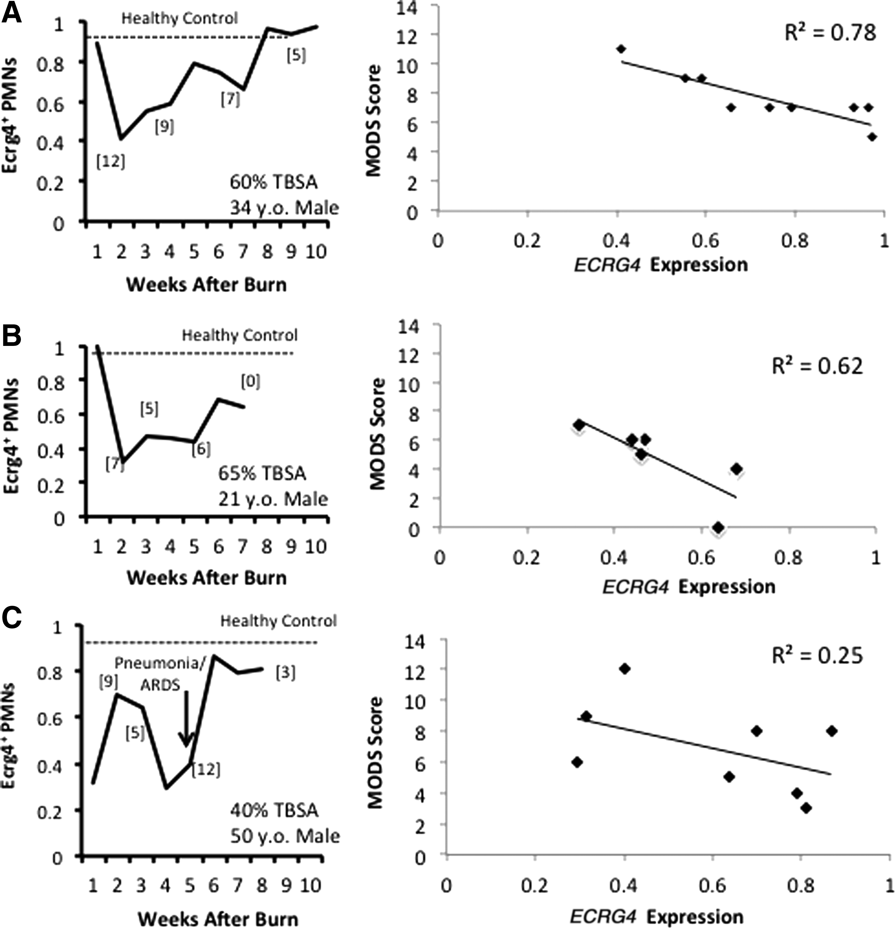
Changes in neutrophil esophageal cancer related gene-4 (ECRG4) expression are associated with changes in clinical status. Representative time-courses demonstrating changes in polymorphonuclear cell (PMN) PMN ECRG4 from individual burn patients correlated with Multiple Organ Dysfunction (MOD) score in brackets. Regression analysis was performed on patients with a length of stay of at least six weeks to compare the concentrations of Ecrg4+ PMNs versus MOD score. (
Discussion
The kinetics of cytokine and cell surface marker expression post-injury have been studied widely in human leukocytes after injury [15–18]. Yet the predictive power of global genetic changes has been disappointing [4,5]. In an effort to address this issue, we used a more targeted approach for gene analyses [6]. First, we mined gene databases [19] for open reading frames that encode proteins that are restricted to the human secretome because we reasoned that if these genes are expressed in leukocytes [6] and regulated by trauma-burn [4,5] they might be involved in maintaining set points of leukocyte homeostasis. In addition, their inclusion in secretome databases [19] would select genes that encode proteins found outside the cell and hence more likely involved in cell–cell signaling. Similarly, their inclusion in the neuropeptidome [20,21] would select genes that encode ligand-candidates and hence more likely to be involved in cell–cell communication. Finally, we reasoned that the presence of CpG islands in their promoter would point to epigenetic regulation by DNA methylation and hence predict that gene expression is affected by both environment [22,23] and aging [24], both of which may alter outcome after trauma-burn. One of the few genes to meet all these criteria is c2orf40, a human open reading frame that encodes the candidate tumor ECRG4.
In this clinical pilot study we analyzed ECRG4, a candidate sentinel factor expressed on the surface of PMNs that is shed on injury from the cell surface. In a series of experiments using peripheral blood from 10 burn-injured patients we demonstrated ECRG4 expression on the surface of PMNs of normal volunteers and after severe burn injury. The decrease of surface ECRG4 protein that was detected by flow cytometry was consistent with our previous in vitro study demonstrating evidence that ECRG4 is expressed highly on normal PMNs and decreased on activation/injury [6,25]. Using regression analyses, we studied the association between ECRG4 expression and MOD score as the basis to correlate gene expression with patient clinical status, and observed that expression of PMN ECRG4 is sensitive to injury and organ dysfunction.
We and others have recently shown that ECRG4 gene expression can be influenced by epigenetic mechanisms [6,22,23], therefore, it is interesting to speculate that differences in the initial expression of ECRG4 and the inflammation response in general have an underlying epigenetic mechanism [26]. If so, the partial recovery of ECRG4+ PMN concentrations during hospitalization in some patients after burn injury might trace back to the epigenetic regulation of ECRG4 gene expression. This is an active topic of investigation that we are pursuing by measuring ECRG4 promoter hypermethylation in patient samples.
Burn and trauma injury share many, albeit not all, inflammatory responses. Accordingly it is noteworthy that a recently published gene micro-array database from trauma patients has been mined for ECRG4 gene expression in leukocytes and similar to burn, ECRG4 was also decreased after severe injury [6]. As in the case of burn patients, the decrease in ECRG4 gene expression is likely a combination of decreased surface ECRG4 protein expression as well as reduced expression of mRNA.
In the studies reported here, the sample size of the patient cohort limits our ability to draw definitive conclusions but the findings are nevertheless consistent with genomic databases, in vitro studies, and the proposed function of cell surface ECRG4 in gauging the response to injury, presumably by modulating the inflammatory response. Larger studies are ongoing to determine whether ECRG4 can be useful in analyzing the clinical trajectory of patients with severe injury or provide insight into why individuals with similar burn-trauma injuries can have significantly different clinical outcomes. In conclusion, our findings support the hypothesis that ECRG4 is candidate sentinel factor that is shed from the surface of PMNs after injury with expression improving to baseline concentrations after recovery from injury. These findings provide further insight into the neutrophil inflammatory response to burn injury.
Footnotes
Acknowledgments
The authors would like to thank Lindsey Prescher, MD, for her assistance in analyzing clinical data and Ann-Marie Hageny for her technical assistance with this project.
Funded by the National Institutes of Health P20 Exploratory Center grant for Wound Healing Research (P20GM078421), American Burn Association/Department of Defense (W81XWH-10-1-0527).
Author Disclosure Statement
No competing financial interests exist.