
Abstract
Abstract
Background:
Ultrasound (US) is the first-line diagnostic study for evaluating gallstone disease and is considered the test of choice for diagnosing acute cholecystitis (AC). However, computed tomography (CT) is used widely for the evaluation of abdominal pain and is often obtained as a first abdominal imaging test, particularly in cases in which typical clinical signs of AC are absent or other possible diagnoses are being considered. We hypothesized that CT is more sensitive than US for diagnosing AC.
Methods:
A prospective registry of all urgent cholecystectomies performed by our acute care surgery service between June 2008 and January 2014 was searched for cases of AC. The final diagnosis was based on operative findings and pathology. Patients were classified into two groups according to pre-operative radiographic work-up: US only or CT and US. The US group was compared with the CT and US group with respect to clinical and demographic characteristics. For patients undergoing both tests the sensitivity of the two tests was compared.
Results:
One hundred one patients with AC underwent both US and CT. Computed tomography was more sensitive than US for the diagnosis of AC (92% versus 79%, p = 0.015). Ultrasound was more sensitive than CT for identification of cholelithiasis (87% versus 60%, p < 0.01). Patients undergoing both tests prior to surgery were more likely to be older, male, have medical comorbidities, and lack typical clinical signs of AC.
Conclusions:
Computed tomography is more sensitive than US for the diagnosis of AC and is most often used in patients without typical clinical signs of AC.
U
Patients and Methods
A prospective registry of all urgent cholecystectomies performed by our acute care surgery service between June 2008 and January 2014 was searched for cases of AC. All patients undergoing cholecystectomy for AC were included. Patients with AC treated without cholecystectomy and patients undergoing cholecystectomy for other indications were excluded. The final diagnosis was based on operative findings and pathology. Demographic and clinical factors were extracted retrospectively from the medical record. We recorded the presence or absence of three medical comorbidities (diabetes mellitus, hypertension, and hyperlipidemia) and four clinical factors (Murphy sign, nausea or vomiting, fever, and white blood cell count >11,000 cells/mcL).
Ultrasound signs of AC were gallbladder wall thickness >3 mm, peri-cholecystic fluid, sonographic Murphy sign, distended gallbladder, and incarcerated gallstone in the gallbladder infundibulum. Computed tomography signs of AC were gallbladder wall thickness >3 mm, peri-cholecystic fluid, distended gallbladder, and peri-cholecystic fat stranding. The presence or absence of these signs was based on the interpretation at the time the studies were performed. A study was considered positive for AC if any of the signs were present and negative if all signs were absent. Patients were classified into two groups according to pre-operative radiographic work-up: US only or CT and US. The groups were compared with each other to identify clinical and demographic factors associated with obtaining CT in addition to US. In patients undergoing both CT and US the sensitivity of the tests was compared. We used χ2 or Fisher exact test as appropriate for comparisons. This study was approved by the Massachusetts General Hospital's Institutional Review Board.
Results
A total of 209 patients underwent cholecystectomy for AC from June 2010 to January 2014. Fifty-seven underwent CT and US and 152 underwent US only. The demographic and clinical characteristics of patients undergoing both CT and US were significantly different from patients US alone (Table 1).
US = ultrasound; CT = computed tomography; WBC = white blood cell count.
To allow more robust comparison between CT and US for the diagnosis of AC, we subsequently identified 44 additional patients with AC undergoing both CT and US between January 2008 and June 2010 and extracted all relevant radiographic parameters. In this larger group of 101 patients undergoing both CT and US, CT was obtained first in 35% of cases and US was obtained first in 65% of cases. Computed tomography was more sensitive than US for the diagnosis of AC (92% versus 79%, p = 0.015). Ultrasound and CT agreed on the diagnosis of AC in 80 cases (79%). In the remaining 21 patients, the diagnosis of AC was missed by US but detected by CT in 13 patients (13%), missed by CT but detected by US in four patients (4%), and missed by both in four patients (4%). Representative CT and US images of a patient with no signs of AC on US and multiple signs of AC on CT are provided in Figure 1. Ultrasound was more sensitive than CT for cholelithiasis (87% versus 60%, p < 0.01).
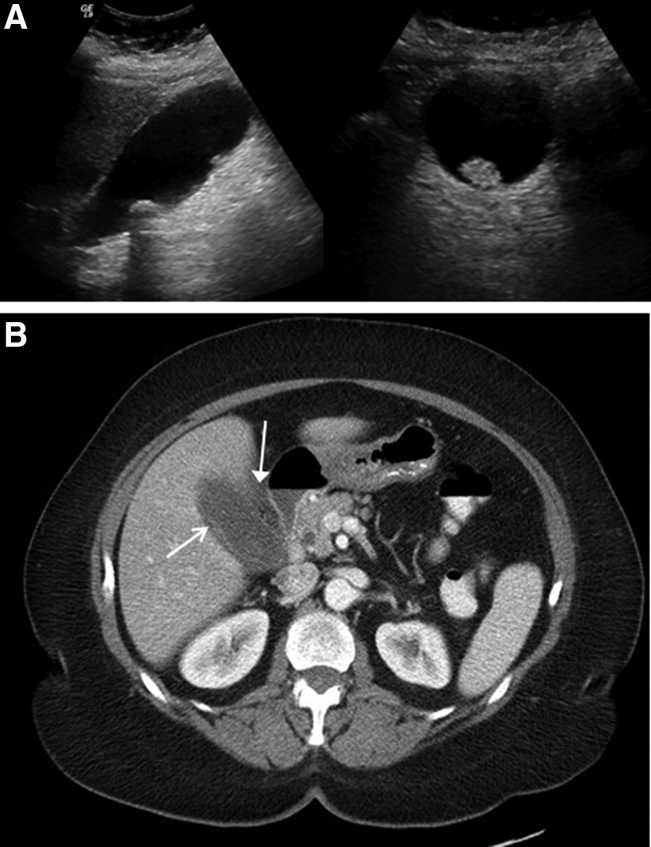
Computed tomographic and ultrasound images of a patient with acute cholecystitis. Ultrasound (
Discussion
Our results show that CT is significantly more sensitive than US for the diagnosis of AC. This is one of the few direct comparisons between the tests in AC. Van Randen et al. [4] compared CT and US directly in a large cohort of patients with acute abdominal pain that included 52 patients who were ultimately diagnosed with AC [4]. The sensitivity of CT and US for AC was identical at 73%. The lack of clear CT criteria for AC (such as for gallbladder wall thickness or size) as well as the heterogeneous CT protocols used at multiple centers may have reduced the sensitivity of CT for AC in their study. Other studies have shown modern high-resolution CT to have higher sensitivity for AC, in line with the 93% we describe in this study but have not provided direct comparison with US [3,10]. Our study yields sensitivities of US and CT for AC that are comparable to other current published results, but by doing so in the largest published cohort of AC patients undergoing both tests, provides the most meaningful basis for comparison between the techniques, and the most credence for the idea that CT is more sensitive than US for the diagnosis of AC.
Generally, US is considered the standard imaging modality for evaluation of AC, but the diagnosis of AC often relies heavily on clinical signs and symptoms [7,8]. In our study, patients undergoing US alone were significantly more likely to manifest typical signs of AC such as right upper quadrant abdominal tenderness or Murphy sign and were also younger with fewer medical comorbidities compared with patients undergoing both CT and US. In younger, healthier patients with a typical clinical presentation, the finding of cholelithiasis on US combined with the clinical picture often provides adequate diagnostic confidence, even when US does not show objective signs of AC. In our study 25% of patients undergoing cholecystectomy after US alone lacked sonographic signs of AC, but nonetheless underwent cholecystectomy and were found to have AC. This illustrates the importance of clinical judgment in the diagnosis AC. We do not believe that CT is necessary in all cases of suspected AC without signs on US, as evidenced by the fact that we undertook cholecystectomy frequently in the setting of a negative US. However, in patients with atypical clinical signs of AC and more comorbidities, US may not provide adequate diagnostic assurance. A number of authors have suggested that CT is useful in this setting [11,12]. We have utilized CT regularly in patients with suspected AC and absent or equivocal sonographic signs of AC and this retrospective study of that experience shows that CT can often confirm a diagnosis of AC when US fails.
The rationale for utilizing both CT and US depended on which test was obtained first. In 35% of patients undergoing both tests, CT was used as the first imaging test in the initial evaluation of abdominal pain. Given the frequency with which CT is utilized as an initial imaging study for patients with abdominal symptoms, the scenario in which a CT scan that is diagnostic of AC precedes an US is a common one. When CT showed AC in our series, US was used to confirm the diagnosis and to identify cholelithiasis. In some scenarios, such as a positive CT with only one sign of AC and no cholelithiasis, this may make sense. Our data suggest, however, that US is not routinely necessary as a confirmatory test after a CT that is diagnostic of AC and does not show other intra-abdominal pathology, a view that has been advocated by others even before our data were available [11]. In 65% of patients undergoing both tests, US was performed first and was either entirely negative or, in combination with equivocal clinical signs, was not considered adequate to confirm the diagnosis of AC. In these cases CT was used to confirm the diagnosis and rule out other intra-abdominal pathology. In light of our findings, this seems the more appropriate reason to perform a second imaging test in the setting of suspected AC.
The main advantages of US in the diagnosis of gallstone disease are its sensitivity for cholelithiasis, lack of ionizing radiation or contrast injection, and relatively low cost. Nonetheless, for AC specifically, we have shown it to be less sensitive than CT. Ultrasound is also limited in its ability to detect other intra-abdominal pathology if gallstone disease does not explain the patient's symptoms. Thus, diagnostic evaluation that begins with US may be more likely to require additional diagnostic tests, which may offset some of the cost savings derived from choosing a less expensive initial test. Although the cost of US is low, in cases in which CT is performed first, any additional cost may be unnecessary. In many facilities US is less readily accessible than CT because trained technicians may not be available to perform the examinations during night and weekend hours, and waiting for US may result in delay. Cholescintigraphy has been recommended in cases of suspected AC not confirmed by US and clinical factors. Whereas both sensitive and specific, its cost is similar to CT, it also uses ionizing radiation, and it shares some disadvantages of US such as lack of widespread immediate availability and inability to evaluate for alternative diagnoses. The most efficient diagnostic algorithm for AC in terms of time and cost is complex and depends greatly on the initial clinical presentation, the pre-test probability of AC, and the likelihood of alternative diagnoses. We believe that CT clearly has a role based on its sensitivity, widespread availability, and ability to rule out many alternative diagnoses, and frequently may obviate the need for US or cholescintigraphy.
Our study has several limitations. As a single-center study, our results may not be generalizable to other practice settings or patient populations, although our sensitivity results are comparable to those reported elsewhere and our use of CT in patients with atypical clinical signs has been suggested by others [10,11]. We only studied patients treated with cholecystectomy because their diagnosis of AC could be verified surgically and pathologically. Patients treated with cholecystostomy tube or with antibiotics alone may be different categorically and therefore CT and US may have performed differently if such patients had been included in our study. Because our study patients were identified retrospectively from a group with confirmed AC, we did not identify patients incorrectly diagnosed with AC by CT or US and we can only compare the sensitivity of the two tests, not the specificity. A prospective comparison of CT and US in patients with suspected AC would be required to define and compare the overall diagnostic performance of the two tests.
In summary, we found that CT is more sensitive than US for diagnosis of AC in the largest series to compare the two techniques directly for the diagnosis of AC. Whereas US may be an adequate diagnostic adjunct in patients with typical clinical signs of AC, CT is especially useful in patients without typical clinical signs or symptoms. Its sensitivity, ready availability, and ability to assess for alternative diagnoses make it a powerful diagnostic tool for AC that can augment or supersede US and cholescintigraphy in many cases.
Footnotes
Author Disclosure Statement
None of the authors have any disclosures relevant to this manuscript.