
Abstract
Abstract
Background:
Cholangitis is the most common complication after the Kasai procedure. It can be life-threatening and may affect long- and short-term outcomes of children with biliary atresia. We summarize our experiences in the prevention of early-onset cholangitis.
Patients and Methods:
From January 2002 to March 2013, children with biliary atresia (BA) who underwent the Kasai procedure in the General Surgical Department were included in a retrospective cohort study. These patients were divided into group A (therapy 1) and group B (therapy 2) depending on the infection prevention protocol and occurrences of cholangitis within the six months after surgery were recorded. Two hundred eighteen children were included in this cohort study. Seventy-six children (35 females and 41 males) were included in group A. One hundred forty-two children (65 females and 77 males) were included in group B. Therapy 1 was our primary protocol and included a third-generation cephalosporin, metronidazole, and human immunoglobulin. Therapy 2 was a modification of therapy 1 that involved imipenem-cilastatin and human immunoglobulin. Statistical analyses were performed. A p value below 0.05 was regarded as significant.
Results:
In group A, 45 children developed cholangitis within the six months after the Kasai procedure. In group B, 14 of these children experienced post-operative cholangitis. A χ2 analysis was used to examine the difference in the incidence of cholangitis between groups A and B. There was a substantial difference in the morbidity of post-operative cholangitis between groups A and B (59.2% vs. 9.9%, p = 0.000).
Conclusion:
Cholangitis in the early period after a Kasai procedure can be prevented effectively with an advanced prophylactic protocol.
C
Patients and Methods
This retrospective study was approved by the ethics committee of our hospital. From January 2002 to March 2013, children with BA who underwent the Kasai procedure in our hospital and were followed up for at least six months were included in this retrospective study and the incidence of early cholangitis was recorded. Two hundred eighteen children were included after 20 children were excluded because of loss to follow-up. These patients were divided into group A (therapy 1) and group B (therapy 2), depending on the protocol for cholangitis prevention. Seventy-six children (35 females and 41 males) were included in group A. One hundred forty-two children (65 females and 77 males) were included in group B.
Therapy 1 was our primary protocol and included the intravenous administration of third-generation cephalosporin and metronidazole during the first two weeks as well as intravenous human immunoglobulin (IVIG) during the first five days, followed by an oral third-generation cephalosporin until six months. Therapy 2 was an improvement on therapy 1 that involved the intravenous administration of imipenem-cilastatin for the first two weeks, IVIG for the first five days, and an oral third-generation cephalosporin given until six months.
The Kasai procedures were performed by the same two pediatric surgeons who had worked together previously treating BA. All patients underwent Roux-en-Y reconstruction procedures with spur anti-reflux intestinal valves (Figs. 1–4). This was a Roux-en-Y anastomosis with a 5-cm spur valve between the hepatic and jejunal limb [6,7]. The length of the Roux-en-Y loop was routinely 60 cm. Post-operatively, we used antibiotic agents, human immunoglobulin, methylprednisolone, and ursodeoxycholic acid (UDCA) in all patients. Methylprednisolone (4 mg/kg) was given intravenously during the first five post-operative days and then orally for the next two months in reduced doses. Ursodeoxycholic acid was given orally from the seventh post-operative day for one year. In group B, we began to administer intravenous imipenem-cilastatin instead of the third-generation cephalosporin and metronidazole during the first two weeks after the Kasai procedure. After two weeks of prophylactic intravenous antibiotic agents, all patients received prophylactic oral antibiotic agents until six months after the Kasai procedure.
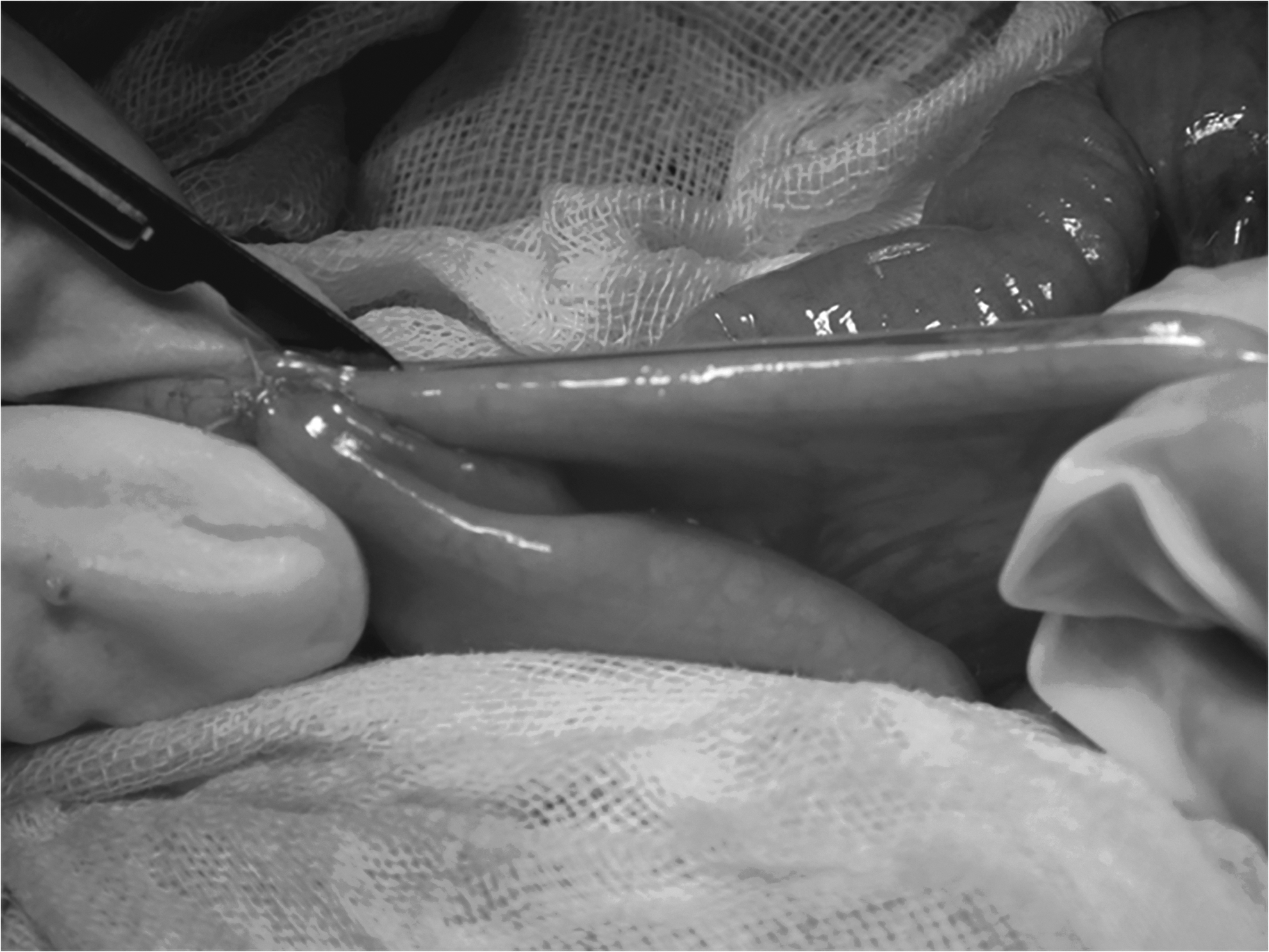
To construct the spur valve, incise the seromuscular layer of the bile loop for 5 cm.
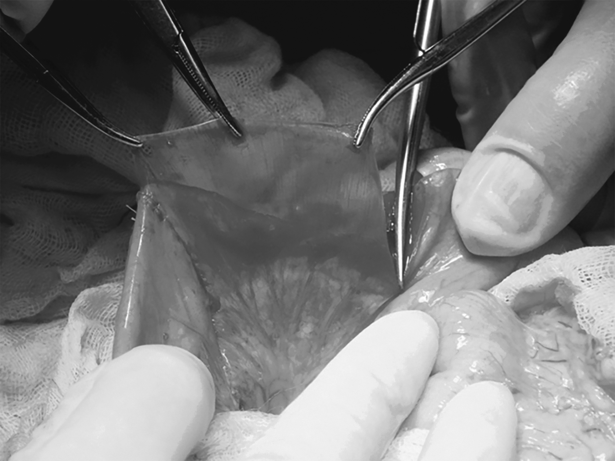
Peel off the medial seromuscular layer.
Cut off the medial seromuscular layer.

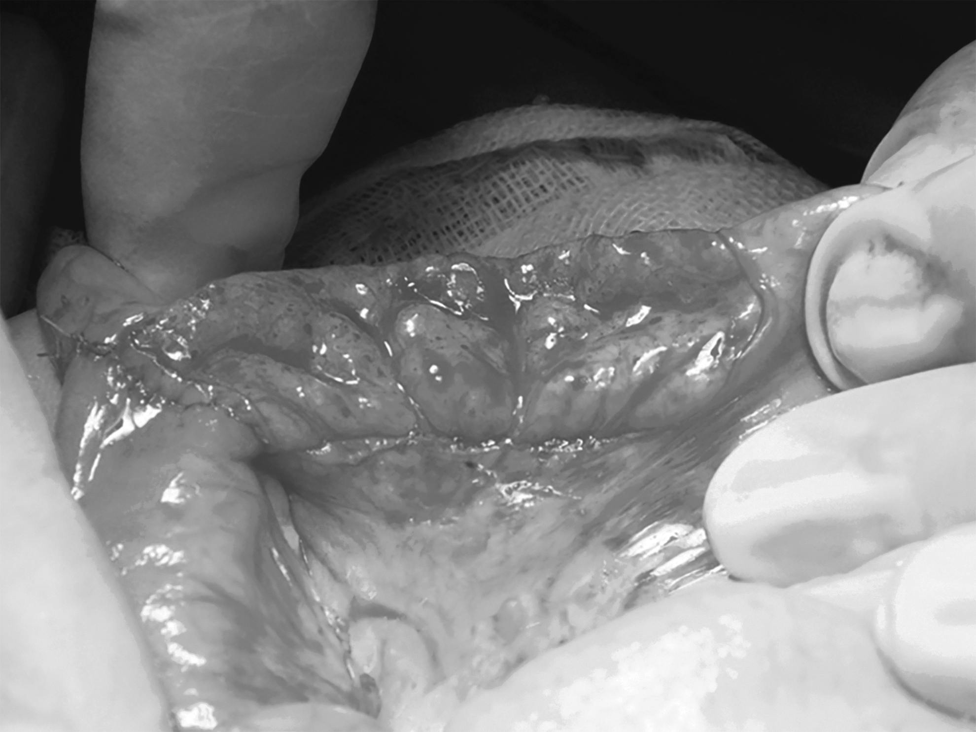
Seam the jejunal loop and bile loop.
The majority of patients was followed in the outpatient department. We drew blood for routine blood tests, tests for C-reactive protein, bilirubin level, and transaminases. For the patients who lived far away, the pediatric surgeons of the regional hospitals were asked to perform the follow-ups, and the information was obtained from their families by mail or telephone. Clinical outcomes, including incidence of early cholangitis, the jaundice clearance rate, and long-term survival rate were compared between the two groups.
Early cholangitis was defined as the development of cholangitis in the first six months after Kasai procedure. Cholangitis was diagnosed based on clinical manifestations such as fever without another identifiable source of infection and/or altered stool color or refractory jaundice, laboratory evidence of elevated C-reactive protein level and/or white blood cell (WBC) count and sudden elevation of serum bilirubin or liver enzymes (alanine transaminase [ALT] or aspartate transaminase [AST]). The blood level of total bilirubin less than 2.0 mg/dL was designated adequate bile drainage. Statistical analyses were performed using the Student t-test or χ2 test, as appropriate. The cumulative survival rates were analyzed using Kaplan–Meier survival curves and the statistical difference was assessed using the log-rank test. A p value below 0.05 was regarded as significant.
Results
There were no differences in terms of age at operation, gender, or body weight between the two groups (Table 1). In group A, 45 of 76 suffered from cholangitis within the first six months after the Kasai procedure. In group B, 14 of 142 experienced post-operative cholangitis. The data of timing of cholangitis in both groups are shown in Table 2.
Patient Characteristics
Data of Timing of Cholangitis in Both Groups
A χ2 analysis was conducted to examine the difference between groups A and B in terms of incidence of early cholangitis after the Kasai procedures. A substantial difference in terms of morbidity of post-operative cholangitis was found between groups A and B (59.2% vs. 9.9%, p = 0.000; Table 3).
Morbidity of Post-Operative Cholangitis in Groups A and B
p = 0.000.
There was no difference in terms of bile drainage before development of cholangitis between the two groups, i.e., 61.7% versus 63.1%, respectively (p > 0.05). The five-year native liver survival rate was 23.7% and 60.6%, respectively (group A vs. group B, p = 0.000; Fig. 5).

The five-year native liver survival rates in groups A and B. The five-year native liver survival rates were 23.7% and 60.6%, respectively. The cumulative long-term survival rate was better in group B than in group A (p = 0.000).
Discussion
Since the first Kasai procedure was conducted in Japan, several therapy protocols involving steroids, antibiotic agents, and UDCA have been introduced [8–12]. Cholangitis is the most common complication, influencing both short- and long-term prognoses.
The mechanism of post-Kasai cholangitis is most likely to be an ascending cholangitis via the Roux loop into the intra-hepatic duct system. Other contributory mechanisms may also be involved, including bacterial overgrowth in the gut, translocation from lymphatics, and hematogenous spread via the portal vein. The common causative organisms have been identified as Klebsiella spp., Escherichia coli, Pseudomonas aeruginosa, Enterobacter cloacae, Acinetobacter baumannii, Streptococcusmitis, and Salmonella typhi [13].
In most case series, more than half of patients have their first episode of cholangitis within the first six months. The first days after the Kasai procedure are the days when bacterial colonization occurs, leading to bacteria in intestine locating to the hepatic porta. The possible mechanism by which prophylactic antibiotic agents prevent cholangitis in BA patients after the Kasai procedure has yet to be determined, however, it is believed to occur in one of two ways: adequate concentrations of antibiotic agents are excreted from blood into the cholangioles or the bacterial concentration within the bilioenteric conduit is diminished [14].
The practice of prescribing prophylactic antibiotic agents in an attempt to reduce the incidence of cholangitis is universal but extremely variable. There is no consensus as to which drug to give or for what duration. Most children present with pyrexia, worsening jaundice, and changes in liver biochemistries; they should be treated aggressively with broad-spectrum intravenous antibiotic agents effective against gram-negative organisms [15]. A third-generation cephalosporin and metronidazole were included in the prevention protocol of group A, and the morbidity of post-operative cholangitis approached 60%. In group A, including children who underwent the Kasai procedure before March 2009, the children who developed cholangitis had difficult experiences. Ceftazidime, cefepime, fluconazole, and meropenem were used as antimicrobial drugs, however, we were unable to identify which drug or combination of drugs was optimal. Meropenem was useful initially but pharmaceutical resistance limited its clinical use. Ultimately, we had to use imipenem-cilastatin and immunoglobulin to control severe cholangitis; treatment with these two agents was found to be optimal.
The morbidity associated with cholangitis is too high and this condition usually lasts for two weeks or more, causing great pain and expense. In group A, the period during which the cholangitis usually developed was the first month after the Kasai procedure. Although not all cases of cholangitis developed during the first two weeks, the most serious cases occurred at this time in group A, always leading to severe sepsis. In group B, there were no such cases of serious cholangitis during the first two weeks after the Kasai procedure, and the subsequent cases of cholangitis were easier to control. There is a saying in traditional Chinese medicine that prevention is always better than a cure, which leads us to believe that the prevention of cholangitis might play a key role in the entire protocol.
In the first days after the Kasai procedure, the time before there is good bile flow, measures should be taken to prevent bacterial colonization as early as possible. Antibiotic agents with an extremely wide spectrum of antibacterial activity should be used, especially specific agents with activity against the described pathogenic bacteria. Imipenem combined with cilastatin is such an antibacterial agent that is Food and Drug Administration-approved for use in children and infants younger than three months of age [16].
The organisms that are commonly isolated in blood cultures are Klebsiella pneumoniae, Enterococcus, Escherichia coli, and Pseudomonas aeruginosa [17], all of which are sensitive to imipenem-cilastatin. Therefore, based on previously described empirical experience, imipenem-cilastatin was given intravenously for the first two weeks after the Kasai procedure. Moreover, the lack of drug resistance and the specificity for bile duct bacterial infections are additional reasons for our ultimate selection of imipenem-cilastatin.
We gave IVIG for the first five post-operative days to regulate and improve the immune abilities of the infants with BA and to allow imipenem-cilastatin to work more effectively [18]. It has been reported that infants with BA always experience immune dysregulation and deficiency in addition to inflammatory conditions. Intravenous human immunoglobulin therapy is known to reduce inflammatory cytokines and to increase anti-inflammatory regulatory T cells. It is an established part of treatment in several autoimmune, immunodeficiency, and inflammatory conditions. Numerous mechanisms for the anti-inflammatory action of IVIG have been proposed, including interference with the cytokine network, neutralization of autoantibodies, modulation of effector functions of T and B cells, and enhancement of regulatory T cells. In addition, human immunoglobulin reduces progressive intra-hepatic bile duct injury. In a recently reported animal experiment, high-dose immunoglobulin G therapy in murine BA dramatically decreased T helper cell-mediated inflammation and biliary obstruction. Fenner et al. [19], using a rhesus-rotavirus model of BA in mice, showed that there were reduced bilirubin levels with lower expression of vascular cell adhesion molectule-1 in the portal epithelium and cytokine production by T cells using high-dose immunoglobulin G therapy. This study supports consideration of IVIG use in infants with BA to diminish the progressive intra-hepatic bile duct injury [19].
Although there has been debate regarding the use of steroids, they play active roles in bile flow after Kasai procedures. Current evidence based on the most recent systematic review of published evidence supports the view that high-dose steroids reduced post-operative bilirubin and cleared jaundice, the two most frequently measured indices of the condition. We used steroids in both groups, however, the effect of steroids may be limited or inhibited by an increasing degree of fibrosis and onset of cirrhosis. Therefore, we suggest reducing the overall age at the time of the Kasai procedure to improve the efficacy of adjuvant therapy [20].
Ursodeoxycholic acid is hydrophilic bile acid found more commonly in bears than in human beings. Exogenous administration has shown biochemical, histologic, and clinical benefits in adults with primary biliary cirrhosis and sclerosing cholangitis, reducing the need for transplant. Enrichment of the bile acid pool with exogenous UDCA is believed not only to increase clearance of toxic endogenous bile acids, but it may also have an immunomodulatory effect on mononuclear cells [15].
There is no debate that cholangitis is the most common risk factor influencing the quality of life in children after the Kasai procedure [21]. The incidence of cholangitis in BA is between 40% and 90%, and the majority of patients experience at least one episode before two years of age [4,5,22]. A similar morbidity was observed in group A (59.2%) in our study; with optimized pharmaceutical treatment, this rate was reduced to 9.9% in group B. Therefore, this advanced prevention protocol was shown to be useful in this study. Although the grade of imipenem-cilastatin is commonly too high limiting its clinical use, we provided evidence supporting the use of this drug in the prevention of cholangitis. In another study that followed the present study, we reduced the time of the administration of imipenem-cilastatin to one week because of drug supervision pressure, and the morbidity of cholangitis increased to 30.3%.
Among the children who developed cholangitis, 38 did not receive regular antimicrobial treatment because of a lack of experience, and the other 21 received imipenem-cilastatin for one to two weeks and human immunoglobulin for five days. Among the latter 21 children with cholangitis, 14 were cured without relapse and seven developed recalcitrant cholangitis, of whom three were cured, three died, and one developed cholestasis. Therefore, a single episode of cholangitis does not predict a poor result, however, recurrent cholangitis might lead to early mortality.
Predictions of long-term prognoses are important for selecting the appropriate management of patients who require life-long follow-up. Thus, in many studies, researchers have assessed clinical parameters to clarify the early risk factors that affect late-presenting liver failure in biliary atresia and have found cholangitis to be the best predictor [20,23–25]. In the latest systematic review of cholangitis, prophylactic antibiotic use was associated with increased transplant-free survival [14]. In our study, the five-year native liver survival rate was higher in group B, and the optimal prophylaxis for cholangitis played an important role. Our study had limitations including potential biases because of the retrospective study design. Furthermore, other possible contributing factors including standardized Kasai procedure [26], consisting of limited and careful transection of the fibrous remnants at the porta hepatis, a long Roux-en-Y loop equipped with a spur valve, and meticulous anastomosis, as well as standardized follow-up were associated with better outcome of Kasai portoenterostomy other than prevention of cholangitis. Cholangitis can lead to cirrhosis, necessitating early transplantation that is not always available to the majority of patients in China, for economic reasons and donor shortages. Thus, we need to make every attempt to ensure that children with BA retain their own livers for as long as possible. Indeed, this is a substantial challenge, but we can derive hope from the work we are currently performing.
Footnotes
Author Disclosure Statement
No competing financial interests exist.