
Abstract
Abstract
Background:
Surgical site infections (SSIs) are a serious problem after abdominal surgery. This study aimed to compare closure of fascia with triclosan-coated monofilament polydioxanone (PDS) or standard PDS in decreasing the incidence of SSIs in patients who underwent abdominal surgery.
Methods:
In this randomized study, a total of 890 consecutive patients undergoing laparotomy for any gastrointestinal pathology were allocated to closure of the fascia with triclosan-coated PDS (treatment group; TG) or standard PDS (control group; CG). Patients were assessed every day during the hospital stay for SSIs and at the first, second, and fourth week after discharge. The surgical site was assessed in terms of superficial, deep incisional, or organ/site SSI.
Results:
The main important finding was that SSIs were reduced as much as 24% by using triclosan-coated PDS. Surgical site infections occurred in 200 patients (22.4%), with 105 being early (in the first week) and 95 occurring late. Eighty five of the SSIs (19.1%) were noted in patients in the TG, whereas 115 of them (25.8%) were in the CG (p = 0.016). The infections were superficial in 126 patients, deep incisional in 48 patients, and organ/site in 26 patients. Most of patients (n = 651) had clean-contaminated sites. In subgroup analysis, SSI rates with triclosan-coated PDS were lower in clean, clean-contaminated, and contaminated incisions (0 in the TG versus 24.2% in the CG; p = 0.009; 13.6% in the TG versus 24.3% in the CG, p = 0.001; and 16.6% in the TG versus 27.8% in the CG; p < 0.0001, respectively).
Conclusions:
Closure of the fascia with triclosan-coated PDS decreased SSI rates as much as 24%. Also, SSIs were decreased significantly at clean, clean-contaminated, and contaminated sites. Therefore, triclosan-coated PDS might be recommended for fascial closure as a means of decreasing SSIs.
Post-operative surgical site infections (SSIs) represent 14% of all nosocomial infections and roughly 5% of all surgical complications. Many methods for preventing SSIs, including antibiotic prophylaxis, hair removal at the incision site, mechanical bowel preparation, skin disinfection, hand decontamination, and the use of sterile dressing gowns, instruments, and gloves, have been explored as a measure to decrease SSIs in the past few decades [1].
Surgical sutures can promote bacterial colonization, and this is related to the structure and chemical composition of the sutures [2]. For this purpose, bioactive glass with silver [3] and antibacterial-coated sutures, such as with triclosan, have been manufactured. In the most recent meta-analysis, antibiotic-coated sutures were reported to reduce infection [4]. Antibacterial-coated biomaterials prevent colonization and adhesion of bacteria in comparison with uncoated biomaterials [5,6]. The aim of antibiotic coating is administering higher local antibiotic concentrations early in the logarithmic growth phase of bacteria. In this context, the idea of reduction of the SSI ratio of the antibacterial-coated sutures has been put forward.
Triclosan (2,2,4′-trihydrochloro-2′-hydroxydiphenylether) is a synthetic, non-ionizing, broad-spectrum antimicrobial agent that has been in use for more than 40 years. Triclosan-coated suture material reduces bacterial growth in vitro and in vivo [2,7]. In a study conducted by Barbolt [8], triclosan-coated suture materials were non-toxic, non-irritating, non-cytotoxic, and non-pyrogenic; and the rate of accumulation in the body was no higher than that of any antimicrobial agent used in the food industry. Moreover, Edminston et al. [2] carried out a contamination study with Staphylococcus aureus, S. epidermidis, and Escherichia coli on suture materials. This study showed that the addition of triclosan decreases bacterial growth significantly in vivo and in vitro. Also, the reliability of triclosan-coated sutures has been demonstrated in clinical studies. The first of these was conducted by Ford et al. [9] without success on SSIs.
Several randomized and non-randomized trials and meta-analyses have evaluated the results of triclosan-coated suture material after laparotomy. However, because of the different patient groups and heterogeneity within these groups, the results are not fully reliable. Therefore, the efficacy of triclosan-coated sutures and their clinical significance is unclear, and further research is needed. The results of this material in relatively homogenous and high-risk patients are promising when it comes to preventing SSIs. The present study was planned because of the lack of data and literature on this topic. In this study, we planned to compare triclosan-coated PDS with uncoated PDS suture for abdominal facial closure after gastrointestinal tract (GI) surgery, which carries a higher SSIs risk for patients.
Materials and Methods
A total of 900 consecutive patients who underwent elective or emergency laparotomy for any type of GI pathology were enrolled in this randomized study between June 1, 2013, and June 1, 2014.
This study was approved by the Human Ethics Committee at the study institution (No: 2013/165). In addition, written informed consent was obtained from all the patients. The patients were randomized to closing the fascia with standard PDS or triclosan-coated PDS after operations using a computer-generated list created by an independent computer consultant.
Inclusion and exclusion criteria
All patients who were 18 years or older and underwent elective or urgent GI surgery for any reason were included initially in this study. But patients who had triclosan allergy, needed re-laparotomy in the first week after the initial operation, were left with an open abdomen, American Society of Anesthesiologists score IV, or who refused randomization were excluded. Finally, two patients from the standard PDS group and four patients from the triclosan-coated PDS group were dropped from the follow-up for different reasons, and three patients from the standard PDS and one patient from the triclosan-coated PDS group died. These patients also were excluded from the study. As a result, 890 patients were enrolled in the study of whom 445 were assigned to the standard PDS group (control group; CG) and 445 to the triclosan PDS group (triclosan group; TG).
Patient follow-up and control tests were done by a blinded researcher, and findings were recorded on the seventh, 14th, and 30th post-operative days.
Surgical technique
The operation site was shaved the day before surgery, and the operative field was cleaned with 10% polyvinylpyrrolidone iodine (polyvidone–iodine). Intravenous antimicrobial prophylaxis was provided with cefazolin 1,000 mg (Cezol®; Deva, Istanbul, Turkey) for aerobic flora and ceftriaxone 1,000 mg (İesef®; I.E. Ulagay, Istanbul) plus metronidazole 500 mg (Flagyl®; Eczacibasi, Istanbul) for anaerobic flora at the induction of anesthesia.
The fascia was closed by standard monofilament polydioxanone 1/0 (PDS II®, Ethicon, Somerville, N.J. U.S.) in the CG or by triclosan-coated monofilament polydioxanone 1/0 (PDS Plus ®, Ethicon) in the TG. Extra suture was not used to close the subcutaneous tissue in either group. The skin was closed with 3/0 polypropylene suture, which was removed on post-operative day 14 if complications had not occurred in the incision.
Infection management
Surgical site infections occurring in the first seven days were accepted as early onset whereas infections occurring thereafter were assessed as late onset. If clinical symptoms of infection such as fever, leukocytosis, purulent drainage, or widespread cellulitis appeared, antibiotic therapy was given after incision debridement. Surgical site cultures were harvested in the cases with definite purulent drainage, and the antibiotic regimen was changed according to the results of the antibiogram in these cases. Sterile culture rods (Sterile Transport Swab®; Copan International, Bresica, Italy) were used for culture. Samples were sent to the microbiology laboratory for culture in the shortest possible time.
The treatment of patients with organ-site SSIs was planned as soon as these infections were diagnosed. Laparotomy or percutaneous drainage was performed, and an appropriate antibiotic was added. Operative sites were divided into four categories: Clean, clean-contaminated, contaminated, and dirty. Patient age, gender, Body Mass Index (BMI), ASA score, target organ for operation, co-morbidities, duration of operation, amount of peri-operative of blood transfusion, operation classification, length of intensive care unit (ICU) stay, length of hospitalization, and occurrence of incisional hernia and SSIs were recorded.
Statistical analysis
Initial power analysis was made using the MedCalc statistics program version 12.7.5.0 to determine the necessary sample size. Approximately 447 patients in each arm was calculated to detect an expected SSIs reaction rate of 30% with a minimum α error of 5% and a β error of 10%. The sample size was established before the study, and 450 patients were to be enrolled in each arm. Analysis of data was carried out with SPSS 17.0.
Differences between the treatment groups were analyzed with chi-square tests. Age, BMI, operation time, incision size, peri-operative blood transfusion, and duration of ICU and hospitalization are presented as mean±standard deviation (SD); and SSI, gender, co-morbidity, ASA scores, and target organ are shown as a percentage in parentheses. P < 0.05 was accepted as significant.
Results
The mean ages of the 890 patients in the two groups did not differ significantly (55.1 ± 16.3 years in the TG and 54.6 ± 16.9 in the CG; p = 0.63) (Table 1). The male/female ratio was 192 (43.1%)/253 (56.9%) in the TG, whereas it was 223 (50.1%)/222 (49.9%) in the CG (p = 0.037). The mean BMI was 26.1 ± 2.9 kg/m2 in the TG and 28.4 ± 3.4 in the CG (p<0.0001). In the TG, 100 patients (22.4%) smoked cigarettes, whereas 169 patients (37.9%) in CG smoked (p < 0.0001). Having a second or greater abdominal operation was true of 96 patients (21.5%) in the TG and 144 patients (32.3%) in the CG (p = 0.043). Urgent surgery was performed for 31 patients (6.9%) in the TG and 74 patients (16.6%) in the CG.
Demographic Characteristics
Age, BMI, duration of surgery, incision length, amount of pre-operative erythrocyte transfusion, length of intensive care unit stay, and length of hospital stay were calculated as mean±standard deviation.
Gender, smoking status, previous abdominal midline incision, co-morbidity, ASA class, and target organ for operation were calculated as percentages.
Anemia (TG = 113 and CG = 101) was the most common co-morbidity in both groups, but there also were instances of hypertension, diabetes mellitus, chronic obstructive lung disease, cancer, chronic renal failure, and cirrhosis; and the difference in co-morbidities in the two groups was significant (p < 0.0001). Also, differences were calculated between the two groups in terms of ASA scores, number of urgent operations, and target organs for surgery (p < 0.0001).
The mean operation time (151 ± 40.5 minutes in the TG and 144 ± 48.9 minutes in the CG; p = 0.038), length of ICU stay (2.98 ± 1.0 days versus 2.69 ± 0.8 days; p = 0.001), and length of hospital stay (7.46 ± 1.7 days versus 6.70 ± 2.2 days; p < 0.0001) were higher in the TG, and the mean incision was longer (23.5 ± 3.3 cm versus 22.6 ± 3.7 cm; p < 0.0001); but the amounts of peri-operative blood transfusion were similar in the two groups. The comparison of the two groups is summarized in Table 2.
Duration of Surgery and Hospital Stay a
Values shown as mean±standard deviation.
Surgical site infection occurred 200 patients (22.4%), 105 of them being early onset and 95 late onset. Most of the SSIs (40%) in the early-onset groups were superficial. Cellulitis around the incision was seen in 126 patients (purulent drainage or microorganisms on site culture), deep incisional infection was found in 48 patients (subcutaneous abscess that led to dehiscence, resulting in debridement or microorganisms cultured from incision site). Organ/site surgical infection was seen in 26 patients (significant purulent drainage from abdominal cavity or intra-abdominal abscess or collection identified radiologically). Some incision healings are shown in Figs. 1 and 2. Despite the fact that organ/site-specific infection was rarely seen early or late, it was dominant among late-onset cases (Table 4). However, the main important finding of this study was that SSIs were reduced as much as 24% by using triclosan-coated PDS. The SSI rate was 19.1% (n = 85) in the TG, whereas it was 25.8% in the CG. This difference was statistically significant (p = 0.016) (Table 3). Furthermore, using triclosan-coated PDS reduced SSIs in all subgroups. Thus, triclosan-coated PDS usage resulted with dramatic reduction in SSIs in clean (p = 0.009), clean-contaminated (p = 0.001), contaminated (p < 0.0001), and dirty (p = 0.001) operations. The details of the subgroup analysis are presented in Table 5.
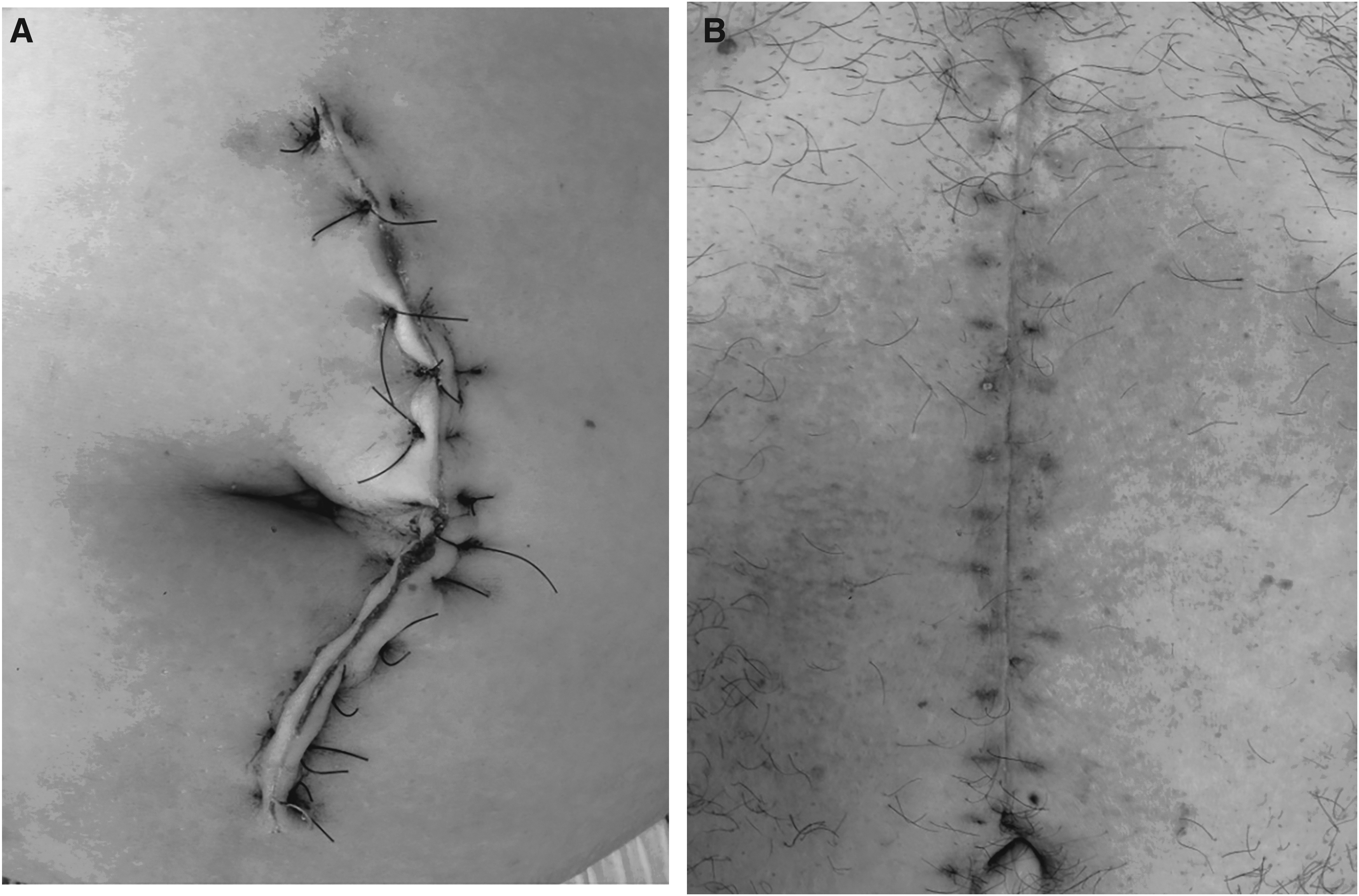
Wound healing. (
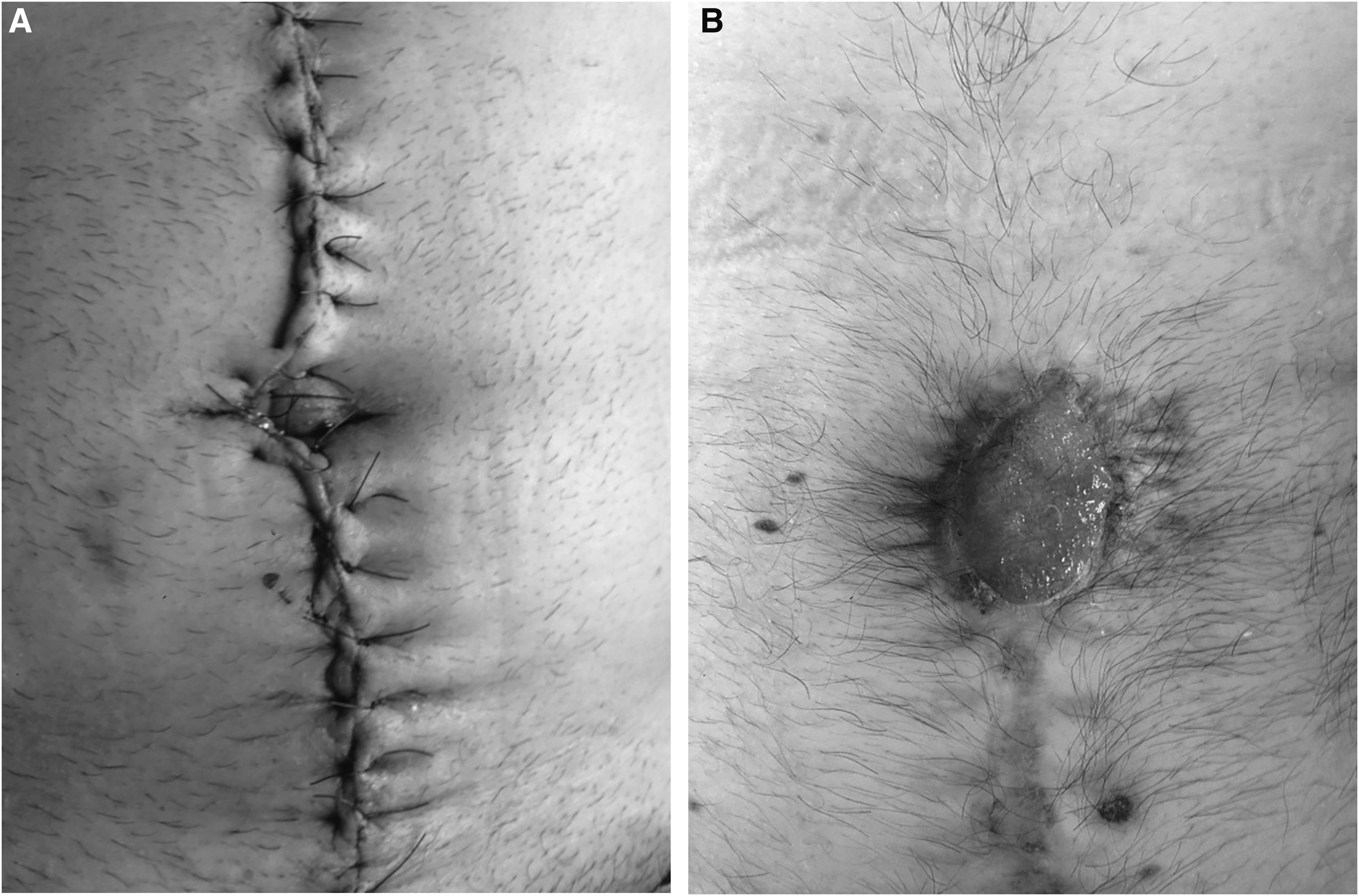
Another example of wound healing. (
Surgical Site Infections and Incisional Hernias
Location of Surgical Site İnfection
Incision Classification
TG = triclosan group; CG = control group.
Isolation of micro-organisms was obtained in 27% of the patients with SSI. Escherichia coli strains were the most often isolated, being recovered in 44%, followed by Klebsiella pneumoniae (18%), Proteus vulgaris (13%), methicillin-resistant S. aureus (11%), and Acinetobacter baumannii and Pseudomonas aeruginosa (7%).
The effect of triclosan coating on the incisional hernia rate also was studied. Incisional hernia was seen in 31 patients (7.0%) in the TG, while it was found in 35 patients (7.8%) in the CG. This difference was not statistically significant.
Discussion
Surgical site infections are one of most important preventable reasons for the increase in morbidity and hospital cost, and yet the rates of SSIs have not been reduced to an acceptable value. The total cost of a patient having an SSI is nearly two times that of a patient without an SSI. At least US$223–275 million was spent for loss of work and persistent morbidity attributable to SSIs in the U.S. [10]. Thus, measures to prevent SSIs have gained importance.
In recent years, studies were intensified on biomaterial designs to prevent micro-organism colonization. Recent studies have shown that biomaterials covered with anti-bacterial agents prevent colonization and adherence of bacteria compared with biomaterials that are not covered [5,6]. In this context, coated anti-bacterial sutures may reduce SSIs. Studies investigating bacterial colonization on surgical suture materials found the colonizing microbial population to be closely related to suture structure and chemical content, which is similar to the findings with other biomaterials [2].
Today, enhanced suture materials with minimal changes in absorption time and better tensile strength, resulting in both reduced tissue reactivity and infection-preventing properties, are being manufactured. As described in previous studies, to prevent colonization of suture materials by bacteria, coating with triclosan has been used since 2003 to create a suitable environment for surgical interventions and wound healing [3].
Recently, a few in vitro and in vivo studies have investigated the effect of triclosan coating on reducing the bacterial load on suture material [7]. The majority of those studies indicated that anti-bacterial-coated suture materials would reduce SSI.
Edminston et al. reported that a significant reduction in bacterial load was produced by adding triclosan to suture materials in fields contaminated by S. aureus, S. epidermidis, and E. coli, which often are isolated from surgical incisions after abdominal surgery [2]. Even if E. coli was the bacterium isolated most frequently in our study, the infection rate decreased dramatically in the triclosan-coated group. This situation might be explained by positing that triclosan reduced the adhesion capability of the organisms, resulting in less infection. In another study, described by Ford et al., the difference was not significant in terms of SSIs between triclosan-coated and classical sutures used in general surgery. However, in the pediatric age group, pain was significantly less on the first day after the operation in the triclosan-coated suture group. This finding was interpreted to mean that there was not a sub-clinical infection, and it was stated that triclosan-coated sutures were promising for high-risk patients in terms of SSI [9]. In the same study, triclosan-coated sutures were easy to use and knotted well and securely. Although our study was not designed for this purpose, we found that we did not have any technical problem with the use of sutures during operations in terms of the ease of creating knotting security.
In a retrospective study that dealt with 479 patients who had sternotomy as part of cardiac surgery, complications related to the incision site were not seen in any of the 103 patients whose incisions were closed by triclosan suture. An SSI was identified in 24 of the 376 patients (6.4%) whose incisions were closed with conventional sutures. In the same study, the cost per patient was US$30 with triclosan-coated suture and US$21 in the other patients, and the mean cost per patient was US$11.20 in the 24 patients with SSI [11]. Cost analysis was not calculated in our study, but the hospital and ICU stay was significantly shorter in the triclosan-coated suture group. This might affect the total cost.
The effect of triclosan-coated suture was investigated in clean operations such as breast surgery. In a randomized clinical trial, triclosan-coated sutures were compared with standard sutures for wound dehiscence and infection rate. Deliaert et al. [12] reported that using triclosan-coated suture was associated with a dramatic decrease in the infection and incision dehiscence rates. Another prospective clinical study also supported using triclosan-coated suture as a means of reducing SSI in breast surgery [13]. According to the literature, the subgroup analysis showed that no SSIs was noted in clean incision operation if triclosan-coated suture was used. In contrast, in a randomized controlled study that involved 328 patients having coronary artery by-pass grafting, there was not any difference between classical and triclosan-covered sutures in terms of SSI rates for the incisions in the leg (10.4% and 10%, respectively) [14].
In randomized study conducted by Nakamura et al., the fascia was closed by triclosan-coated sutures in 206 of the 410 patients undergoing elective colorectal surgery, and the SSI rate was 4.3%, whereas the SSI rate was 9.3% in the control group (p = 0.047) [15]. The same study showed that triclosan-coated suture reduced the cost. Our study confirmed the results of Nakamura et al., because we found that the use of triclosan-coated suture significantly reduced SSIs in elective colorectal surgery of the clean-contaminated class (p = 0.001).
On the other hand, in a randomized study by Chang et al. including a meta-analysis covering 836 patients, no significant difference was noticed in SSIs or wound dehiscence in the two groups [16]. In the meta-analysis of Wang et al., which was published in 2013 after that study, a total of 3,720 patients were evaluated in 17 randomized studies. The results showed that triclosan-coated sutures reduced the rate of SSIs as much as 30% [4]. This study also found that triclosan-coated sutures reduced the risk of SSIs in all subgroups but especially in adult patients having abdominal surgery who had clean-contaminated incisions.
According to the multicenter study by Diener et al., which was published in 2014, the use of triclosan-coated suture to close the fascia of patients having elective laparotomy did not result in a reduction of SSIs. It has been emphasized that the development of multifactorial strategies, not simply the use of drug-coated suture, is essential in order to reduce SSI [17].
Even if different results were obtained in various studies, it is accepted that the use of triclosan-coated sutures may lower the rate of SSIs. We found that triclosan reduced the likelihood of SSI. Improvement of these strategies has gained much importance. Antibiotic resistance has increased recently, and risk factors, especially those specific to patients, are important to the amelioration of SSI.
Many in vitro and in vivo studies defend the view that triclosan-coated sutures have many advantages over classical sutures to prevent SSIs. Previously, in some randomized studies in small series, no significant difference was found regarding the number of patients and the inefficiency of workforce [4]. Our study included 890 patients in contrast to other studies from a single center, all of which had fewer patients than we did.
Surgical site infections are an important and expensive healthcare problem around the world. This study found that using triclosan-coated sutures to close the fascia after laparotomy reduced the SSI rate by as much as 24%. Although the SSIs had multiple causes, and some of them might have been preventable. In this situation, surgical innovations and clinical research should be prioritized. The use of triclosan-coated suture for closing laparotomy incisions may reduce the rate of SSIs in all subgroups of incision classifications in a single-center experience. We demonstrated that using triclosan-coated suture reduced SSIs in patients with clean, clean-contaminated, and contaminated wounds, especially in colorectal operations, which have a high infection rate.
Footnotes
Author Disclosure Statement
The authors have no financial conflicts of interest related to this manuscript.