
Abstract

To the Editor:
N
The best initial imaging examination is computed tomography (CT) scan showing enlargement of the affected muscle with edema or gas between muscle planes [5]. Diagnosis of necrotizing infection is confirmed by surgical exploration, allowing for evaluation of the scope of necrotized tissue and for the debridement of nonviable tissue. Surgical exploration should not be delayed when there is clinical suspicion for a necrotizing infection, because its rapid progression can lead to sepsis, limb loss, and death [3]. Furthermore, early debridement is associated with better health outcomes [6].
Here, we describe a rare occurrence of necrotizing myositis in which diagnosis was delayed secondary to low clinical suspicion for infection because of leukocytosis in a patient with chronic myelogenous leukemia (CML) and lack of cutaneous involvement.
The patient is a 38-year-old male with a medical history of CML, presenting with left thigh pain, swelling, and difficulty ambulating, with onset a few days after a fall. He was admitted for management of presumed blast crisis after CBC found leukocytosis elevated to 580,000 per microliter. His thigh pain was suspected to be secondary to his fall, because its overlying skin appeared normal and the possibility of a necrotizing infection was overlooked. After persistent left thigh pain, a radiograph of the left lower extremity was performed five days after presentation and showed a localized area of cystic radiolucencies in the anterolateral thigh suspicious for soft tissue gas (Fig. 1), that was further supported with CT scan with contrast (Fig. 2), showing an enlarged left vastus lateralis with muscular gas, highly concerning for necrotizing myositis.

Radiograph of patient's left leg demonstrating localized area of cystic lucencies in the lateral aspect of the leg.
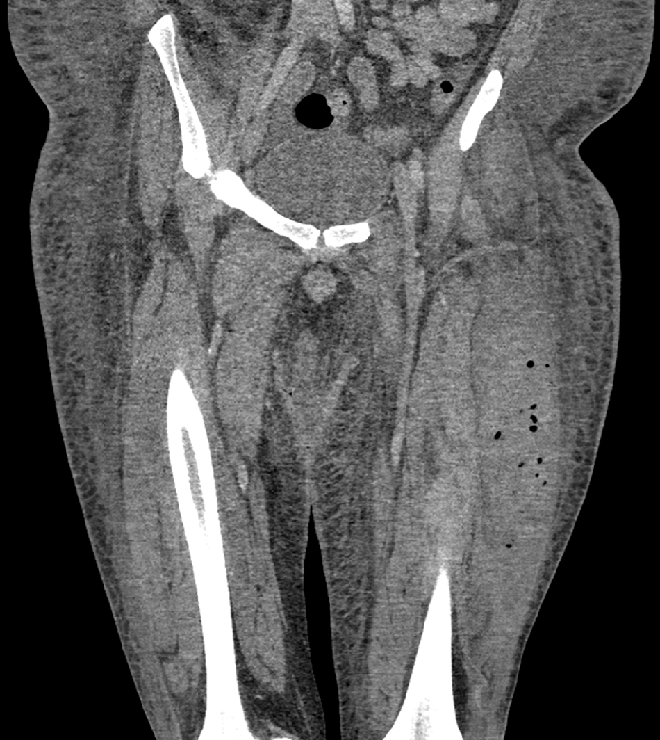
Computed tomography with contrast demonstrating localized left vastus lateralis gas accumulation and enlargement in the left lower extremity.
After emergent surgical exploration, the patient was found to have a non-viable left vastus lateralis in the form of an infected hematoma that was debrided, and its drainage fluid showed evidence of GAS colonization. Furthermore, the bone marrow biopsy and flow cytometry results showed no increase of blasts, confirming that he was in the chronic phase of CML. He was diagnosed with necrotizing myositis and treated with intravenous penicillin G. On post-operative day six, the left thigh was healing well with no signs of infection, and he was expected to have some remaining left quadricep function.
Our case is representative of the rare occurrence of necrotizing myositis that can be overlooked in patients with leukemias who present with leukocytosis and acute limb pain. A high index of suspicion is required for necrotizing myositis that does not typically present with cutaneous findings, because early diagnosis and emergent surgical intervention reduce patient mortality substantially [6].