
Abstract
Background:
Infection is the leading cause of death after thermal injury. Optimal prevention and treatment of burn wound infection is enabled by an in-depth understanding of burn wound treatment modalities not only from a technical standpoint, but also from the standpoint of the clinical context in which these modalities were originally developed.
Methods:
A review of the historical literature on the topical antimicrobial care of burn wounds was performed.
Results:
As our understanding of post-burn infection evolved, and as new products were developed for the prevention of post-burn wound infection, major advances in post-burn survival occurred. Ultimately, improvements in anesthetic, surgical, and critical care management have permitted early excision and grafting of the burn wound, decreasing but not eliminating the importance of topical antimicrobial care, and shifting much of the burden of wound infection prevention to the post-operative period.
Conclusions:
The development of effective topical antimicrobial agents for wound care was, arguably, the single most important advance in the care of the burn patient. Still, many gaps in our ability to treat complicated burn wounds remain. Fungal infection is an unusual but daunting challenge. Patients with impaired wound healing and those with advanced age or medical comorbidities may not benefit from early excision, and the benefits of early excision may not be available in austere or remote locations. For these reasons, research on optimal topical treatment continues.
According to Basil A. Pruitt, Jr. (1930–2019), burns should not be viewed as a unique phenomenon but rather as the “universal trauma model” that exemplifies many of the body's responses to injury, inflammation, and infection [1]. Indeed, the development of topical antimicrobial agents for burn care is a microcosm of the larger campaign to understand and treat surgical infections. The development by Pruitt and colleagues of effective topical antimicrobial agents for the prevention of invasive gram-negative burn wound infection was the single most important step in the history of burn care and led to important and sustained improvements in post-burn mortality. This achievement was marked by the introduction to the bedside of topical mafenide acetate cream (Sulfamylon®, Mylan, Inc., Canonsburg, PA) in January 1964 [2]. This pivotal event was grounded in years of integrated laboratory and clinical research and was followed by continued efforts to address the ever-changing epidemiology of burn wound infection [3]. These efforts are incomplete and continue to this day.
Antisepsis
The modern history of topical antimicrobial wound treatment begins with the English surgeon Joseph Lister (1827–1912), who recognized that micro-organisms, rather than “miasma,” were the cause of wound infection. In 1867 he described “the antiseptic principle in the practice of surgery”:
I arrived…at the conclusion that the essential cause of suppuration in wounds is decomposition, brought about by the influence of the atmosphere upon blood or serum retained within them, and, in the case of contused wounds, upon portions of tissue destroyed by the violence of the injury (…) When it had been shown by the researches of Pasteur that the septic property of the atmosphere depended not on the oxygen, or any gaseous constituent, but on minute organisms suspending in it…it occurred to me that decomposition in the injured part might be avoided…by applying as a dressing some material capable of destroying the life of the floating particles [4].
For this purpose, he selected carbolic acid (phenol), because it was used in the town of Carlisle to reduce the stench of sewage applied as a fertilizer to pasture land, with the additional effect of preventing parasitic infections in the cattle who grazed there [5]. Lister applied it topically to compound fractures and abscess cavities, he used it to cleanse the instruments and the surgeon's gloved hands, and he had an assistant spray it into the air during surgery [6]. Lister avoided the ignominious fate of his Hungarian predecessor, Ignaz Semmelweis (1818–1865), in part because of the contemporaneous work of Louis Pasteur (1822–1895), and in part because of his travels to communicate his findings to surgical audiences in Europe and America. Thus, for example, Lister was able to convert Henry Bigelow (1818–1890), professor of surgery at Harvard University, from a skeptic who referred to Lister as a practitioner of “medical hocus-pocus” [6] to a devoted proponent:
But after two years' experience, I have accepted the new doctrine with most of its details. I have learned that…the duty of the surgeon is to act as if all the particles made visible by a sunbeam were noxious, falling like snow-flakes during every operation and every dressing…His aim should be to destroy the actual intruders, and effectually to exclude their thronging companions [7].
Even so, it is worth noting that Lister in later life discarded the practice of spraying carbolic acid during surgery [8].
The onset of World War I challenged surgeons with injuries of unprecedented severity and number, caused by machine-gun fire and artillery shells during trench warfare. Massive wounds, contaminated field conditions, and delayed evacuation led to a high rate of death from necrotizing wound infections [9]. Under these circumstances, listerian principles were questioned. Antiseptic solutions, applied to the surface of a wound, were incapable of eradicating infection from septic penetrating injuries. Rather, immunologist Almroth Wright (1861–1947) argued that hypertonic saline (5%) dressings should be applied to septic wounds in order to “attract water” from the depths of the wound, thus producing an “outflowing current of water” and “drawing into the tissues from the blood stream lymph inimical to microbial growth.” Thus, his idea was to enhance the body's own antimicrobial processes [10].
Meanwhile, French surgeon Alexis Carrel (1873–1944) and English chemist Henry Drysdale Dakin (1880–1952) developed a refinement of the antiseptic technique. Dakin tested a number of chemicals, settling on 0.5% sodium hypochlorite solution, buffered with boric acid, as the agent of choice. He described the antimicrobial properties of the solution and asserted that clinicians had found it non-irritating to tissues [11]. Carrel tested the solution as one component of what we would nowadays call a “bundle” of care, which included a three-week training program for physicians and nurses, wide incision and debridement of wounds, implantation of tubes into the wound to permit the instillation of the solution, frequent (every two to four hours) infusion of the solution, bacteriologic analysis of wound contents, and delayed wound closure to correspond with resolution of infection [12,13]. Development, documentation, and popularization of the method was accelerated by Carrel's relationship with leading Belgian surgeon Antoine Depage, at whose hospital at Compiègne Carrel oversaw an 80-bed “experimental clinic” [14]. Carrel's method was, however, subject to numerous pitfalls requiring precision in its implementation, and was logistically and technically demanding [15,16].
Antimicrobial Agents
The role of antisepsis in wound care began to be eclipsed during the inter-war years by the development of antibacterial drugs. Paul Ehrlich (1854–1915), the pioneering immunologist and biochemist, had discovered dyes that preferentially stained tissues such as axons in living organisms and other dyes that identified the different categories of granulocytes (neutrophils, basophils, and eosinophils). Perhaps the same concept could be used to target micro-organisms? Methylene blue was only somewhat effective against malaria, and trypan red against Trypanosoma equinum. Ehrlich et al. [17] then turned to arsenicals, synthesizing and testing approximately 1,000 compounds. Their compound 606, arsphenamine (salvarsan), was found to be effective against Treponema pallidum in a rabbit model of syphilis, the first synthetic antibiotic.
Gerhard Domagk (1895–1964], a German pathologist and bacteriologist, built on Ehrlich's work, exploring the antibacterial properties of various dyes at IG Farbenindustrie. He noted that, “For coccal infections, there have been no reasonable effective chemotherapeutants known.” He found that a red crystalline powder related to the azo dyes and synthesized by others in 1932, Prontosil (4′-sulfanamid-2,4-diaminoazobenzene; Bayer AG, Leverkusen, Germany), was curative when given subcutaneously or orally in a lethal mouse model of intra-peritoneal streptococcal infection. (Interestingly, this drug was effective only in vivo but not in vitro; this is because it is what we now call a pro-drug and is metabolized to sulfanilamide.) Also, Prontosil had differential efficacy against different organisms; it was effective against Streptococcus, somewhat against Staphylococcus, and not at all against Pneumococcus. He referred to this selectivity as “elektive Wirkung” [18].
After performing murine studies of streptococcal peritonitis similar to those done by Domagk [19], English physician Leonard Colebrook (1883–1967), a student of Almroth Wright, conducted an uncontrolled clinical trial of Prontosil in puerperal sepsis in 1936. The use of Prontosil resulted in a decrease in the death rate in this disease from 16.6%-31.6% in previous years to 4.7% [20]. This stunning advance heralded the beginning of the age of antibiotic agents.
Another inter-war development was growing interest in improving the care of thermal injuries, but application of the recent advances in microbiology and debridement was delayed by competing theories on burn pathophysiology. The toxemia theory held that the eschar released a toxin into the circulation. The identity and effects of this toxin were ill-defined, which did not deter Davidson [21] from writing in 1925 that the concept was “most strongly supported.” Although some authors described the “early and complete removal of the burned tissue,” Davidson [21] proposed the use of tannic acid to precipitate proteins and other “poisonous materials” in the burn wound. This became a standard first aid treatment for burns through the middle of World War II.
On the other hand, Aldrich claimed that tannic acid merely delayed the onset of infection [22]. He, and others, performed bacteriologic studies of burn wounds, demonstrating that wound cultures became positive for streptococci beginning at approximately the eighteenth hour post-burn [23,24]. Aldrich [22] used a dye-based topical antimicrobial, gentian violet, to prevent streptococcal infection. Later, he added acriviolet and brilliant green to address gram-negative bacteria, thus constituting “triple dye” therapy. He concluded “that the conception of a burn as an infected surgical lesion is correct, and that it is infection rather than the absorption of a split protein which causes death” [23].
When the United Kingdom began experiencing large numbers of burned aviators during the Battle of Britain in 1940, Archibald McIndoe assumed the care of these casualties at the Queen Victoria Hospital in East Grinstead [25]. Among his numerous contributions to the nascent field of burns was condemnation of tannic acid treatment. Not only did it fail to prevent infection, but also “the hard inelastic crust” led to “totally crippled hands and severe facial deformities with loss of vision” [26].
During 1940–1941, the U.S. government stood up two organizations to support medical preparations for war: the Advisory Committees to the Surgeon Generals of the National Research Council (NRC) and the Committee on Medical Research. The NRC's Subcommittee on Surgical Infections funded eight U.S. civilian hospitals to establish wound study units. The original project called for a multicenter controlled study of sulfa drugs for the prevention of infection in burn and non-burn wounds. These plans changed after the attack on Pearl Harbor of December 1941 [27]. Approximately 60% of the more than 500 casualties at the naval hospital had burns, and their treatment varied widely. Despite McIndoe's rejection of tannic acid, it was liberally used there. For example, Flit guns (normally used to deliver insecticide) were used to spray tannic acid onto wounds (Fig. 1). Gentian violet or triple dye, with or without silver nitrate, was also used. In some cases, sulfanilamide powder was mixed in with these substances. Sulfa drugs were also given orally if infection was suspected [28].
Use of a Flit gun to apply a topical agent to a burn patient following the attack at Pearl Harbor, 1941. (Source: Getty Images.)
I.S. Ravdin and Perrin H. Long visited Pearl Harbor after the attack to report back to the NRC on the medical response. They derived an overly optimistic impression of the “incalculable value of sulfonamide therapy,” both oral and topical [29]. As a result, they recommended that, “You cannot withhold from these patients the benefit of the sulfonamide drugs,” and the use of the controls in the eight-center civilian study became optional. The results of that study ultimately were less sanguine and found no clear advantage to sulfa drugs. Referring to staphylococci and gram-negative organisms in the burn wounds, Frank Meleney concluded: “Something must be sought which will be effective at halting the growth of these organisms in the presence of the dead and damaged tissue of burns” [27]. Furthermore, the wound study units found that “tanned burns showed a high incidence of infection,” leading to a recommendation in October 1942 that procurement of tannic acid be discontinued [30].
The limitations of tannic acid and of sulfa drugs gave impetus to the development of penicillin. Alexander Fleming discovered penicillin in 1929, tested it in vitro, and suggested that it could serve as an “efficient antiseptic” when applied topically onto or injected into infected wounds [31]. Eleven years later, the Oxford team of Chain and Florey [32] and others succeeded in producing enough penicillin to permit in vivo studies of efficacy in murine models, and clinical use in a handful of patients [33]. With this evidence in hand, Florey traveled to the United States in 1941 to gain support for mass production and clinical trials. One such clinical study was ready to go under the leadership of Champ Lyons at the Massachusetts General Hospital (MGH) at the time of the Cocoanut Grove Nightclub fire in November 1942 [30,34]. Topical treatment of these patients at the MGH consisted of boric ointment strips and sterile gauze under a pressure dressing. Intravenous sulfadiazine was given prophylactically, and penicillin as a treatment to 13 patients with clinical signs of infection, albeit at doses subsequently recognized as subtherapeutic [34].
As penicillin production ramped up, it was first studied in U.S. combat casualties by Lyons (now an active duty Army officer) at Bushnell General Hospital, Brigham City, Utah, and then at Halloran General Hospital, Staten Island, New York [35]. It was then distributed to British and U.S. hospitals in the Mediterranean theater. Lyons reported from that theater in 1944 that the proper role of antibiotic agents was as an adjunct to, not as a substitute for, surgical management. Furthermore, he rejected the listerian concept of topical antisepsis: “Experience in wound management justifies the abandonment of local use of any chemical agent in a wound” [36]. Despite that caveat, the extraordinary collaboration among military, commercial, academic, and government entities that delivered this new drug to the battlefield is certainly one of the most important achievements of the war, and prepared the way for further post-war antibiotic development [37].
Topical Antimicrobial Agents
The exposure method
In January 1947, Edwin Pulaski moved from the Halloran General Hospital (where early clinical studies of penicillin had been conducted during World War II) to Brooke General Hospital, Fort Sam Houston, Texas, to establish a new surgical research unit (SRU; later the U.S. Army Institute of Surgical Research [ISR]). This unit's mission was “evaluation and interpretation of new antibiotics, chemotherapeutic agents and surgical technics” [38]. Patients admitted to this unit included not only those with burns, but also those with a variety of infected wounds. After 1949, the unit focused increasingly on burns in anticipation of possible nuclear war. Pulaski traveled to Birmingham, United Kingdom, to learn about topical treatment with penicillin cream from Colebrook, but ultimately adopted the “exposure method” from A.B. Wallace at Edinburgh, who claimed that allowing burns to dry out prevented infection and enhanced healing [39–42]. Results of the exposure technique at the SRU were less encouraging. Careful documentation of clinical and microbiologic data in burn patients at the SRU showed that during 1953–1963, there was a continued high prevalence of bacteremia as the cause of death, and a gradual shift in the causative organism from Staphylococcus aureus to Pseudomonas. Control of Staphylococcus aureus was attributed to the successful use of penicillin derivatives. On the other hand, intravenous antibiotic agents were ineffective at controlling Pseudomonas wound infection or bacteremia. Furthermore, 90% of “burn wound sepsis” cases in 1963 had burn wound cultures positive for large amounts of Pseudomonas.
Two divergent approaches were proposed to address this problem. On the one hand, SRU surgeons noted that half of septicemic patients during 1950–1954 had been treated with the exposure method, and half with occlusive dressings. Thus, the method of topical treatment did not seem to matter—particularly in larger burns. This “focuses attention on the need for wound closure at the earliest possible time.” But the “heroic” decision to “excise eschars boldly in stages during the immediate post-burn period is difficult to make.” On the other hand, they developed a model of invasive Pseudomonas aeruginosa burn wound infection in rats, in which they proved the wound origin of fatal post-burn septicemia [43]. The failure of systemic antibiotics to prevent or treat these infections was explained by the avascular nature of the eschar [44].
Mafenide
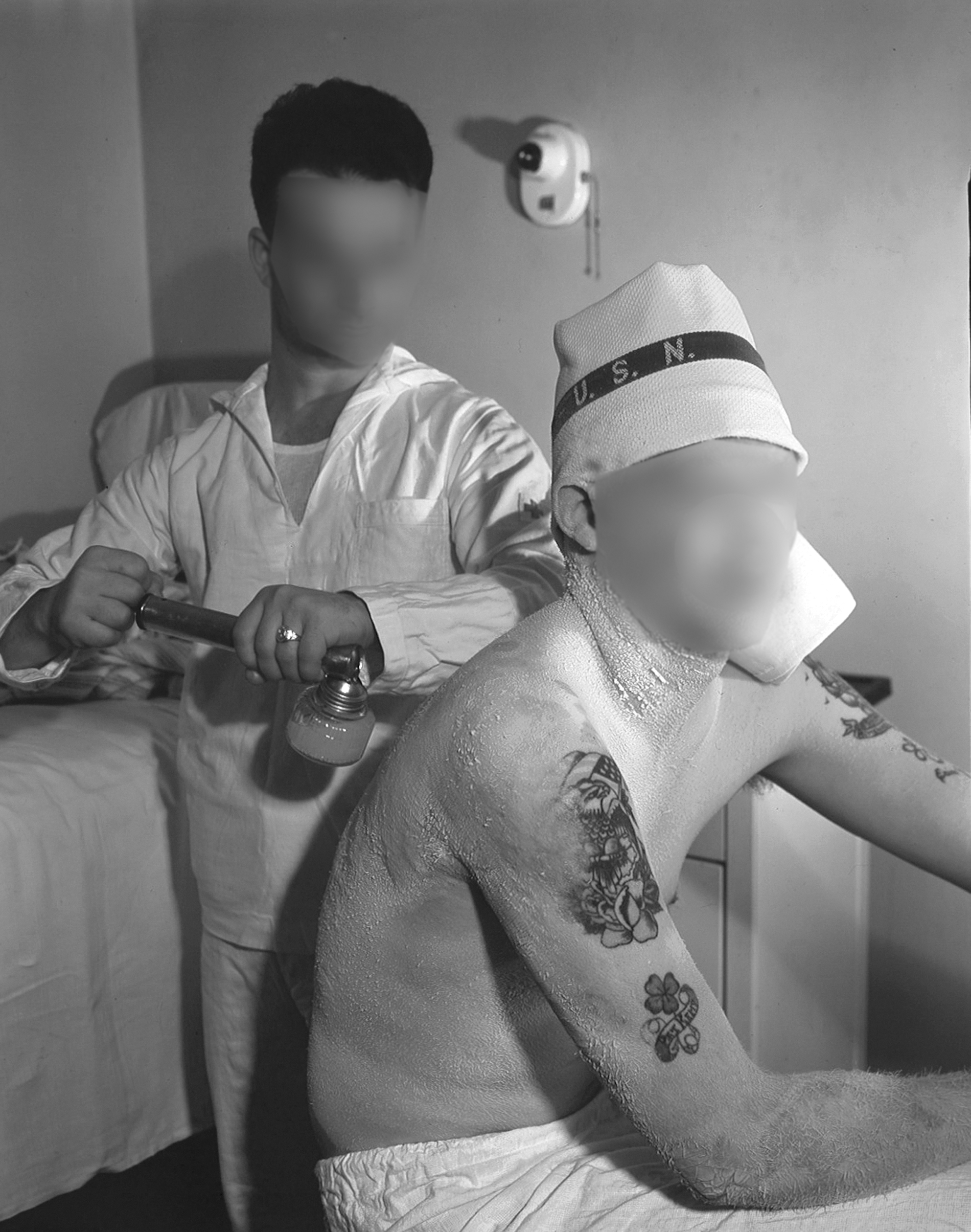
An answer to this problem was suggested by Janice Mendelson, an Army surgeon doing research on blast injury in goats. Fortuitously, a supply of mafenide hydrochloride (Sulfamylon or marfanil; p-aminomethylbenzenesulfonamide) seized from the Germans during World War II was discovered in an Army warehouse in Maryland [45]. This antibiotic had been synthesized in the United States but abandoned because of a report of low efficacy against streptococci [46]. (The murine model used to test efficacy was intra-peritoneal injection of Streptococcus pyogenes, followed by oral treatment [47].) However, it was shown to have better activity against Clostridium perfringens [48]; the Germans issued it to their troops for topical and oral use on the Eastern Front in 1941, and more widely in 1943, mainly in order to prevent gas gangrene [49]. Mendelson and Lindsay [50] demonstrated its efficacy when applied topically (by spray) in a goat model of undebrided blast injury complicated by a (naturally occurring) Clostridium perfringens infection and related these findings to the SRU group. At the SRU, Robert Lindberg and Arthur D. Mason, Jr. (1928–2013) applied Sulfamylon in the form of a cream in the rat burn wound infection model. When applied either immediately or at 24 hours post-burn, the treatment produced a 100% survival rate. Even as late as 72 hours post-burn, there was a mortality effect [44]. This dramatic finding motivated immediate translation to burn patients in January 1964 (Fig. 2) [2]. The results were similarly dramatic: a reduction in burn wound sepsis as the cause of death from 59% (during 1962–1963) to 10% (during 1964–1966) [2].

Col. John Moncrief watches Maj. Basil A. Pruitt, Jr. apply Sulfamylon burn cream to a patient, 1969. (Source: U.S. Army Institute of Surgical Research.)
Despite its structural similarity to sulfanilamide, mafenide's mechanism of action is different. Unlike sulfa drugs, mafenide does not act on bacteria to inhibit the folate precursor, para-aminobenzoic acid (PABA). Furthermore, PABA does not inactivate mafenide, which is important because PABA may be present in large quantities in wounds. Unlike silver preparations, it readily diffuses through the avascular eschar, or through avascular tissues such as cartilage [51]. The latter property makes it ideal for prevention of chondritis in patients with ear burns. On the other hand, this means that it needs to be re-applied twice daily to maintain an effective concentration in the wound [51]. Once absorbed systemically, it is metabolized rapidly to an inactive metabolite, p-carboxybenzenesulphonamide, a compound with no antibacterial activity [52]. However, this metabolite is a carbonic anhydrase inhibitor. Thus, some patients with extensive burns receiving twice-daily Sulfamylon cream treatment may develop metabolic acidosis and compensatory respiratory alkalosis [53].
Silver compounds and dressings
While SRU researchers were evaluating mafenide in the early 1960s, Carl Moyer and colleagues performed studies of silver nitrate (AgNO3) aqueous solution for topical burn wound treatment. Silver nitrate had been used at concentrations of 5%–10% as a tanning agent and had been discarded. Here, however, a 0.5% solution was used. This was based on personal experience by one of the authors with this solution in the treatment of problem wounds such as necrotizing fasciitis. They observed that a 0.5% solution, but not a 1% solution, was safe from the standpoint of not impeding epithelialization. They presented a case series, demonstrating a reduction in wound colonization with pathogens like Pseudomonoas aeruginosa and Staphylococcus aureus. The primary indication of efficacy was a reduction in mortality, from a predicted 41% to an observed 14% [54].
Moyer et al. [54] emphasized the importance of using a thick gauze dressing and of keeping the dressing moist continuously. That is, as the water evaporates from the dressing, the concentration of AgNO3 remaining at the wound surface was thought to increase past the safe level. The cause of death while receiving AgNO3 was related to electrolyte abnormalities. As a hypotonic solution, silver nitrate “leeches” electrolytes across the wound surface. This can cause life-threatening levels of hyponatremia, hypokalemia, and hypocalcemia, mandating frequent laboratory determinations (at least every six hours). Also, use of this solution can cause hypothermia caused by evaporation [54]. The primary limitation of AgNO3 is its inability, unlike mafenide, to penetrate the eschar. However, this limitation does not apply in the treatment of superficial injuries, or of non-burn desquamating skin diseases such as toxic epidermal necrolysis syndrome; the latter is the primary indication for its use at the U.S. Army Burn Center at this time.
The technical challenges associated with the use of silver nitrate led Fox to develop silver sulfadiazine (SSD), a complex of silver nitrate and the antibiotic sulfadiazine. When mixed with chloride-containing fluids on the surface of a burn wound, this complex releases the silver cation. In scalded mice with otherwise lethal Pseudomonas aeruginosa wound infections, SSD appeared to reduce mortality compared to both silver nitrate and mafenide [55]. The primary mechanism of action of SSD relates to the silver, rather than to the sulfadiazine. In contrast to silver nitrate solution, release of silver from the silver sulfadiazine complex is slow and steady [56]. Sometimes, SSD is associated with leukopenia, which resolves within a few days despite continuation of therapy [57].
The first silver textiles used as dressings in the modern era were made from nylon and were originally intended to serve as flexible electric shields or radar reflectors [58]. Deitch et al. [59] demonstrated in vitro efficacy of silver nylon against Pseudomonas aeruginosa, Stapylococcus aureus, and Candida albicans. Chu and McManus [58] at the ISR embarked on a multiyear study of silver nylon dressings in rat models. Interestingly, they demonstrated greater efficacy when the dressing was connected, as an anode, to a weak direct current source, causing increased release of silver cations onto the wound. Under these conditions, silver nylon was as effective as SSD [60]. Today, silver nylon is used extensively in burn care, especially in superficial burns and in clean, deeper burns of limited extent [61]. A variety of other silver dressings have been developed, to include those made with nanocrystalline silver (Acticoat, Smith+Nephew, Fort Worth, TX) [62], soft silicone combined with foam (Mepilex Ag®, Molnlycke, Gothenburg, Sweden) [63], moisture-retentive spun fibers of sodium carboxymethyl-cellulose (Aquacel Ag, ConvaTec, Oklahoma City, OK) [64], and ceramic silver (Milliken Assist, Milliken, Spartanburg, SC).
The mechanism of action of silver is “oligodynamic,” meaning that small amounts (parts per million) of silver are required for efficacy. Elemental silver is biologically inert; only the cation (Ag+) is antimicrobial. Proposed mechanisms of action include: (1) blocking the microbial electron transport chain; (2) rupturing the cell membrane or wall; (3) binding to and damaging bacterial DNA (vulnerable because of its location in the cytoplasm); and (4) destroying the cell by silver free radicals. Antimicrobial resistance to silver is uncommon, thus, the mechanism of action is likely multifactorial, such that evolution of multiple resistance strategies would be required. A disadvantage of silver therapy is the fact that silver cations can be precipitated on the wound surface by anions such as chloride. This means that the effective concentration in vivo is many times higher than that in aqueous solutions. It also means that silver, unlike mafenide, does not penetrate deeply into a wound. Systemic silver toxicity (argyria) is extremely rare in burn care [65]. An advantage of silver over mafenide is that the former has some antifungal activity, particularly against yeasts, whereas the latter has none.
Cerium
In 1977, Monafo, who with Moyer had described the use of silver nitrate, treated patients with a combination of cerium nitrate and SSD. Cerium is a rare-earth metal of low toxicity. Monafo originally stated that cerium is an antimicrobial, although subsequent studies have not substantiated that claim [66]. Rather, cerium nitrate results in a firm eschar that protects against bacterial ingress [67]. The eschar is tightly adherent without a tendency to spontaneously separate from the burn wound for at least six weeks [68,69]. Cerium also appears to exert a beneficial effect on host immune function. In animal studies, cerium nitrate treatments decrease local and systemic inflammation [70,71], limit burn wound progression [72], and restore the helper-to-suppressor T-cell ratio [73,74]. Moreover, cerium nitrate plus silver sulfadiazine treatment—but not mafenide, silver nitrate, or silver sulfadiazine alone—mitigates cell-mediated immunity suppression in a mouse model [75].
It is not clear why cerium nitrate has these immune benefits. Martin Allgöwer (1917–2007), a Swiss surgeon best known as the co-inventor of the AO system for internal fixation of fractures, proposed (in a manner reminiscent of the toxemia theory), that burn wounds release lipid-protein complexes (“pernicious effectors”) that cause immunosuppression and systemic illness. Allgöwer and colleagues stated that cerium nitrate binds to these complexes and mitigates these effects. Other authors demonstrated superficial connective tissue calcification [69], which may result from cerium displacing calcium from pyrophosphate to allow calcium to deposit, analogous to the pyrophosphate-calcium interaction within cancellous or cortical bone [67]. At the ISR, Kai Leung and colleagues have shown in the Walker-Mason full-thickness scald-burn model that cerium nitrate solution (40 mM) treatment, as compared to a control solution, reduces circulating levels of pro-inflammatory damage-associated molecular patterns (DAMPs) such as high-mobility group box protein 1 (HMGB1), hyaluronan, and xanthine oxidase (XDH) on post-burn day one. On post-burn day seven, circulating DAMP levels were on average halved by cerium nitrate treatment, as were levels of cytokines interleukin (IL)-1β and IL-10 and chemokines GRO-KC and MIP-1α in wound tissue [70]. At present, cerium nitrate with SSD is available in several countries as Flammacerium (Derma UK Limited, Newcastle upon Tyne, UK), and has received orphan drug status in the United States (it is not yet Food and Drug Administration-approved).
Honey
Recently, there has been growing interest in the antimicrobial properties of honey. Honey has been used since ancient times as a topical antimicrobial and has been proposed as an alternative under austere or battlefield conditions [76]. Medical-grade honey is now available throughout the world. The mechanism of action of honey as an antimicrobial is multifactorial. Honey is hyperosmolar and has a low pH. Most honeys, because of the action of glucose oxidase, produce hydrogen peroxide when diluted with water. Some honeys contain specific antibacterial agents. For example, manuka honey (from the flowers of the Leptospermum tree of New Zealand) contains methylglyoxal, an alpha-oxoaldehyde that reacts with DNA, RNA, and proteins. This variety of honey has a broad range of antibacterial activity, low mammalian toxicity [77], and (to date) low likelihood of inducing resistance [78]. Honey also shows promise as an antifungal agent [79]. A recent Cochrane Review found that the studies of honey for burns treatment are of low or very low quality. The studies are also difficult to interpret because the comparator treatment is often one that is not widely used [80]. Despite the limitations of these studies, we now commonly use medical honey and honey dressings, particularly in the care of problem wounds.
Clinical Experience and Conclusions
A review of burn patient data from 1950–1999 permitted analysis of the effects of topical mafenide and SSD on survival at the ISR Burn Center (Fig. 3) [81]. The introduction of mafenide into burn care in 1964 resulted in a decrease in age- and burn-size-adjusted mortality risk. This was followed by an increase in mortality in the latter half of the 1960s, as resistant strains of Providencia stuartii and Enterobacter cloacae appeared, and sepsis became more common [82]. In 1973, SSD was introduced, and improved control of sepsis and mortality were gradually achieved. Briefly, SSD was used as the sole antimicrobial agent; later, it was alternated every 12 hours with mafenide (a practice called alternating agents). Throughout this period, typical burn treatment was as follows. Daily hydrotherapy and topical antimicrobial care were performed until the burns either healed or the eschar separated. Then, cadaver allografts were applied, and finally autografting was performed. Starting in 1978 and following the pioneering work on the tangential excision by Janzekovic [83], excision of extensive burns was introduced. A new burn unit was built, and better isolation procedures were instituted in 1983 [84], resulting in further improvements in mortality.

Age- and burn-size adjusted log-odds of mortality following thermal injury at the U.S. Army Institute of Surgical Research (U.S. Army Burn Center), 1950–1999 (Source Reference [81].)
The role and relative importance of topical antimicrobial agents in burn care has since evolved. Today, patients with smaller burns are readily treated with silver dressings or with synthetic bilaminar skin substitutes (Biobrane®, Smith+Nephew; Permeaderm, Milliken). The latter have been shown to hasten healing in comparison with SSD [85]. Total-body excision of patients with large burns is completed as soon as possible from a physiological standpoint, often within days of injury [86]. Usually, these wounds are immediately closed with autograft, or where insufficient donor sites are available, a combination of widely meshed autograft covered with an allograft overlay. Under these conditions, topical antimicrobial treatment is a temporizing intervention. The major problem becomes the care of the post-operative wound while it heals, and the prevention or early detection and treatment of post-operative infections. In this regard, fungal wound infections remain a particular menace which independently increase post-burn mortality [87], and for which no good preventative measures exist.
On the other hand, some patients are not candidates for early excision and grafting. These include those with concomitant medical diseases or advanced age. Other patients may live in countries where modern surgical techniques are not readily available. In a mass-casualty or military setting, rapid excision may be logistically impossible. In the military setting, we continue to recommend the use of alternating agents (mafenide and SSD) for the treatment of unexcised wounds [88]. Furthermore, Flammacerium or other cerium-based treatments may be applicable as well. In conclusion, effective management of the burn wound, to include prevention of infection and successful wound closure, remain the keys to survival in burn patients [89].
Footnotes
Acknowledgments
The author gratefully thanks Mr. Glen E. Gueller, Records and Knowledge Manager (retired), and Ms. Susan Reyna, Library Assistant, both of the Information Management Division, U.S. Army Institute of Surgical Research.
Funding Information
The author reports no funding for this work.
Author Disclosure Statement
The opinions or assertions contained herein are the private views of the author, and are not to be construed as official or as reflecting the views of the Department of the Army or the Department of Defense.