
Abstract
Background:
It is widely acknowledged that pathogenic germs delay wound healing to some extent. To explore factors influencing the wound healing process, the current study was conducted to evaluate the antibacterial effect of topical application of copper sulfide nanoparticles (CuS NPs) in vitro and on infected wound healing process in the rat model.
Materials and Methods:
In this study, the morphology and size of CuS NPs were detected. Staphylococcus aureus and Escherichia coli were used so that the antibacterial ability of CuS NPs could be evaluated better. In addition, a 2-cm circular full-thickness wound infected with a solution of 107 colony forming units (CFU) Staphylococcus aureus was created on the back of each rat. The rats were divided into four groups including the control group, the 100 mcg/mL CuS NPs group, the 250 mcg/mL CuS NPs group, and the 500 mcg/mL CuS NPs group. Tissue bacterial count and histologic assessment were evaluated.
Results:
The results indicated that CuS NPs had antibacterial activity against Staphylococcus aureus and Escherichia coli. Moreover, they could decrease the incidence of bacterial colonization and promote wound healing through re-epithelialization and collagen deposition. Furthermore, CuS NPs could maintain Cu2+ continuous release and inhibit the viability of Staphylococcus aureus through lipid peroxidation.
Conclusions:
This study found that CuS NPs have fine antibacterial properties, and particularly, the 500 mcg/mL CuS NPs had better effects, without increase of side effects. They could promote infected wound healing, the prospective clinical application of which was further confirmed in the treatment of wound infection.
Wound healing is quite a well-orchestrated and regulated process involving the following four interrelated stages: hemostasis, inflammation, proliferation, and remodeling [1]. It is commonly known that these stages overlap because of several anatomic and physiologic factors in vivo. The physiology of the healing process may be affected by both external and internal factors [2]. Furthermore, the most frequently encountered issue in wound closure is colonization and replication of bacteria in the wound site. Therefore, dealing with an infected wound is a challenging clinical problem associated with high costs of medical treatment and increased risk for life [3,4]. Until now, it has been proved that antibiotic agents are effective to prevent bacteria from breeding, however, overuse, misuse, and improper disposal of antibiotic agents could lead to an increase in antibiotic resistance [5,6]. Moreover, treatment of drug-resistant bacteria often occurs with a higher dose of antibiotic agents as well as various expensive drugs that may be less effective, even more toxic, and cause undesirable side effects [7,8]. Thus, it is important to develop an alternative therapeutic modality to decrease the incidence of infection and promote wound healing.
Currently, nanomaterials have received extensive attention because of their unique properties, which are recognized as powerful materials to kill germs. Previously, some researchers have reported the positive effects of copper nanoparticles (Cu NPs) in wound healing, such as the stabilization of the expression of hypoxia-inducible factor-1α, extracellular skin proteins, and the secretion of vascular endothelial growth factor [9-11]. In addition, they are also known as effective antimicrobial materials, which could promote wound healing through the reduction of the incidence of infections. However, what is rarely available to people is their highly toxicity in vivo [9]. Despite this disadvantage, as an easily prepared and stable material, copper sulfide nanoparticles (CuS NPs) appear to have good biocompatibility, low toxicity, and fairly antibacterial activity [12]. Thus, CuS NPs have been considered as a prospective agent in the fields of photothermal cancer therapy, molecular imaging, and biomolecule sensing [13]. When taking an overall view of research about CuS NPs, it was found that there were some reports regarding the antibacterial activity of CuS NPs, but the most of them are about basic evaluations [12,14]. Until now, the effect of CuS NPs on infected wound healing and its safety have been rarely reported. Therefore, a study from this perspective appears to be essential and beneficial.
In this study, CuS NPs were synthesized to evaluate antibacterial activities in vitro and in vivo. At the same time, the effect of CuS NPs on infected wound healing was investigated. Furthermore, the antimicrobial mechanism was studied.
Materials and Methods
Reagents
Copper chloride dehydrate (CuCl2·2H2O) was purchased from BBI Life Science Corporation (Shanghai, China). Sodium sulfide (Na2S) was obtained from Sigma-Aldrich (St. Louis, MO). Sodium citrate (C6H5Na3O7) was obtained from Adamas (Shanghai, China).
Synthesis of CuS NPs
Copper sulfide nanoparticles were prepared according to a previously reported method [15] Fifty milligrams of CuCl2 ·2H2O and 50 mg of C6H5Na3O7 were mixed with 30 mL of deionized water in a beaker under stirring for five minutes and then 1 mL of Na2S (50 mg) was added drop by drop, and the mixed solution was incubated for 12 hours under stirring at room temperature to prepare CuS NPs. After centrifugation and washing, the product CuS NPs were freeze-dried for further use.
Characterization of CuS NPs
The hydrodynamic diameters and the morphology of CuS NPs were evaluated by dynamic light scattering spectrometer (DLS; Nano-ZS90, Malvern Panalytical, Malvern, UK), scanning electron microscopy (SEM; S-4800II, Hitachi, Tokyo, Japan), and transmission electron microscopy (TEM; Tecnai 12, Philips, Eindhoven, The Netherlands), respectively. The structure and composition of the CuS NPs were respectively detected through high-resolution TEM (HRTEM; Tecnai G2 F30 S-TWIN, FEI, Hillsboro, OR) and x-ray photoelectron spectroscopy (XPS; ESCALAB 250Xi, Thermo Fisher Scientific, Waltham, MA). In addition, the microstructure of CuS NPs was observed by using x-ray diffraction (XRD; D8 Advance, Bruker-AXS, Karlsruhe, Germany).
Bacterial culture
In this study, Staphylococcus aureus and Escherichia coli were used to evaluate the antibacterial property of CuS NPs. The lysogeny broth medium (5 g/L yeast extract, 10 g/L tryptone, and 0.5 g/L sodium chloride) was used to culture bacterial cells, which was done under shaking for approximately 12 houre at 37°C to achieve a preliminary bacterial suspension. The bacterial cells were then entered into log phase by re-seeding the bacterial suspension into the fresh medium at a 1:100 proportion, and were cultivated for two to three hours in a shaking incubator at 37°C until the optical density reached 0.7–0.8 at 600 nm (OD600).
Antibacterial effect of CuS NPs
The antibacterial abilities of the CuS NPs were determined via the number of colony-forming units (CFU) by using the plate counting method. In this study, four groups of bacteria were tested: bacteria; bacteria plus CuS NPs (100 mcg/mL); bacteria plus CuS NPs (250 mcg/mL); and bacteria plus CuS NPs (500 mcg/mL). Briefly, monocolonies of Escherichia coli (CMCC (B) 44102) and Staphlococcus aureus (ATCC 29213) were cultured following the above methods, then were diluted 10 times, with 100 mcL of diluted bacteria then added to four 1.5-mL centrifuge tubes, respectively. The final concentration of bacteria was 1.0 × 10⁷–1.0 × 10⁸ CFU/mL in the sodium acetate buffer (0.1 M, pH 4.5). The mixed suspension was then incubated under shaking for one hour at room temperature, and 100 mcL of the above suspension was spread evenly on solid agar medium to determine CFU.
Determination of internal malondialdehyde
As a natural product of lipid oxidation in organisms, malondialdehyde (MDA) can be used to detect the level of lipid oxidation. Bacteria were treated with CuS NPs for one hour, and then were lysed by lysozyme and proteinase K for MDA measurement at 4°C. After lysation, the supernatant was centrifuged at 10,000g to 12,000g for 10 minutes to determine lipid peroxidation by using a Micro-MDA Assay Reagent Kit (KeyGEN Biotech, Nanjing, China).
Cu2+ release from CuS NPs in vitro system
Five milligrams of CuS NPs were separately dispersed in 3 mL of pH 4.5 sodium acetate buffer (0.1 M), and were then placed into a dialysis bag (cutoff molecular weight: 3,500 Da). The dialysis bag was placed in 30 mL sodium acetate buffer under stirring conditions (37°C; 100 rpm). Partial release medium (3 mL) was removed for inductively coupled plasma mass spectrometry testing at given times (Elan DRC-e, PerkinElmer, Waltham, MA), followed by the addition of free-release medium (3 mL) for continuous observation of release behavior.
Cell culture and cytotoxicity evaluation
Human dermal fibroblasts were cultured in Dulbecco modified Eagle medium with 10% fetal bovine serum, 100 U/mL penicillin, and 100 mcg/mL streptomycin, which were maintained at 37°C in a humidified incubator with a 5% carbon dioxide atmosphere. Cells were seeded in triplicates in 96-well plates (1 × 10⁴ cells in 100 mcL medium per well) and were treated with different concentrations of CuS NPs for 24 hours. Then, 10 mcL CCK8 was added to each well and incubated at 37°C for two hours. Optical density at 450 nm was measured by using a TECAN Infinite 200 (Tecan Group, Männedorf, Switzerland).
Animals
Animal experiments were carried out following the guidelines for the care and use of experimental animals issued by the National Institutes of Health. Ethical approval was approved by the Ethics Committee of the Clinical Medical College of Yangzhou University (Yangzhou, China).
Male Wistar rats (n = 32; with body weight of 200–220 g) were purchased from Yangzhou University (Yangzhou, China). The rats were allocated equally into four groups (control group; 100 mcg/mL CuS NPs group; 250 mcg/mL CuS NPs group; and 500 mcg/mL CuS NPs group) and were transferred to the laboratory one week before the study. We found that obvious bactericidal effect was produced by using 100 mcg/mL CuS in our pre-experiment, so we chose 100, 250, and 500 mcg/mL to conduct further research.
More specifically, the rats were divided into four groups: deionized water; CuS NPs (100 mcg/mL); CuS NPs (250 mcg/mL); and CuS NPs (500 mcg/mL) with eight rats in each group. Considering that Staphylococcus aureus is the main pathogenic bacteria in clinical wound infection, we chose the wound model infected by Staphylococcus aureus. In brief, a wound was surgically created on the back of each rat and covered with 1 × 10⁷ CFU of Staphylococcus aureus after anesthesia to establish an infected wound model. After infection for 24 hours, 100 mcL deionized water or CuS NPs solutions were dropped onto the wound area in the corresponding groups once per day until day seven. Death and wound size were recorded at different points. After three days of treatment, two rats were chosen randomly from each group and euthanized with pentobarbital overdose. The infected wounds tissues were collected and homogenized to evaluate their antibacterial activities in vivo by detecting bacterial amounts.
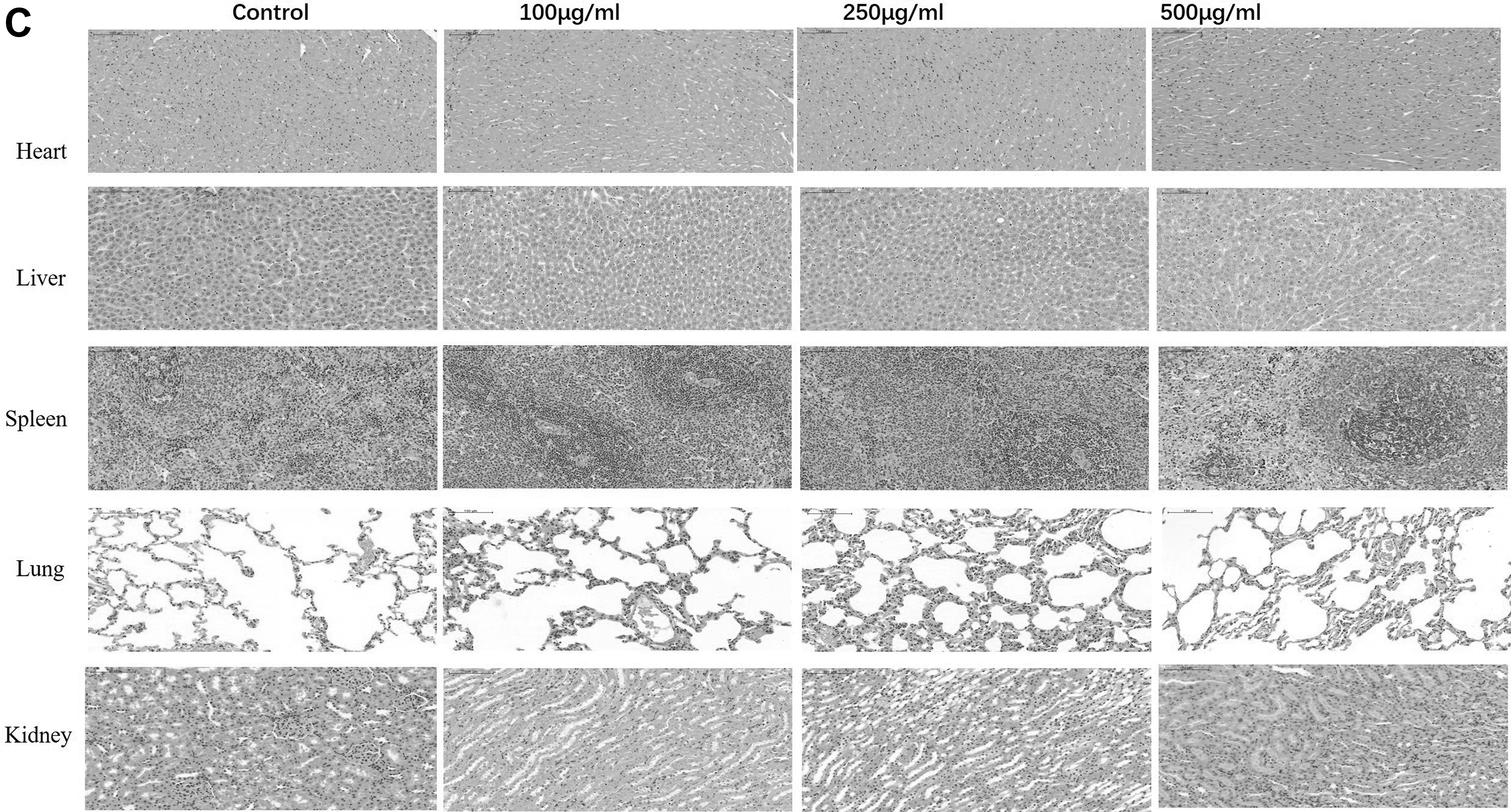
On day 21, blood tests of aspartate transaminase (AST), alanine transaminase (ALT), blood urea and nitrogen (BUN), and serum creatinine (SCr) were performed on rats from all groups to assess the toxicity of the CuS NPs to the liver and kidneys. For histologic analysis, the remaining rats were euthanized by intravenous injection of pentobarbital. The wound and the samples of heart, liver, spleen, lung, and kidney were fixed in formalin and paraffin was used to embed the tissues. A 4-mcm continuous cross section was taken and stained with hematoxylin and eosin. Next, the collagen density of wound tissue slices was observed by Masson trichrome. Finally, the tissue sections were detected by a Nikon Eclipse Ci microscope (Nikon, Tokyo, Japan) in a bright-field mode.
Statistical analysis
Data were shown as mean ± standard deviation. Statistical analysis was performed using t test, and one-way analysis of variance (ANOVA; *p < 0.05, **p < 0.01, ***p < 0.001, and ****p < 0.0001.
Results and Discussion
Morphologic and structural characterization of CuS NPs
As shown in Supplementary Figure S1, the morphology and size distribution of CuS NPs were detected by using TEM (Supplementary Fig. S1A) and SEM (Supplementary Fig. S1B). It shows that dispersed nanoparticles are more or less spherical and some aggregate of small grains. The size of the particles ranges from 20 to 60 nm.
The DLS data showed that the CuS NPs had an average diameter of approximately 68.1 nm, as shown in Supplementary Figure S1C. The diameter of CuS NPs detected by DLS was larger than that observed by TEM and SEM. It is believed by us that CuS NPs trend to aggregate to form larger nanoparticles with more stability. To verify the copper phase on the CuS NPs, XRD was conducted, as shown in Supplementary Figure S1D. The CuS NPs were indexed to standard CuS [16].
Using XPS spectra (Supplementary Fig. S1E) it was demonstrated the presence of copper, sulfur, carbon, oxygen, and nitrogen elements in the CuS NPs that were mainly composed of carbon (18.01 atom %), oxygen (23.32 atom %), nitrogen (3.39 atom %), sulfur (26.39 atom %), and copper (28.89 atom %). The carbon, nitrogen, and oxygen peaks indicated successful coating of sodium citrate on the surface of the CuS NPs. The Cu 2p peaks, because of Cu 2p3/2 and Cu 2p1/2, appeared at 931.06 and 950.92 eV (Supplementary Fig. S1F), respectively, corresponding to Cu (II) [17]. The composition of the CuS NPs was observed by HRTEM as shown in Supplementary Figure S2.
Antimicrobial bioassays in vitro
To evaluate the antibacterial activity of CuS NPs against gram-positive (Staphylococcus aureus) and gram-negative (Escherichia coli) bacteria, both bacteria were cultured with different concentration of CuS NPs for 12 hours at 37°C. As shown in Figure 1A, with the concentration of CuS NPs increased, the antibacterial efficacy of CuS NPs was also increased correspondingly. Figure 1B and 1C illustrate the impact log reductions.
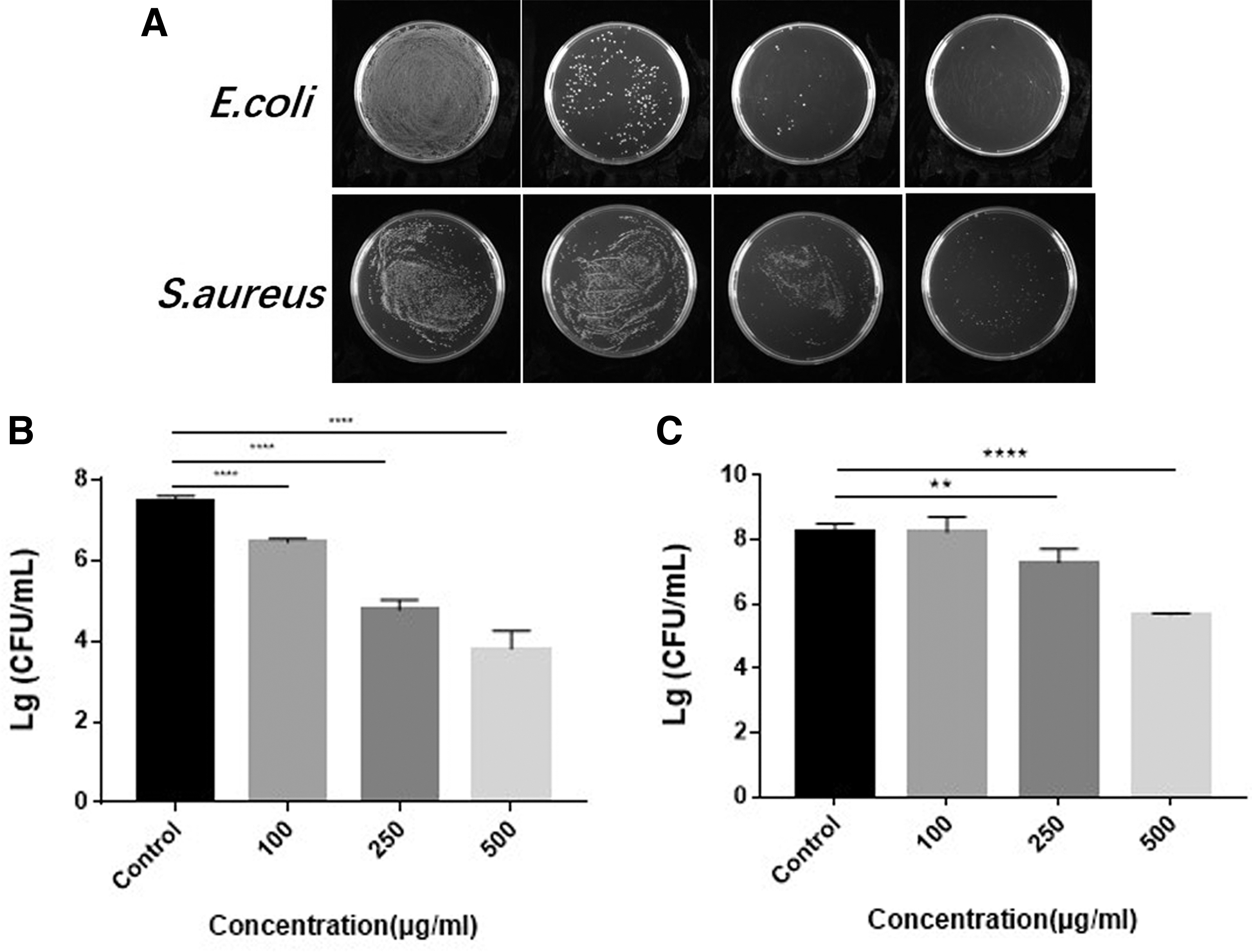
Antibacterial effect in vivo of copper sulfide nanoparticles (CuS NPs) cultivation overnight. (
Determination of internal MDA
Lipid peroxide radical is one of the proposed antibacterial mechanisms of metal nanoparticles. For an intensive study on the interaction between CuS NPs and bacteria, the production of lipid peroxide radical was investigated by measuring the production of MDA. The MDA content in the 500 mcg/mL group was 6.32 ± 0.14 mcg/mL, which was higher compared with the 250 mcg/mL group (2.38 ± 0.03 mcg/mL; p < 0.05), the 100 mcg/mL group (1.88 ± 0.04 mcg/mL; p < 0.05), and the control group (0.17 ± 0.02 mcg/mL; p < 0.05). The MDA content in the 250 mcg/mL group was higher than those in the 100 mcg/mL group (p < 0.05) and the control group (p < 0.05). The MDA content in the 100 mcg/mL group was also higher compared with the control group (p < 0.05). It was also found that the MDA content increased in a dose-dependent manner. The results of one-way ANOVA are shown in Table 1, and the statistical analysis of MDA contents of each group is shown in Figure 2A.
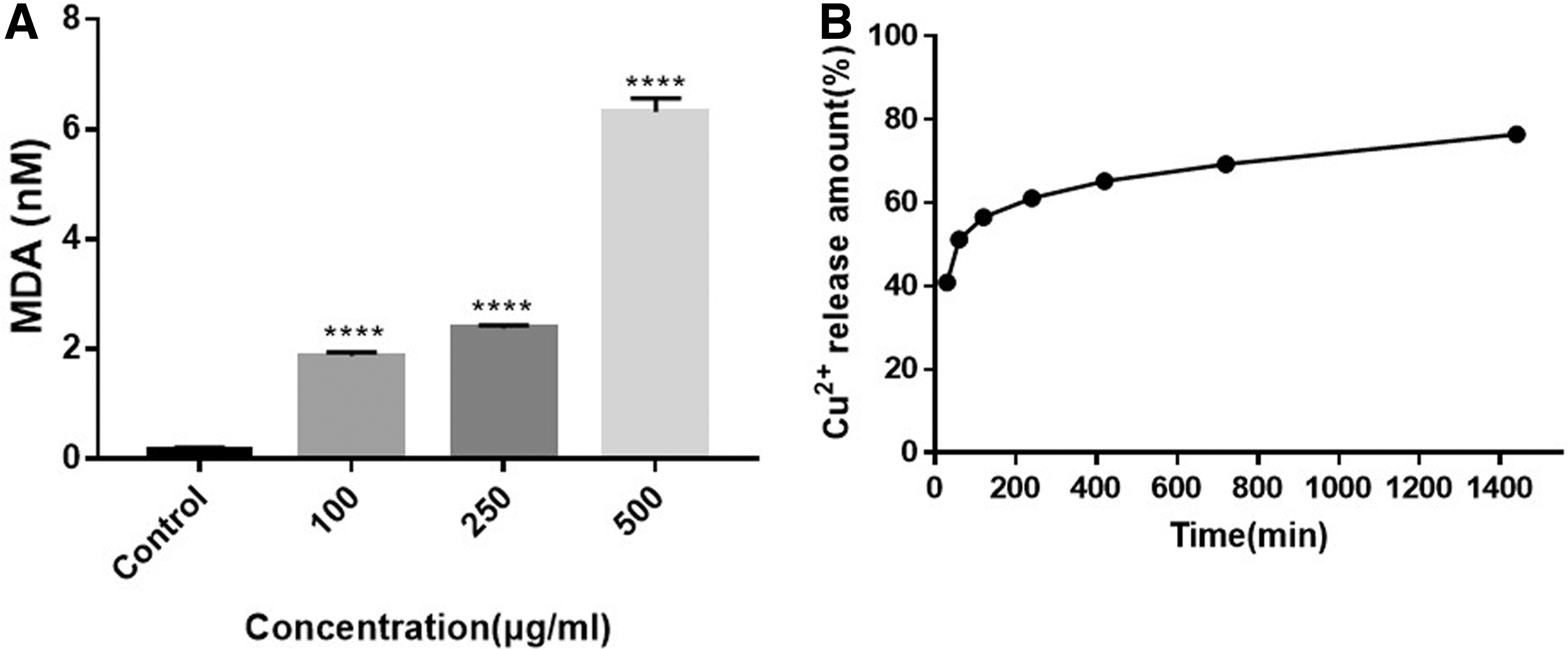
Bacteria-killing mechanism of copper sulfide nanoparticles (CuS NPs). (
Results of One-Way ANOVA
ANOVA = analysis of variance; SS = sum of the squares of the data; DF = degree of freedom; MS = mean sum of the squares of the data; F = F-statistic; DFn = degrees of freedom error; DFd = degrees of freedom in the denominator.
Cu2+ release
The previous study showed that Cu2+ release could damage the structures of DNA and enzymes, thus, it could disrupt the cell membrane and effect biochemical property [18]. Therefore, we initially dispersed 5 mg of CuS NPs in 3 mL of sodium acetate (0.1 M, pH 4.5), and then placed it into a dialysis bag (cutoff molecular weight: 3,500 Da). The concentration (0.917 mg/mL) of Cu2+ species in buffer at pH 4.5 after contact for 24 hours with CuS NPs is shown in Figure 2B. A sustained release of Cu2+ was observed over 24 hours, and the percentage released result was approximately 76%. The antibacterial activity may be attributed to release of Cu2+, which corresponds with previous studies [18,19].
In vivo animal experiments
After the verification of the antibacterial activity of CuS NPs, we set out to investigate its therapeutic efficacy on Staphylococcus aureus infection in the rat model. To evaluate the antibacterial effects in vivo, different treatments were conducted in rats with infected wounds on their back. No accidental death occurred during this experiment. We photographed the wound on days zero, one, three, five, seven, 14, and 21 to determine the change in the wound size and the status of wound healing, and potential side effects were evaluated through histologic methods on day 21. Figure 3A shows wound closure at different time points (days zero, one, three, five, seven, 14, and 21). During the experiment, all groups showed severe bacterial infection on day one. Despite the fact that the wound was infected, the 500 mcg/mL CuS NPs and the 250 mcg/mL CuS NPs groups had faster wound closure than the 100 mcg/mL CuS NPs group and the control group. As shown in Figure 3B, wound size treated with 500 mcg/mL CuS NPs and 250 mcg/mL CuS NPs became smaller and healed better than other groups on day 21. However, the wound size in the 100 mcg/mL CuS NPs group was almost the same as that in the control group. To determine whether CuS NPs prevented the rats from infection, the infected wound tissues of the rats were homogenized and appropriately diluted in phosphate-buffered saline and then spread on agar plates. The agar plates were incubated at 37°C overnight and then the number of bacterial colonies in each group was measured. The quantitative analysis of in vivo antibacterial effects is presented in Figure 3C and Figure 3D. Compared with the control group, the 100 mcg/mL CuS NPs group, and the 250 mcg/mL CuS NPs group, the number of bacteria in the wounds of the 500 mcg/mL CuS NPs group were inhibited substantially. This indicates that CuS NPs could reduce the microbes and save the rats from infection.
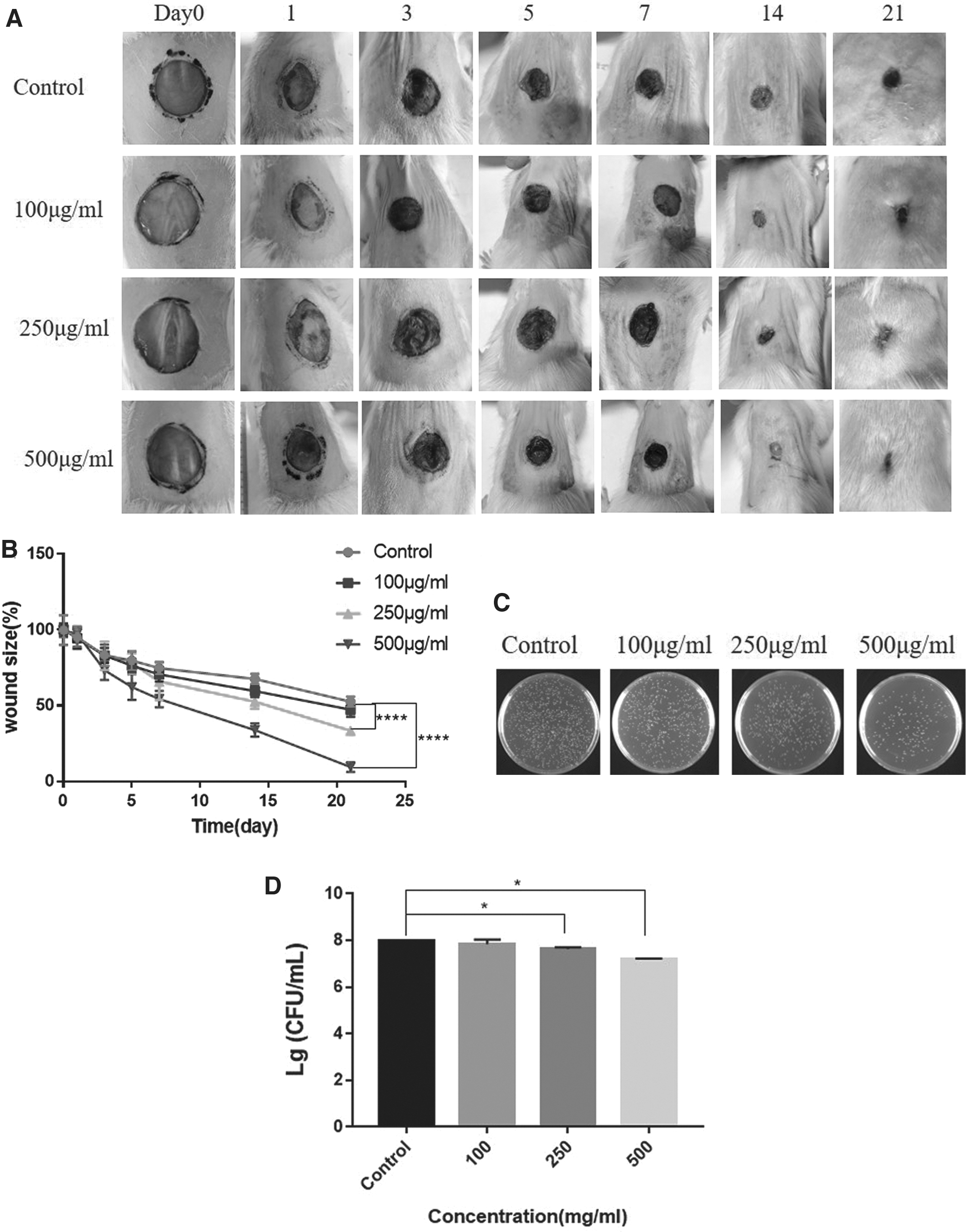
The effects of copper sulfide nanoparticles (CuS NPs) on Staphylococcus aureus-infected wounds and wound healing in rats. (
On day 21, wound recovery was observed, and tissue samples were taken from the wound to prepare pathologic analysis. Epithelial tissues formed with a papillary structure and collagen deposition, which was always used to investigate wound healing [20]. Hematoxylin and eosin-stained sections (Fig. 4A) demonstrated that most wounds had some degree of epidermal covering. The 500 mcg/mL CuS NPs-treated wounds displayed mature epithelialization with rete pegs and the 250 mcg/mL CuS NPs-treated group showed moderate epithelialization. However, epithelialization was slightly observed in the 100 mcg/mL CuS NPs group and the control group, and the wounds in these two groups appeared to contain numerous inflammatory cells.
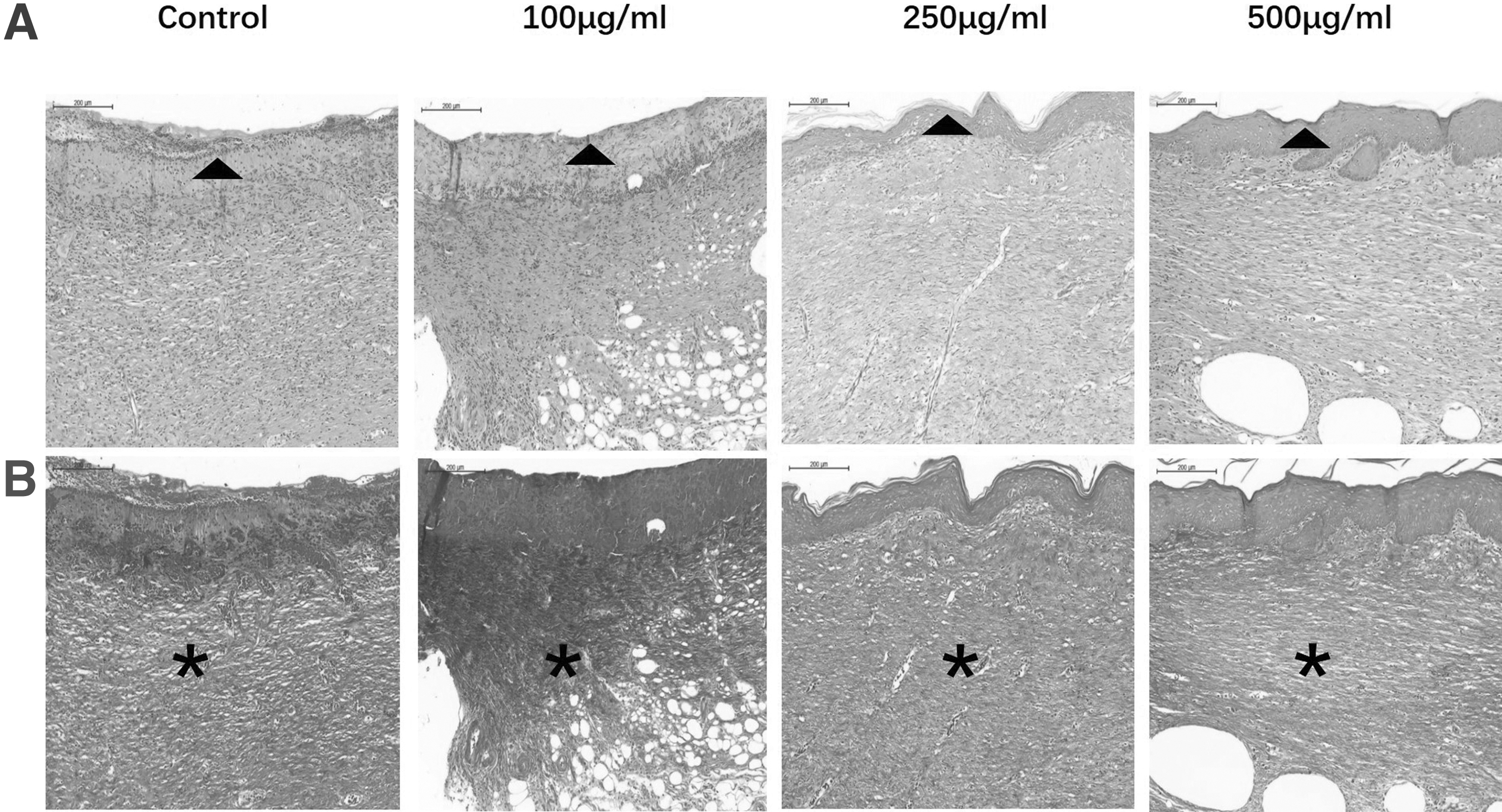
Histologic analysis of wound healing of different groups at day 21 (scale bar = 200 mcm). (
The dermis of nature skin consists of large, organized fibers. As shown in Figure 4B, the dermis of the 100 mcg/mL CuS NPs-treated and the control group wounds had some unorganized large fibers present in the dermis by morphologic appearance. The dermis of the 250 mcg/mL CuS NPs-treated wounds showed increased presence of organized fibers. The 500 mcg/mL CuS NPs-treated wounds with highly organized fibers and minimal unorganized small fibers seemed to be similar to the healthy skin.
Pilot toxicity study
Regarding cytotoxicity of CuS NPs in vitro, the CCK8 assays revealed that after 24 hours of treatment, the rate of inhibition reached 33% of the dose (500 mcg/mL) used. In addition, the cell viability of the 250 mcg/mL group has no significance with the control group (Fig. 5A).
(
Regarding the potential toxicity of CuS NPs in vivo, our results indicated that the liver (AST and ALT) and kidney BUN and SCr) maintained normal functions (Fig. 5B). Furthermore, the histologic analysis indicated that CuS NPs did not show any substantial changes of the major organs at the end of this study (Fig. 5C).
Conclusions
In this study, it is indicated that CuS NPs possessed the ability to kill bacteria and accelerate wound healing, and 500 mcg/mL CuS NPs had better effects without increase of side effects. In addition, Cu2+ released from the CuS NPs may be the critical factor in killing the bacteria and accelerating wound healing. Furthermore, the CuS NPs demonstrated less toxicity.
It should be noted that we only evaluated the effect of three different concentrations of CuS NPs on infected wound healing in this study. Considering the aggregation of CuS NPs, it is necessary to find and use better stabilizing agents to control the size and shape of particles. More importantly, more experiments are needed to verify its specific mechanism, safety, and potential toxicity in treating infected wounds before medical application.
Footnotes
References
Supplementary Material
Please find the following supplemental material available below.
For Open Access articles published under a Creative Commons License, all supplemental material carries the same license as the article it is associated with.
For non-Open Access articles published, all supplemental material carries a non-exclusive license, and permission requests for re-use of supplemental material or any part of supplemental material shall be sent directly to the copyright owner as specified in the copyright notice associated with the article.