
Abstract
To the Editor:
A
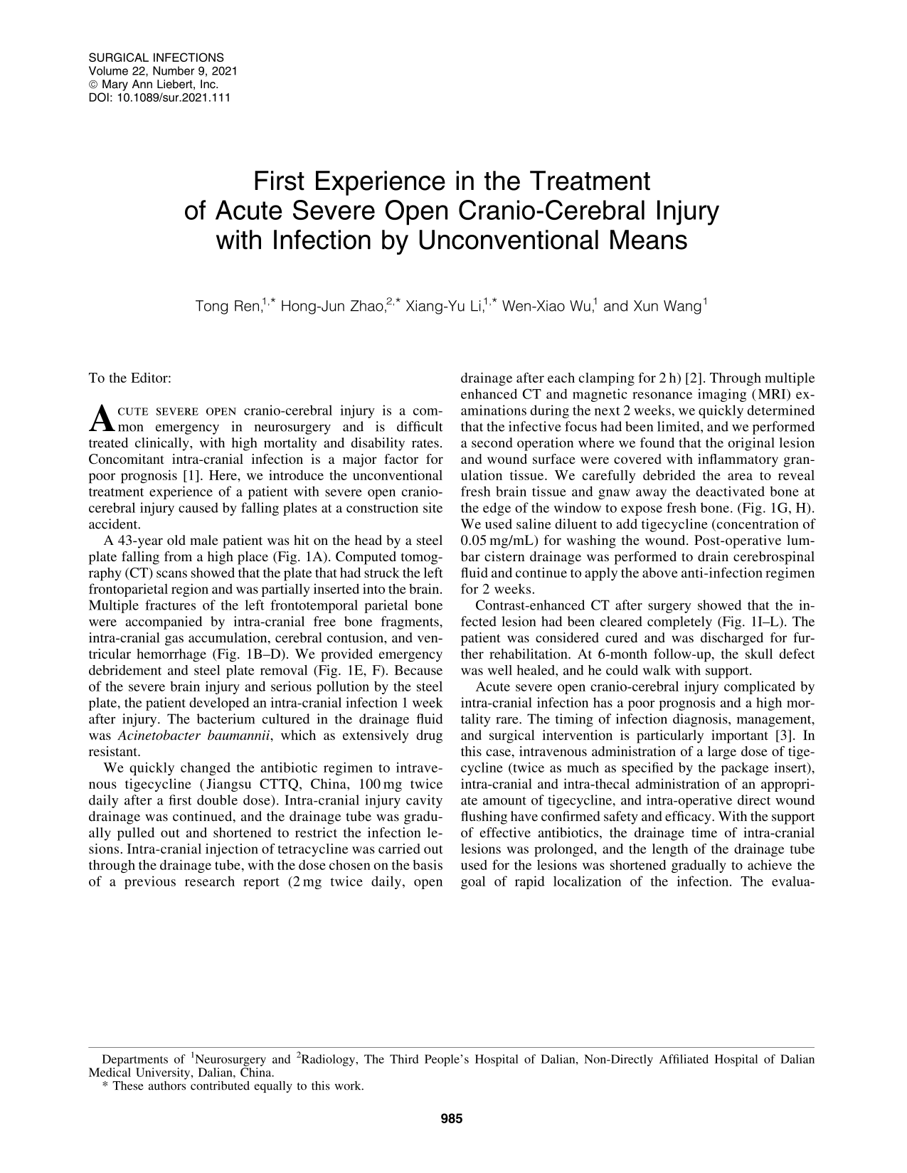
A 43-year old male patient was hit on the head by a steel plate falling from a high place (Fig. 1A). Computed tomography (CT) scans showed that the plate that had struck the left frontoparietal region and was partially inserted into the brain. Multiple fractures of the left frontotemporal parietal bone were accompanied by intra-cranial free bone fragments, intra-cranial gas accumulation, cerebral contusion, and ventricular hemorrhage (Fig. 1B–D). We provided emergency debridement and steel plate removal (Fig. 1E, F). Because of the severe brain injury and serious pollution by the steel plate, the patient developed an intra-cranial infection 1 week after injury. The bacterium cultured in the drainage fluid was Acinetobacter baumannii, which as extensively drug resistant.

Images from case history. (
We quickly changed the antibiotic regimen to intravenous tigecycline (Jiangsu CTTQ, China, 100 mg twice daily after a first double dose). Intra-cranial injury cavity drainage was continued, and the drainage tube was gradually pulled out and shortened to restrict the infection lesions. Intra-cranial injection of tetracycline was carried out through the drainage tube, with the dose chosen on the basis of a previous research report (2 mg twice daily, open drainage after each clamping for 2 h) [2]. Through multiple enhanced CT and magnetic resonance imaging (MRI) examinations during the next 2 weeks, we quickly determined that the infective focus had been limited, and we performed a second operation where we found that the original lesion and wound surface were covered with inflammatory granulation tissue. We carefully debrided the area to reveal fresh brain tissue and gnaw away the deactivated bone at the edge of the window to expose fresh bone. (Fig. 1G, H). We used saline diluent to add tigecycline (concentration of 0.05 mg/mL) for washing the wound. Post-operative lumbar cistern drainage was performed to drain cerebrospinal fluid and continue to apply the above anti-infection regimen for 2 weeks.
Contrast-enhanced CT after surgery showed that the infected lesion had been cleared completely (Fig. 1I–L). The patient was considered cured and was discharged for further rehabilitation. At 6-month follow-up, the skull defect was well healed, and he could walk with support.
Acute severe open cranio-cerebral injury complicated by intra-cranial infection has a poor prognosis and a high mortality rare. The timing of infection diagnosis, management, and surgical intervention is particularly important [3]. In this case, intravenous administration of a large dose of tigecycline (twice as much as specified by the package insert), intra-cranial and intra-thecal administration of an appropriate amount of tigecycline, and intra-operative direct wound flushing have confirmed safety and efficacy. With the support of effective antibiotics, the drainage time of intra-cranial lesions was prolonged, and the length of the drainage tube used for the lesions was shortened gradually to achieve the goal of rapid localization of the infection. The evaluation of the focus of infection was made as soon as possible, as was the decisive second operation to clear the focus of infection.
This is the first time these unconventional methods have been adopted, but they not only demonstrated safety but also achieved satisfactory results. Therefore, we should try some unconventional treatments for this kind of case, which is highly worthy of attention and research.