
Abstract
Background:
Pyoderma gangrenosum is a scarce ulcerating inflammatory skin disease, which requires excluding other causes of ulceration such as infections, malignancies or connective tissue diseases.
Case Report:
We report the case of a 38-year-old woman who developed a progressive bilateral breast skin ulcer after breast plastic surgery, suspected initially with an early postoperative infection. The lack of improvement despite adequate antimicrobial drugs conducted to perform a skin biopsy, concluding to an ulcerated neutrophil dermatosis which led to the diagnosis of postoperative pyoderma gangrenosum. The clinical course was favorable with a systemic treatment based on steroids.
Conclusions:
In order to prevent debridement and extension of local complications, this case report illustrates the importance to suspect pyoderma gangrenosum as differential diagnosis of infection after surgery.
Pyoderma gangrenosum (PG) is an uncommon ulcerating inflammatory skin disease with an incidence of three to 10 patients per million, affecting slightly more females than males in the second through fifth decades. 1 It has been described in association with systemic diseases such as inflammatory bowel disease, rheumatologic conditions, and hematologic disorders. 2 Pyoderma gangrenosum is often considered as a diagnosis of exclusion, which requires excluding other causes of ulceration such as infections, malignancies, or connective tissue diseases. We report an unusual presentation of bilateral breast PG in a French young patient after breast plastic surgery.
Observations
In March 2020, a 38-year-old female with no significant medical history sought care for fibroadenoma of the right breast in the plastic and reconstructive surgery department. After surgical removal, the patient presented with necrotizing fasciitis of the right breast treated successfully with adequate antibiotic agents and an enlarged resection with directed healing. The diagnosis was based on clinical presentation and histologic examination. The favorable outcome observed after antimicrobial drugs and surgery ruled out inflammatory skin diseases, such as neutrophilic dermatosis. Six months later (September 2020), the patient was hospitalized to undergo a resection of the inflammatory scar with a total skin graft from the contralateral breast and its provide symetry.
On day three of hospitalization, the patient presented with a fever of 39°C, chills, and breast pain. Clinical examination revealed purulent discharge from the scar of the left breast with a partial graft failure of the right breast. Laboratory tests showed an elevated C-reactive protein (CRP) at 412 mg/l. Direct examination and culture of cutaneous sample were negative. Peripheral blood culture was also negative. Empiric treatment began with antimicrobial drugs (piperacillin-tazobactam, 12 g, intravenous infusion per day) and clindamycin (600 mg orally four times per day).
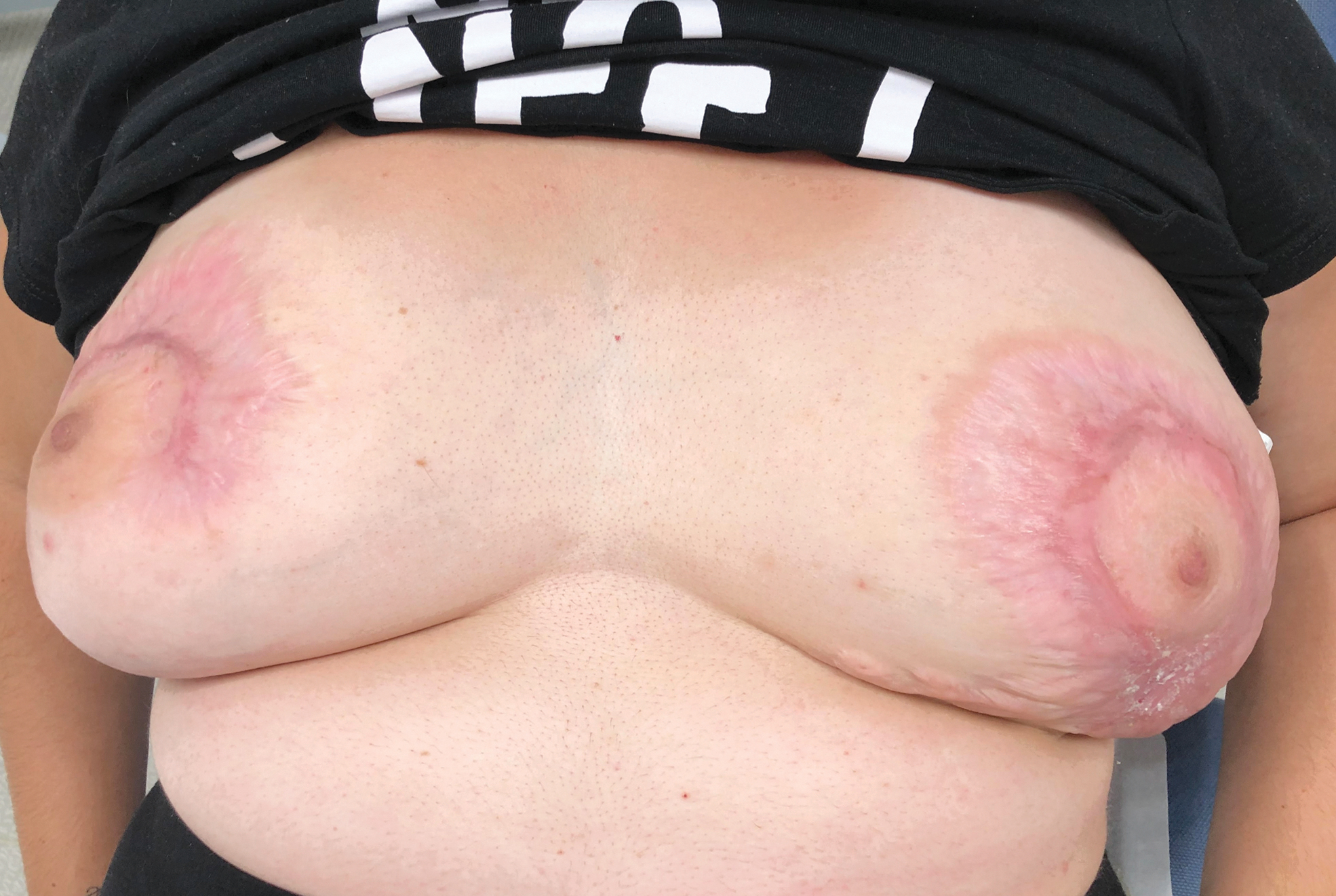
The patient developed a progressive necrosis of the total graft in the right breast and of the skin and subcutaneous peri-scar tissue of the left breast with the sutures coming off (Fig. 1). The hypothesis of an early post-operative infection was discarded and it was decided to perform a skin biopsy. This examination revealed an ulcerated neutrophil dermatosis (Fig. 2) and pyoderma gangrenosum was diagnosed. We started a treatment based on steroids (prednisone 1 mg/kg orally per day with progressive decrease) and doxycycline (100 mg orally two times per day). The clinical course was favorable, and the patient was discharged (Fig. 3).

Bilateral breast pyoderma gangrenosum, with erythematous necrotic ulcer associated to violaceous undermined borders.

Light microscopy using hematoxylin and eosin stain ( × 10) showed a neutrophilic inflammatory infiltrate in the dermis and hypodermis, which are also present in exocytosis in the epidermis and grouped pustules.

Follow-up at one month after treatment started (steroids), with substantial improvement of the necrotic ulcer.
Three months later (January 2021), the patient was seen as an outpatient regularly. Examinations showed an appropriate outcome and improvement of the local presentation and skin scarring (Fig. 4), with no evidence of relapse afterwards.
Follow-up at three months after after treatment started (steroids), with complete healing.
Regarding the etiologic diagnoses, colonoscopy and computed tomography scan of chest, abdomen, and pelvis were normal. Biologic and immunologic investigations were also normal and no monoclonal gammopathy, rheumatic inflammatory disease, or an inflammatory bowel disease was found.
Discussion
Pyoderma gangrenosum is a neutrophilic dermatosis, defined by a pathologic abundance of neutrophils in the absence of infection and characterized by recurrent skin ulcerations with mucopurulent or hemorrhagic exudate. 1 Classic PG commonly presents as an extremely painful erythematous lesion that rapidly progresses to a blistered or necrotic ulcer with a violaceous undermined edge. 2
Other differential diagnoses that cause cutaneous ulcers should be considered.2,3 These include infectious diseases, malignancy, vasculitis, insect bites, venous or arterial insufficiency, and above all, factitious ulcerations. In our case, the biopsy of ulcer edge demonstrating neutrophilic infiltrate confirming the diagnosis of PG, without any arguments for infection or other etiology (excluded by microbiologic testing and further investigations). The pathogenesis of PG is multifactorial and involves neutrophilic dysfunction, inflammatory mediators, and genetic predisposition. 3 Pathergy has been described in roughly 20%–30% of patients with PG and defined as the provocation of new lesions by trauma. The increased activity of polymorphonuclear leukocytes has been suggested in the underlying cause of pathergy. 1
Post-operative PG is an unusual clinical identity involving pathergic response. 4 At first, it is usually diagnosed as an infection, but followed by a failure of antimicrobial therapy and wound debridement, which leads to rapid ulcer enlargement. Furthermore, clinical appearance of discrete wound with irregular, violaceous, and undermined borders involving surgical sites, especially with nipple sparing, should raise the suspicion of post-operative PG rather than infection or ischemia, even with concomitant fever and leukocytosis.5,6 Apart from this cutaneous semiology specific to post-surgical PG, Touil et al. 6 highlighted that severe pain, rapid progression of the lesions, negative peripheral blood culture, and no response to antimicrobial drug indicates post-surgical PG rather than necrotizing fasciitis.
Tolkachjov et al. 7 presented a clinical review about this unusual presentation. First, they emphasized that post-operative PG has a lower association with systemic diseases than other forms of PG. Second, they concluded that the breast and abdomen were the most frequently involved sites of post-operative PG. In a large review of the literature, Tuffaha et al. 8 emphasized that post-operative PG is usually bilateral after bilateral breast plastic surgery, such as in our case. It is also important to know that patients can develop PG in a post-operative context after previous surgical interventions in the same site, as in our case. 9 Currently, this entity should not be misdiagnosed, especially with the increased breast reconstructions performed, which can also increase the incidence of post-operative PG. 10
Conclusions
We highlight a rare presentation of PG after breast plastic surgery, as a problematic differential diagnosis to post-operative infection, for which making the diagnosis has proven difficult. Indeed, early recognition of post-operative PG may prevent debridement and extension of local complications. Systematic biopsy of the ulcer can help clinicians in this context.
Footnotes
Authors' Contributions
V.C. and S.Z. drafted the manuscript. All authors revised and approved the final manuscript.
Funding Information
This research did not receive any specific grant from funding agencies in the public, commercial, or not-for-profit sectors.
Author Disclosure Statement
All authors declare no competing interests.