
Abstract

To the Editor:
P
A 66-year-old female presented to our hospital with a palpable and painless breast mass. She denied having a fever, night sweats, or weight loss. Denial of history of infection and exposure to tuberculosis. No abnormalities were found in relevant laboratory tests. Ultrasonography of the breast revealed a 4.7 × 2.6 × 2.1 cm uneven echoic mass in the left breast with irregular shape, unclear boundary, and enhanced echo in surrounding tissue. Breast ultrasonography was considered as a possible breast tumor (Fig. 1).

Ultrasonography of the breast revealed a 4.7 × 2.6 × 2.1cm uneven echoic mass in the left breast with irregular shape, unclear boundary, and enhanced echo in surrounding tissue.
Because the possibility of malignancy was not ruled out, the patient then underwent a routine aspiration biopsy of the breast mass. After biopsy, the histopathology revealed granulomatous inflammation with caseolecrosis, and the final diagnosis was primary breast tuberculosis (Fig. 2). The patient was subsequently referred to the infectious diseases department for specialized treatment, and the prognosis was good.
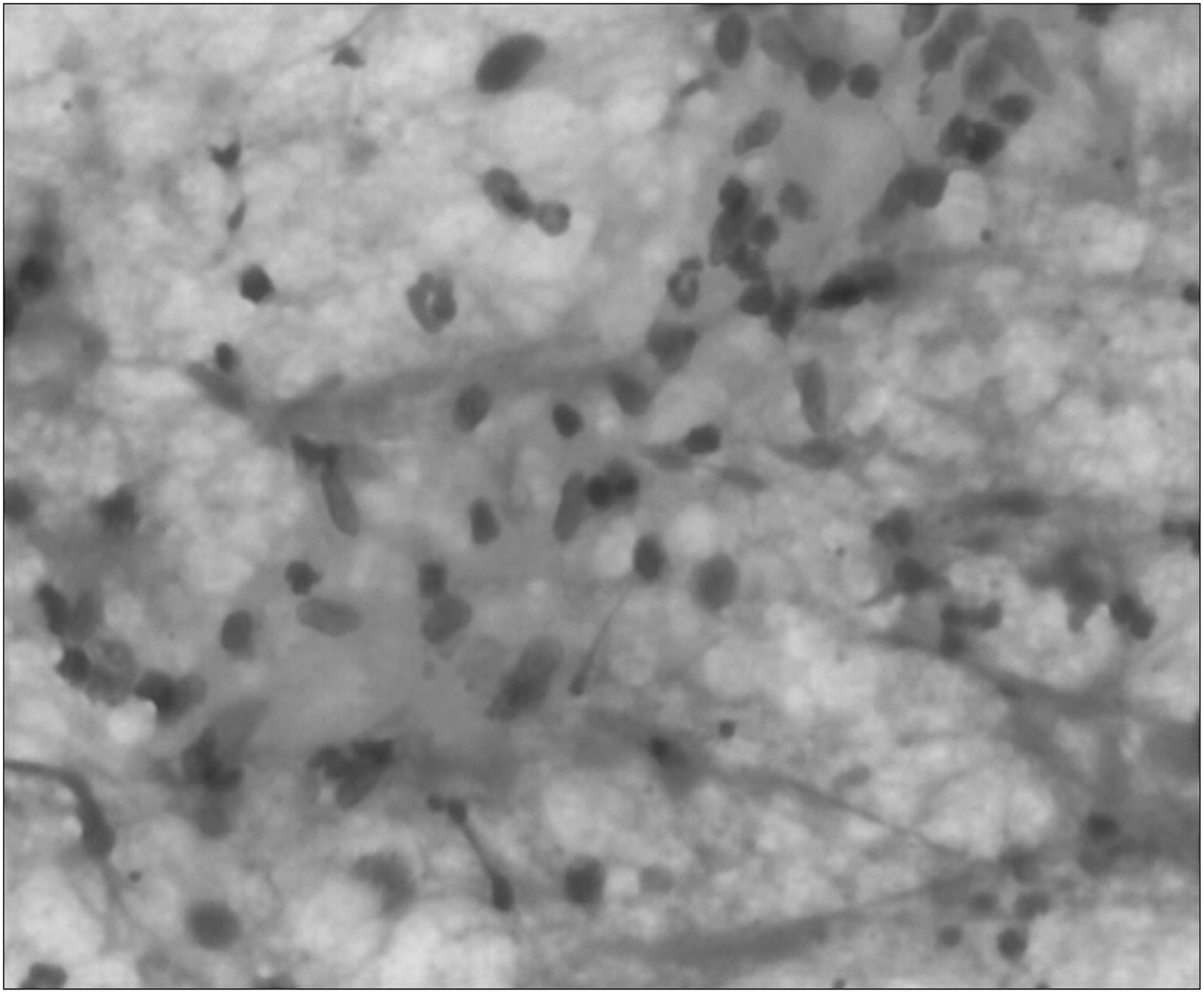
Biopsy histopathology revealed granulomatous inflammation of the breast with caseous necrosis.
Primary breast tuberculosis is considered an unusual disease with an estimated incidence of 0.1% of all breast lesions. Most patients have no previous history of tuberculosis, which often leads to their misdiagnosis as breast tumors.2,3 Because the imaging and histologic features of primary breast tuberculosis are similar to those of various other granulomatous mastitis and breast tumors, the differential diagnosis is often difficult. Diagnosis is usually based on histopathology and needle biopsy. Because of the good prognosis of this disease, the correct diagnosis of primary breast tuberculosis is extremely important. Formal antituberculosis therapy is recommended after diagnosis. Because breast tuberculosis may be primary or secondary, once diagnosed, further tests should be conducted to determine if a primary lesion is present. The possibility of breast tuberculosis should be considered for painless masses of the female breast in endemic areas. The prognosis of this disease is good, once diagnosed, regular antituberculosis therapy should be performed.