
Abstract

To the Editor:
S
A 36-year-old male presented to our hospital with a right neck mass. The mass gradually increased in size over a period of one month and was accompanied by hoarseness and pain. He denied dyspnea and arm numbness. He had no other specific medical history. Physical examination revealed a soft mass on the right side of the neck of approximately 5 × 4 × 1 cm with regular morphology, well-defined borders, poor mobility, significant pressure pain and the skin temperature of the lump increased. Magnetic resonance of the neck showed an abnormal signal mass in the right sternocleidomastoid muscle with T1 low signal and T2 high signal (Fig. 1).
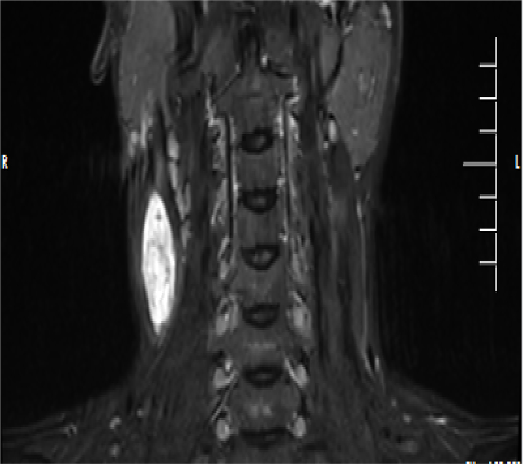
Magnetic resonance scan of the neck showed a deep abnormal signal mass in the right sternocleidomastoid muscle.
The patient then underwent a right neck mass removal. During the procedure, the mass was completely removed. The post-operative histopathologic background was blood vessels and mucus, with stele and spindle cells in the mucus, which was diagnosed as superficial angiomyxoma (Fig. 2). There was a little white pus in the mass, and the bacterial culture of the pus showed Staphylococcus aureus infection. A drug sensitivity test showed sensitivity to cefuroxime antibiotic agents. The patient recovered well after one week of cefuroxime symptomatic anti-infection.
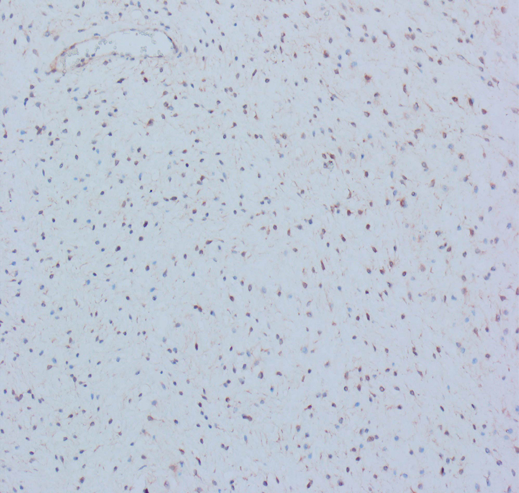
Post-operative histopathology showed a background rich in blood vessels and mucus, with stellate and spindle cells in the mucus.
Superficial angiomyxoma is an extremely rare mucinous soft-tissue tumor that is most common in males and usually occurs on the trunk, limbs, and genital region. 2 The lesions are located in the reticular layer of the dermis and often involve the subcutaneous layer, superficial angiomyxoma originating in the muscle is extremely rare. 3 Clinically, the lumps are slow-growing and painless. Clinical manifestations include skin papules, nodules, or polypoid masses, and larger lesions may be fluctuant. Accurate diagnosis is very challenging because of the lack of clinical and imaging specificity. Confirmation of the diagnosis often relies on post-operative pathologic histology. The treatment of superficial angiomyxoma is complete surgical resection. In patients with a neck mass that increases in size over a short period of time and is accompanied by painful symptoms, the possibility of superficial angiomyxoma with infection should be considered.