
Abstract

To the Editor:
Oral tuberculosis is a rare manifestation of extrapulmonary tuberculosis, accounting for about 0.05%−5% of TB patients. 1 Due to atypical symptoms, it is easy to be missed and misdiagnosed clinically. Herein, we report a case of oral tuberculosis masquerading as an oral neoplasm.
A 59-year-old male presented with a chief complaint of pain in the floor of the mouth and tongue for six months. Physical examination showed the formation of a tough mass on the left lingual margin and tongue abdomen, ulcerative lesions on the mucosal surface of the bottom of the mouth, and submental palpation of an enlarged lymph node with hard quality and poor mobility. Computed tomography of the chest showed a thick-walled cavity in the posterior segment of the upper lobe of the right lung, about 1.3*1.4 cm in size, and the lungs were diffusely covered with small nodular, patchy, and cord shadows (Fig. 1).
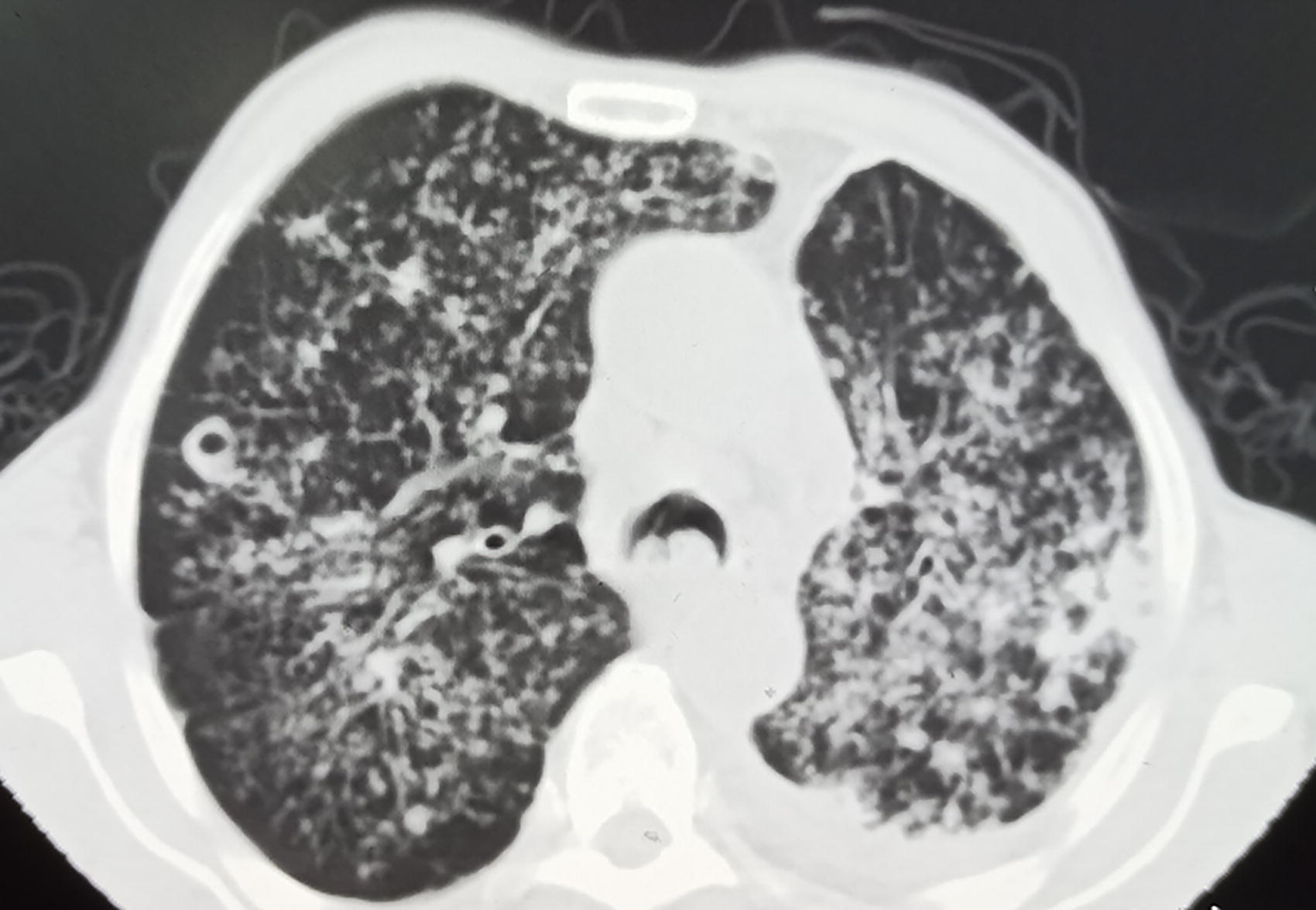
Computed tomography of the chest showed a thick-walled cavity in the posterior segment of the upper lobe of the right lung, about 1.3*1.4 cm in size, and the lungs were diffusely covered with small nodular, patchy, and cord shadows.
Owing to concern about oral cancer, a biopsy of the tongue mass tissue was performed. Post-operative histopathological examination revealed granulomatous inflammation with necrosis (Fig. 2), Mycobacterium tuberculosis was not found by acid-fast staining, but TB-DNA was positive. Therefore, oral tuberculosis secondary to pulmonary tuberculosis was considered a high probability.

Post-operative histopathological examination revealed granulomatous inflammation with necrosis.
We then gave him systemic anti-tuberculosis treatment, the specific regimen was based on isoniazid, rifampicin, ethambutol, pyrazinamide, and isoniazid nebulized local therapy. However, renal function was impaired during this period, so pyrazinamide was discontinued and moxifloxacin was replaced. At the same time, he repeatedly coughed and coughed up sputum, infectious indicators showed an upward trend, so piperacillin-tazobactam was given anti-infective treatment. After the above treatment, the oral and respiratory symptoms were effectively relieved.
Tuberculosis is currently the world’s second leading cause of death from a single infectious pathogen after the novel coronavirus (COVID-19). 2 Oral tuberculosis is categorized as primary or secondary and is usually dominated by oral lesions that appear secondary to pulmonary tuberculosis. Studies have shown low levels of M. tuberculosis in the oral cavity, which may be related to the cleansing effect of saliva, the relative lack of lymphoid tissue in the tongue, and antibodies in the saliva.3,4 The intact oral mucosa provides barrier protection against M. tuberculosis infection, and when the oral mucosa is ruptured, it can enter the tissues through the wound and cause disease.
Once oral tuberculosis is diagnosed, systemic anti-tuberculosis therapy should be carried out promptly, without the need for surgical treatment. Histopathological changes, diagnostic support of molecular biology, and imaging evidence can be used to make a clear diagnosis, providing a perspective of thinking about the differential diagnosis of other diseases, thus improving the diagnostic rate of the disease.
Footnotes
Author Disclosure Statement
The authors declared no potential conflicts of interest with respect to the research, authorship, and/or publication of this article.
Funding Information
No funding was received for this article.