
Abstract

To the Editor:
Hydatid disease is still a serious public health concern in many countries around the world, notably in the Middle East and the Mediterranean. Although the liver and lungs are the most commonly involved organs, hydatid cysts can be found in almost any organ or tissue in the body. 1 In hydatid endemic regions, hydatid cysts are among the differential diagnoses of numerous diseases, and can give rise to a wide variety of clinical symptoms. Given the rarity of this disease in preschool children, the diagnosis and treatment of hydatid cysts can be challenging, especially when they occur in an unusual site or in a complicated form. 2
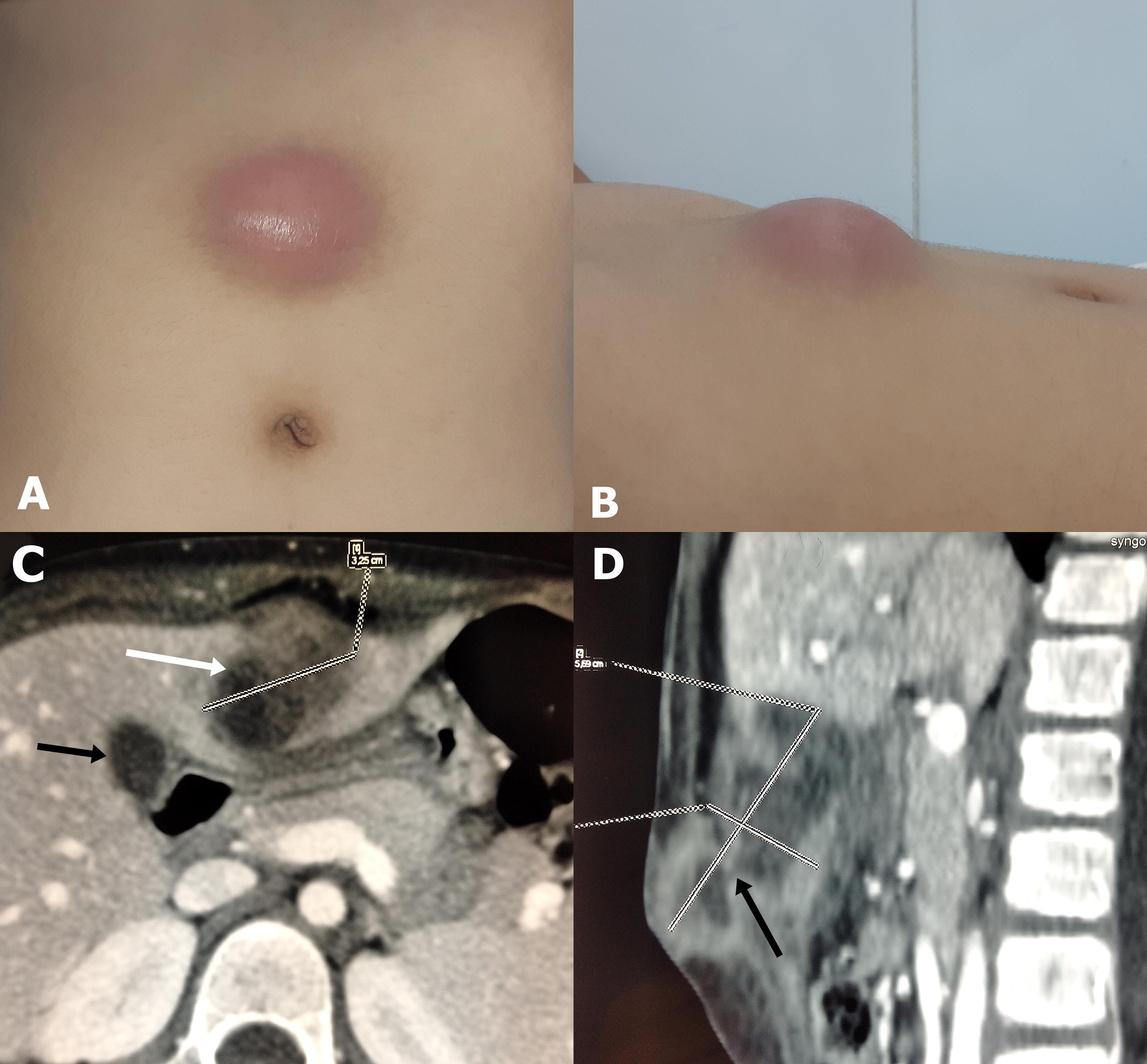
A previously healthy 5-year-old boy living in a rural area presented to the emergency department with supraumbilical abdominal swelling and abdominal pain. Physical examination revealed a fluid-like swelling above the umbilicus, with local signs of inflammation, suggestive of an abdominal wall abscess (Fig. 1A, 1B). There was no fever. Laboratory work-up showed anemia, normal leukocyte count of 8740 × 109/L, and a high C-reactive protein (CRP) level of 39 mg/L. Abdominal computed tomography scan showed an infected hydatid cyst in segment III of the liver with peripheral enhancement and perilesional inflammatory changes. The cyst measured 3.5 cm in long axis and showed cutaneous fistulization. A second small cyst was found in segment IV of the liver (Fig. 1C, 1D). The patient underwent laparotomy with initial aspiration of the cutaneous swelling contents, followed by excision of the hydatid cyst and closure of the fistulous tract. The post-operative period was straightforward, and the child was discharged on the 7th postoperative day on albendazole (10 mg/kg/day). The pathological exam of the operative specimen confirmed the hydatid cyst diagnosis. Currently, with a 2-year follow-up, the child has not developed a recurrence.
Echinococcosis or hydatid disease is a parasitic zoonotic infection caused by the larval form of Echinococcus granulosus. This disease is endemic in the Middle East, South America, Russia, Australia, Eastern Europe, India, and China. The incidence of echinococcosis is higher in rural areas, where agriculture, regional climate, low socioeconomic status, and unhygienic animal slaughtering all play a role. 3 Hydatid disease progresses very slowly and may remain silent for several years. Occasionally, the disease can be complicated by infection, rupture or, more rarely, fistulization into other organs or tissues. 4 Surgical treatment of hydatid cysts can be challenging, especially in complicated forms. This treatment is not only associated with significant morbidity but also generates considerable healthcare costs in countries where the disease is still endemic. 5 Improving the management of this disease will certainly require educating people in endemic regions, especially rural areas, about the rules of hygiene and prevention.
Although cutaneous fistulization is a rare complication of hydatid cysts in children, this diagnosis should be considered in the presence of any abdominal swelling in a child living in a hydatid endemic area.
Footnotes
Authors’ Contributions
M.Z. was responsible for conceptualization, project administration, writing (original draft), and writing (review). M.B., N.B.K., E.K., and A.I. contributed to data curation, methodology. M.B.D. and R.M. performed supervision, and validation. M.Z. is the guarantor.
Author Disclosure Statement
The authors, have no financial or personal relationships with other people or organizations that could potentially and inappropriately influence our work and conclusions.
Funding Information
The authors have not declared a specific grant for this research from any funding agency in the public, commercial, or not-for-profit sectors.