
Abstract
Abstract
A new type of synthetic hydrogel scaffold that mimics certain aspects of structure and function of natural extracellular matrix (ECM) has been developed. We previously reported the conjugation of collagen mimetic peptide (CMP) to poly(ethylene oxide) diacrylate (PEODA) to create a polymer-peptide hybrid scaffold for a suitable cell microenvironment. In this study, we showed that the CMP-mediated microenvironment enhances the chondrogenic differentiation of mesenchymal stem cells (MSCs). MSCs were harvested and photo-encapsulated in CMP-conjugated PEODA (CMP/PEODA). After 3 weeks, the histological and biochemical analysis of the CMP/PEODA gel revealed twice as much glycosaminoglycan and collagen contents as in control PEODA hydrogels. Moreover, MSCs cultured in CMP/PEODA hydrogel exhibited a lower level of hypertrophic markers, core binding factor alpha 1, and type X collagen than MSCs in PEODA hydrogel as revealed by gene expression and immunohistochemisty. These results indicate that CMP/PEODA hydrogel provides a favorable microenvironment for encapsulated MSCs and regulates their downstream chondrogenic differentiation.
Introduction
Cell–extracellular matrix (ECM) interactions play numerous roles in all biological cell behaviors, including differentiation, homeostasis, and regeneration of a tissue.2–4 Therefore, scaffolds with various native ECM components have been created for tissue repair. Interactions between chondrocytes and collagen gels has been shown to specifically maintain cell phenotype and induce proliferation.5–7 MSCs have also been encapsulated in collagen matrix and shown to form hyaline-like cartilage in osteochondral defects of patients. 8 Collagen has various cell and growth factor–binding domains as well as enzyme-sensitive cleavage sites, which may provide suitable microenvironments for MSC differentiation and subsequent cartilage matrix production, but collagen alone as a scaffold has several problems: weak mechanical strength, large porosity, shrinkage upon implantation, and immune response in the host body.9–12
Previous studies have shown that poly(ethylene oxide) diacrylate (PEODA) hydrogel scaffolds provide three-dimensional (3D) structural support for in vitro and in vivo chondrogenic differentiation of stem cells, but PEODA gels are bio-inert like most synthetic scaffolds and non-adhesive to cells and proteins.13,14 Therefore, recent years have witnessed a surge of interest in using synthetic- bioactive hybrid materials as tissue-engineering scaffolds by integrating principles of cell and molecular biology; such scaffolds mimic certain aspects of structure and function of natural ECM, creating a suitable cell microenvironment. Collagen mimetic peptides (CMPs), -(Pro-Hyp-Gly)7- (Hyp = hydroxyproline), have a unique collagen-like triple helical conformation and have been shown to associate with collagen molecules and fibers via a strand invasion process.15,16 As an effort to develop a bioactive 3D PEODA hydrogel suitable for encapsulated cells, we produced a PEODA scaffold conjugated with CMP (CMP/PEODA). 17 In this study, we demonstrate that CMP/PEODA provides a suitable bio-active microenvironment, facilitating the efficient chondrogenic differentiation of MSCs.
Materials and Methods
Synthesis and purification of acryloyl-poly(ethylene glycol)–CMP
CMP (Pro-Hyp-Gly)7-Tyr) and acryloyl (ACRL)-poly(ethylene glycol) (PEG)-CMP were synthesized as described previously. 17 Briefly, CMP was synthesized using Fmoc-mediated solid-phase peptide coupling methods using an automated peptide synthesizer (Symphony Quartet, Protein Technologies, Tucson, AZ), and high-performance liquid chromatography (C18 Vydac column) was used to purify the peptide. ACRL-PEG-N-hydroxysuccinimide (NHS) (Nektar, Huntsville, AL) (molecular weight: 3400 g/mol) was crosslinked to CMP, and the product was purified using Sephadex G-25 size-exclusion column (GE Healthcare, Piscataway, NJ). The molecular weight of the target product was confirmed using matrix-assisted laser-desorption/ionization time-of-flight (MALDI-TOF) mass spectrometry.
MSC culture and photoencapsulation
Bone marrow–derived MSCs were isolated and cultured as previously described. 18 Control PEODA macromer solution was prepared by mixing 0.05% (w/v) photoreactive initiator (Irgacure 2959; Ciba Specialty Chemicals, Tarrytown, NY) and 10% (w/v) PEODA (Nektar, molecular weight: 3400 g/mol) in sterile phosphate buffered saline (PBS). A macromer solution of CMP/PEODA hydrogel composed of 2% (w/v) ACRL-PEG-CMP and 8% (w/v) PEODA, instead of 10% PEODA was polymerized. Expanded MSCs were resuspended in the macromer solution (100 μL) at a concentration of 20 × 106 cells per mL. Then the solution was photopolymerized using ultraviolet exposure (EXFO Acticure 4000; wavelength, 365 nm; intensity, ∼5 mW/cm2). The hydrogel construct was transferred to a well in a 12-well culture plate containing 2 mL of chondrogenic differentiation medium composed of 10 ng/mL transforming growth factor beta-1 (TGFβ-1), 100 nM dexamethasone, 40 mg/L proline (Sigma, St. Louis, MO), 50 mg/L ascorbate-2-phosphate (Sigma), 100 mg/L sodium pyruvate (Gibco, Carlsbad, CA), 50 mg/mL insulin-transferrin-selenium Premix (Collaborative Biomedical, San Jose, CA), and 1% penicillin-streptomycin in high-glucose Dulbecco's modified Eagle medium (DMEM). Constructs were cultured at 37°C for 3 weeks in 5% carbon dioxed atmosphere, and medium was replaced every 2 to 3 days.
Cell viability test
Viability of encapsulated cells in PEODA hydrogel was determined based on the integrity of cellular membrane using the Live/Dead Viability/Cytotoxicity Kit (Molecular Probes, Eugene, OR), which contains calcein-AM (Live dye) and ethidium homodimer-1 (Dead dye). Dye solution was made with 0.5 μL of calcein-AM dye and 2 μL of ethidium homodimer-1 dye in 1 mL DMEM. Constructs were washed twice with PBS. A slice of the construct was incubated in the Live/Dead dye solution for 30 min. Fluorescence microscopy was performed, and the percentage of viable cells was determined by comparing the number of live cells, which were stained with green dye, with the the total number of cells presented in three different fields.
Histology and immunostaining
After 3 weeks of culture, MSC-encapsulated PEODA constructs were fixed overnight in 10% formalin solution and stored in 70% ethanol. The fixed constructs were embedded in paraffin, sectioned, and stained with Safranin-O. Immunohistostaining was performed using rabbit polyclonal antibody against type I, type II, or type X collagen (Research Diagnostics, Flanders, NJ) using a 1:100, 1:100, or 1:40 dilution factor, respectively. After extensive washing, Texas Red or fluorescein isothiocyanate–conjugated anti-rabbit antibodies (Jackson ImmunoResearch Laboratory, West Grove, PA) were used for fluorescent detection of the specific collagen. Finally, DAPI solution (0.1 mg/mL, Millipore, Temecula, CA) was applied to dye cell nucleus. Confocal microscopy (LSM 510 Meta Confocal Microscope, Zeiss, Maple Grove, MN) was performed to collect images. In order to quantify the overall immunostaining result for a section, the percentage of type II or type X positive cells was determined by counting the cells with the specific collagen in their periphery, relative to the total cells stained by 4',6-diamidino-2-phenylindole (DAPI) in three separate fields.
Biochemical assays of MSC-encapsulated hydrogel
Hydrogels were harvested after 3 weeks and lyophilized for 48 h. Water contents of the hydrogels was calculated using the following equation:
One unique feature of collagen is hydroxyproline. This exclusive amino acid has been used to quantify the amount of total collagen content of the gel because its ratio to the total collagen is known to be approximately 1:7.46. 22 Hydroxyproline assay was conducted using papain-digested solution after overnight hydrolysis reaction in 6 N hydrochloric acid at 115°C. Hydrolyzed samples were reacted with p-dimethylamino benzaldehyde and chloramines-T hydrate. 23 Absorbance was measured at 550 nm on a UV-Vis spectrophotometer. A standard curve was generated using pure trans-4-hydroxy-L-proline (Sigma-Aldrich, St. Louis, MO). The DNA, GAG, and collagen contents from the biochemical assays were normalized against the construct dry weights. The values are presented as means ± standard deviations. Statistical significance was determined using the unpaired Student t-test and set as p < 0.05.
Reverse transcription polymerase chain reaction and real-time polymerase chain reaction analysis
Total RNAs were isolated from three hydrogels per group using TRIzol (Invitrogen, Carlsbad, CA) following the manufacturer's instruction. The first-strand cDNA synthesis was performed using SuperScript First-Strand Synthesis System (Invitrogen). Real-time polymerase chain reaction (PCR) was preformed on the ABI Prism 7700 Sequence Detection System (Perkin Elmer/Applied Biosystems, Rotkreuz, Switzland) using SYBR Green PCR master mix and specific primer sets, as listed in Table 1. The level of expression of each target gene was calculated as −2ΔΔCt, as described previously. 24
Results
Preparation of CMP/PEODA macromer
As characterized in a previous study, CMP sequence (Pro-Hyp-Gly)7 maintains the unique triple helix conformation of collagen with an approximate 37°C melting temperature. 17 ACRL-PEG-(Pro-Hyp-Gly)7-Tyr (CMP), a photopolymerizable CMP derivative, was synthesized and purified using Sephadex gel permeation chromatography, and its molecular weight was confirmed using MALDI-TOF mass spectrometry. MALDI-TOF mass spectrometry also indicated that the purity of ACRL-PEG-CMP was greater than 80% (data not shown). ACRL-PEG-CMP: mass calcd: 5452.16 [M + H]+; average mass found: 5441.01 [M + H]+
MSC encapsulation in CMP/PEODA hydrogels
To determine the effect of CMP/PEODA on the chondrogenic differentiation of MSCs, cells were encapsulated in the CMP/PEODA hydrogels using photopolymerization of macromer solutions with or without 2% (w/v) ACRL-PEG-CMP. The ACRL end of the CMP derivative or PEODA was cross-linked to another ACRL end of PEODA via a free-radical polymerization reaction, which resulted in the formation of a hydrogel, as shown in Figure 1. Live/Dead fluorescence staining results after 24 h of culture in chondrogenic medium indicated that most of the MSCs remained viable and homogeneously distributed throughout the PEODA constructs. MSCs in CMP/PEODA hydrogels showed comparable viability with that of control hydrogels, indicating that conjugated CMP did not have any toxic effect on cells (Fig. 2).

Poly(ethylene oxide) diacrylate (PEODA) was photopolymerized to form a cross-linked polymer network (
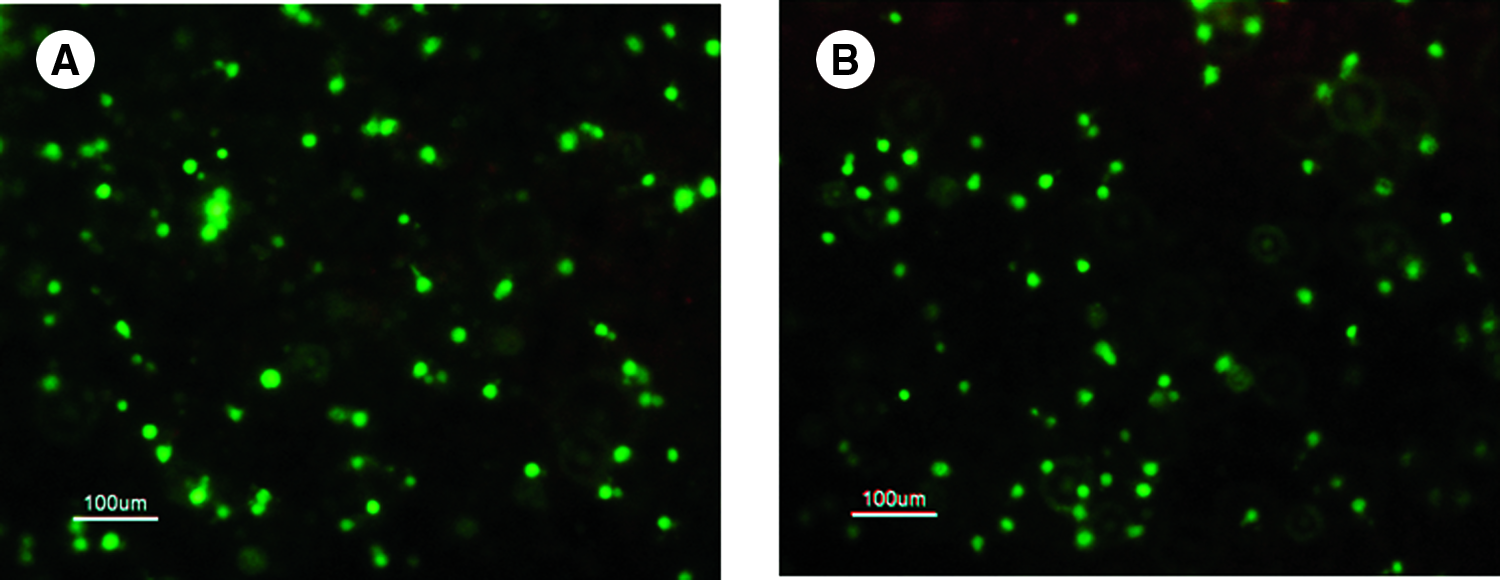
Fluorescence micrographs of mesenchymal stem cells encapsulated in poly(ethylene oxide) diacrylate (PEODA) (
Histology and immunohistochemistry of MSC-encapsulated hydrogels
Chondrogenic differentiation of MSCs in both hydrogels was evaluated after 3 weeks of culture. Histological analysis demonstrated that CMP-containing constructs enhanced chondrogenic differentiation and matrix secretion. More Safranin-O–stained areas were observed in CMP/PEODA hydrogels than in control hydrogels. Cells in CMP/PEODA hydrogel showed round cellular morphology and produced well-defined cartilaginous ECMs in the periphery of the cells, as indicated by intense Safranin-O staining (Fig. 3A).
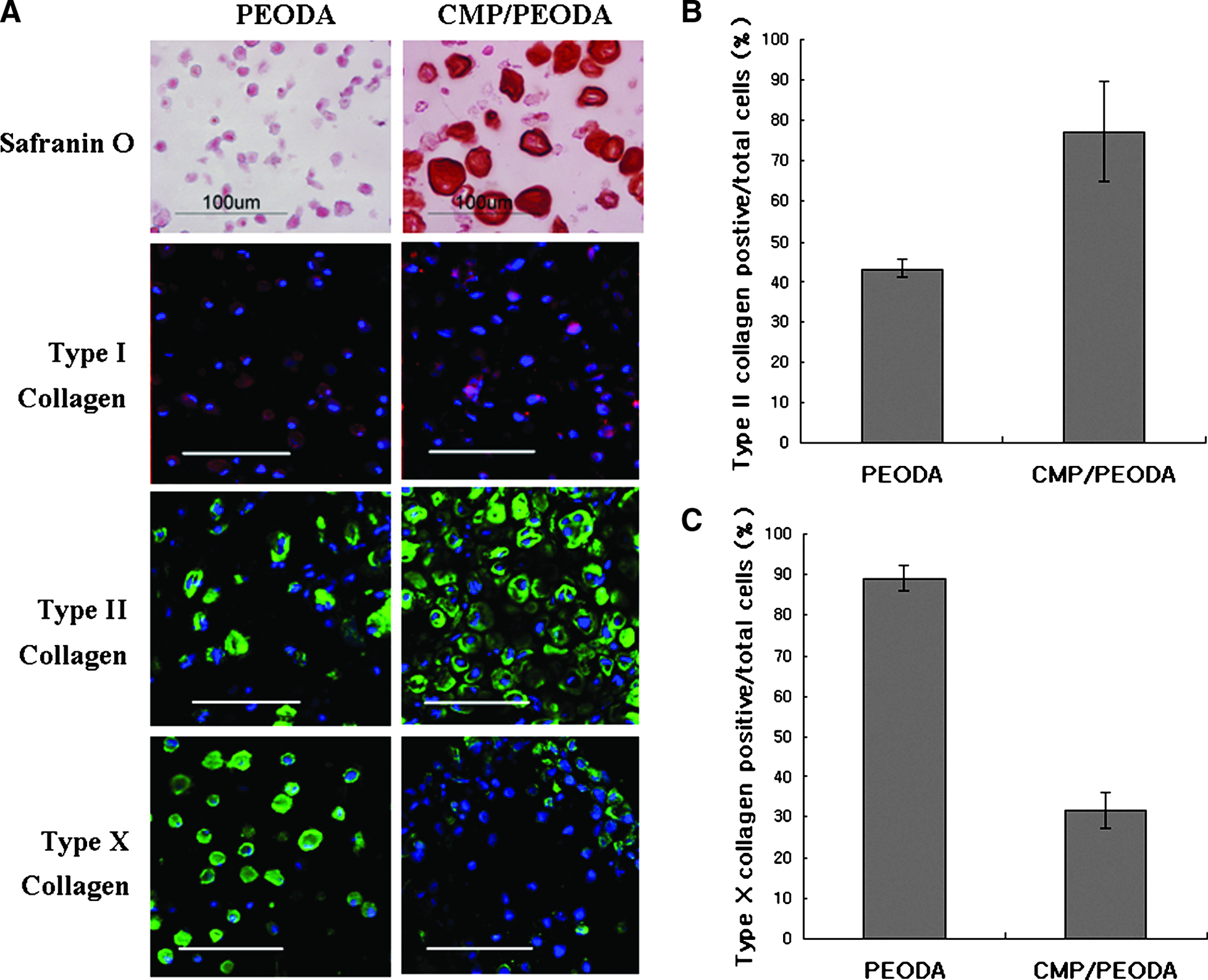
Histological sections of hydrogels seeded with mesenchymal stem cells were evaluated using Safranin-O staining and collagen immunostainings after 3 weeks of culture (
Immunostaining for types I, II, and X collagen also showed significant differences between cells in PEODA and CMP/PEODA hydrogels (Fig. 3A). MSCs in CMP/PEODA hydrogel showed significantly more positive type II collagen than the cells in PEODA hydrogel (Fig. 3A). A quantitative evaluation indicated that 77% of cells in CMP/PEODA hydrogels were positive for type II collagen, significantly more than the 43% in PEODA gel (Fig. 3B). CMP/PEODA hydrogel had significantly less type X collagen: 32% type X collagen–positive cells compared with 89% in PEODA hydrogel (Fig. 3A, C). CMP/PEODA and PEODA constructs had comparable but minimal amounts of type I collagen (Fig. 3A).
Matrix production of MSCs in CMP/PEODA hydrogels
After 3 weeks of culture, the water content of engineered tissues in the CMP/PEODA and PEODA hydrogels were similar (approximately 93%) (Fig. 4A). The cell proliferation profile, quantified according to DNA measurement, indicated that the resulting number of cells in CMP/PEODA is similar to the number of cells in control PEODA hydrogels (Fig. 4B). Biochemical assays were employed to quantify GAG and collagen produced by MSCs encapsulated in hydrogels. The biochemical assays for GAG and hydroxyproline supported the previous histology results, confirming that the conjugation of CMP to the PEODA gel resulted in MSC hydrogel cultures with higher levels of GAG and total collagen than the control PEODA hydrogel (Fig. 4C, D). GAG and total collagen contents in the CMP/PEODA hydrogels were more than twice as high as those of the control PEODA hydrogel. After 3 weeks of culture in chondrogenic medium, MSCs in CMP/PEODA hydrogel produced 7.4% GAG and 4.0% collagen per dry weight, as opposed to 3.0% and 2.0%, respectively, for MSCs in control PEODA hydrogel.

Water contents of engineered tissues were similar for both hydrogels (
Gene expression
Chondrogenesis of MSCs was further evaluated using real-time PCR to monitor gene expression. After 3 weeks of culture, greater chondrogenic gene expressions and lower hypertrophic gene expression in CMP/PEODA constructs than in control PEODA confirmed the previous histology and biochemical assay results. Expressions of aggrecan and type II collagen in CMP/PEODA hydrogel were more than twice as high as those in PEODA hydrogel (Fig. 5A, B), although CMP/PEODA hydrogels had approximately half as much core binding factor alpha 1 (cbfa1), a major transcription factor responsible for promoting hypertrophy (Fig. 5C). There was also two-thirds as much type X collagen, another hypertrophic marker, confirming previous immunostaining results (Fig. 5D).

Real-time polymerase chain reaction analysis indicated more chondrogenic markers but fewer hypertrophic markers of encapsulated cells in collagen mimetic peptide–conjugated poly(ethylene oxide) diacrylate (PEODA_ hydrogel than in PEODA hydrogel (n = 3, p < 0.001): (
Discussion
PEODA-based hydrogels enable homogeneous and non-invasive in situ delivery of encapsulated cells.14,25 PEODA hydrogels provide 3D support for encapsulated cells.13,26,27 These hydrogels can also be easily modified, and one such modification is conjugation of a peptide. For example, attachment of integrin-binding peptide, RGD, has been extensively investigated to compensate for PEODA's inert properties that prevent interaction with proteins and cells. Hern et al. seeded fibroblasts on RGD-modified PEODA surface and illustrated significantly more adherence and spreading. 28 Studies by Burdick et al. and Yang et al. found greater bone formation of encapsulated osteoblasts and MSCs, respectively, in 3D PEODA scaffold conjugated with RGD.29,30 Although natural proteins are subject to denaturation and degradation, and their chemical reactivity is unpredictable, peptides, in general, are considered more stable and easier to conjugate to synthetic hydrogels while preserving functions of proteins.
Recently, a synthetic CMP, (Pro-Hyp-Gly)x, that mimics collagen's periodic amino acid sequence was shown to recognize and bind to natural collagens.15,16 The binding affinity originates from CMP's strong propensity to form a triple helix. It is believed that the observed adhesion between CMP and collagen arises from a single-stranded CMP invading structurally unstable domains of collagen molecule and forming a stable product secured by a triple helix assembly. We hypothesized that, when the CMP is conjugated to synthetic tissue scaffold, it could help sequester the collagens secreted by the cells and allow fast assimilation of their microenvironment. When chondrocytes were encapsulated and cultured in the CMP/PEODA hydrogel, there was markedly more GAG and collagen content in the hydrogel construct. 17 In the present study, we encapsulated MSCs in CMP/PEODA hydrogel and, for the first time, evaluated the potential of a CMP-mediated microenvironment to promote the proliferation and differentiation of MSCs into the chondrogenic lineage.
Collagen has been explored extensively as a scaffold for cartilage regeneration.7,12,31 Chondrocytes have been cultured in various types of collagen gels and shown to accumulate ACRL and maintain their round morphology.32,33 Furthermore, Bosnakovski et al. cultured MSCs in alginate hydrogels with exogenous type I or type II collagen and showed significantly greater GAG and type II collagen production in 3 weeks. They also demonstrated that the addition of type II collagen to a scaffold promoted chondrogenic differentiation of encapsulated cells even in the absence of TGFβ-1 and dexamethasone. 34 Despite their biological contribution to MSC differentiation, collagen-based hydrogels lack mechanical strength. Therefore, a synthetic biological hybrid scaffold with good mechanical strength and biological activities have been proposed. CMP/PEODA hydrogel provides similar biological benefits while using the mechanical properties of PEODA hydrogels. Moreover, a mixture of collagen and other material for structural strength has been studied, but a physical network of collagen and a synthetic material without chemical cross-linking results in heterogeneous distribution of collagen throughout the scaffold. Advantages of a synthetic peptide–conjugated scaffold are a more-uniform and -controllable environment for cell encapsulation. 6 After 3 weeks of culture, MSCs in PEODA hydrogel produced only 3.0% GAG per dry weight and 2.0% total collagen per dry weight, which were significantly less than those of native cartilage (10-15% and 55-85%, respectively). 35 Therefore, in addition to chondrogenic medium, an improvement in MSC chondrogenesis using a new approach (CMP/PEODA hydrogel) was pursued. MSCs in CMP/PEODA hydrogel with 3 weeks of chondrogenic medium culture produced 7.4% GAG per dry weight and 4.0% total collagen per dry weight, significantly more than in the control PEODA hydrogels. Histology and immunostaining results further supported this trend. Additionally, prolonged culture of MSCs might have further improved ECM accumulation, because Williams et al. previously cultured MSCs in PEODA hydrogel for 6 weeks and demonstrated significantly greater ECM accumulation over time. 18
One major limitation of the use of MSCs in a cartilage tissue-engineering application is their further maturation. In vitro culture of MSCs eventually leads to hypertrophic chondrocytes and differentiates toward osteogenic lineages.36,37 In particular, gene expression of cbfa1 has been used to determine the degree of hypertrophy in chondrocytes. For example, Inada et al. performed histology and in situ hybridization on tibias and femurs of embryos and showed greater cbfa1 expression in chondrocytes according to their maturation. 38 Expression of cbfa1 was down-regulated in CMP/PEODA hydrogel more than in the control as shown using real-time PCR. 39 Moreover, gene expression levels of another hypertrophic marker, type X collagen, was also lower in CMP-conjugated hydrogels. Our results indicated that the CMP-mediated microenvironment inhibited or delayed the further differentiation of MSCs into hypertrophic phenotype.
We previously observed significant collagen retention in CMP/PEODA hydrogel. 17 In addition to direct CMP–cell interactions leading to greater chondrogenic differentiation, CMP-based hydrogels may have also improved retention of MSC-secreted collagen by physical association and, thereby, provided a more-suitable collagen-rich environment for chondrogenic differentiation of MSCs. In summary, this novel synthetic biological hybrid scaffold provided encapsulated MSCs with a suitable microenvironment for their chondrogenic differentiation.
Footnotes
Acknowledgments
This work was supported by National Institutes of Health (GM-74812 and EB05517-01), the Arthritis Research Foundation, and Whitaker Biomedical Engineering. We acknowledge Hopkins A.B. Mass Spectrometry Proteomic Facility for the use of the MALDI-TOF spectrometer.