
Abstract
Abstract
An acellular xenogeneic scaffold derived from the bovine vocal fold lamina propria has shown some promise for in vitro vocal fold tissue engineering. To further explore the potential of the scaffold for cellular attachment, migration, and infiltration, as well as the transport of oxygen, proteins, and nutrients in vivo, this study examined the architecture of pores in the scaffold in terms of several key parameters. Porosity was determined using a standard fluid replacement method with a pycnometer. Average pore size and the pore size distribution were assessed using digital image analysis of scanning electron micrographs. The intrinsic permeability to water was measured using a custom-built hydrostatic pressure apparatus as an estimation of the overall porous nature of the acellular scaffold. The results indicated that the bovine acellular scaffold has a reasonably high porosity (90.49 ± 4.33%), a proper pore size distribution (>60% of the pores with equivalent diameters ≥10 μm and < 100 μm) that could facilitate cellular attachment and infiltration, as well as a relatively high intrinsic permeability (0.21–3.21 darcy) for the transport of soluble factors. These findings offered preliminary support of the potential of the scaffold for facilitating functional extracellular matrix remodeling in vocal fold reconstruction.
Introduction
In contrast to scaffolds for tissue-engineering applications for musculoskeletal structures, heart valves, blood vessels, bladder, and skin, the pore architecture of scaffolds for the reconstruction of the human vocal fold lamina propria remains largely unknown. Current tissue regeneration approaches in laryngology and phonosurgery involve two major categories of scaffolds. The first category includes synthetic biomaterials such as injectable homologous collagen matrix6,7 and hyaluronic acid (HA) and its derivatives.8–10 The porosity of these materials varies widely according to the methods of synthesis, including the different types and fractions of reagents used, the chemical reactions involved, and the extent of cross-linking.6,8 The second category includes acellular biological extracellular matrix (ECM) scaffolds such as those derived from the porcine small intestinal submucosa (SIS) or urinary bladder submucosa (UBS) 11 and an acellular xenogeneic scaffold derived from the bovine vocal fold lamina propria. 12 Only limited aspects of the pore architecture of these acellular biological scaffolds have been reported. 12
Unlike with synthetic biomaterials, it is difficult to precisely control the pore architecture of acellular scaffolds derived from biological tissues. Table 1 is a summary of some key pore properties of several acellular biological scaffolds published in the literature. The pore architecture of an acellular scaffold depends critically on the donor; the tissue source of the ECM; the method of decellularization; the geometry, thickness, and orientation of the scaffold; and the extent of enzymatic and chemical treatment, among other factors.13- 17 Previous studies have also shown that, with proper treatment, the pore architecture of the acellular scaffolds could be improved. For example, Wei et al. 16 demonstrated that the pore size of acellular bovine pericardia more than tripled when treated with acetic acid or collagenase, and the porosities increased from 63.4% to greater than 90%. Besides chemical treatment, physical procedures such as lyophilization can also change the pore structures. It was reported that the permeability of the porcine bladder ECM decreased dramatically after lyophilization at –70°C and a vacuum of 121 millitorr followed by rehydration. 18
N/A = no data available.
This measurement was reported as the “porosity index” in the studies, yet it represented an estimate of the permeability to water. It is more accurate to describe it as flow per unit area.
In our previous study, an acellular ECM scaffold fabricated from the bovine vocal fold was shown to have the potential for regenerating the human vocal fold lamina propria in vitro. 12 Porosity of the scaffold as assessed according to image analysis was reported, but other key parameters of pore architecture such as pore size, pore size distribution, and pore interconnectivity were not examined. In this study, a more-precise measurement of the scaffold porosity was obtained using the standard fluid-replacement method with a pycnometer.19,20 Measurements of the average pore size and its distribution were made using digital image analysis on scanning electron micrographs of three major regions of the bovine acellular scaffold, corresponding to the superficial, intermediate, and deep layers of the bovine vocal fold lamina propria. The intrinsic permeability of the scaffold to water was also determined using a custom-built hydrostatic pressure apparatus. The primary objective of the present study was to measure, analyze, and report the pore architecture of the bovine acellular scaffold in terms of these key parameters, to infer whether the pore structure of this particular biological scaffold may be conducive to functional vocal fold ECM remodeling.
Method
Fabrication of acellular scaffolds
Thirty-two bovine acellular scaffolds were fabricated from native bovine vocal fold lamina propria specimens using a saline-based osmotic approach of decellularization as described in Xu et al. 12 Sixteen bovine larynges were excised from heifers and bulls (∼30 months old; body weight ranging from ∼273 kg to 591 kg) from an abattoir, and the lamina propria was dissected from each vocal fold using phonomicrosurgical instruments. The U.S. Department of Agriculture Meat and Poultry Inspection Program approved the animal euthanization protocol, which was in accordance with the U.S. Public Health Service Policy on Humane Care and Use of Laboratory Animals, the National Institutes of Health Guide for the Care and Use of Laboratory Animals, and the Animal Welfare Act (7 U.S.C. et seq.). During the process of decellularization, a natural, constant static tension corresponding to the in situ length (resting, cadaveric length) of each bovine vocal fold was applied to each specimen using a custom-built plastic mounting frame. 12 After decellularization and upon release from the mounting frame, the dimensions of the bovine acellular scaffolds, measured using digital calipers, were approximately 14.0 mm × 6.0 mm × 0.36 to 0.74 mm (length by width by thickness).
Analysis of scaffold porosity
The porosity of the bovine acellular scaffold was estimated based on the standard fluid replacement method, with which the density of a material under investigation can be directly measured using a pycnometer or density bottle.19,20 Seven sample scaffolds of the reported dimensions were obtained from seven animals and were measured. The procedure involved three major steps (Fig. 1). A standard 25-mL plug-type specific-gravity bottle (Model S41187, Thermo Fisher Scientific Inc., Waltham, MA) was first filled with the replacement fluid (phosphate buffered saline (PBS) solution with 0.9% sodium chloride and a density of ρF). PBS solution was used as the replacement fluid instead of water because PBS was the buffer throughout the decellularization procedure as well as the solution for storage of the resulting acellular scaffold. PBS solution also mimics the physiologic in vivo osmotic environment that the scaffold will be subjected to upon implantation into a host. The total mass of the density bottle and the PBS solution (m1) was measured using an analytical balance (Pinnacle Series Model P-214, Denver Instrument, Denver, CO), with an accuracy of 0.1 mg. Each sample scaffold was immersed in the density bottle and underwent a series of approximately eight liquid–air exchange cycles along with mechanical agitation until PBS solution replaced all air bubbles in the sample (no more air bubbles emerged upon vigorous mechanical agitation). Each liquid–air exchange cycle was defined as subjecting the bottle to a low pressure of approximately −60 kPa (40 kPa absolute pressure) followed by atmospheric pressure. The cyclic change of air pressures induced penetration of the fluid into the entire volume of the sample until no more trapped air bubbles emerged from the sample. After all air bubbles in the sample were removed, the volume of the PBS solution lost in the density bottle was refilled, and the total mass of the density bottle, the sample, and the PBS solution was measured (m2). Next, the sample filled with the fluid was removed from the density bottle, and the total mass of the density bottle together with the PBS solution remaining inside was measured (m3). Finally, the sample was freeze-dried, and its mass was measured (mS). Throughout the entire experiment, the density bottle was kept in a water bath at 30°C except when it was put on the balance for the measurement of mass. Figure 1 shows the schematic illustration of the mass of the density bottle with and without the sample and the replacement fluid (0.9% PBS).

Schematic illustration of the fluid replacement method for the measurement of porosity with a pycnometer (density bottle). Step 1: The total mass of a density bottle filled with phosphate buffered saline (PBS) solution (m1) and the mass of a lyophilized bovine acellular scaffold sample (mS) were measured. The sample was immersed in the density bottle. Step 2: After air bubbles in the sample have been removed, the density bottle was filled with PBS solution. The total mass of the density bottle, the sample, and the PBS solution was measured (m2). Step 3: After the sample filled with PBS solution was removed from the density bottle, the mass of the density bottle together with the PBS solution remaining inside was measured (m3).
To compute the porosity of the sample, m1 − m2 + mS represents the mass of the displaced fluid with a volume equal to that of sample scaffold skeleton (VS). VS can be determined by (m1 − m2 + mS)/ρF. The mass of the sample with its pores filled with the fluid is m2 − m3, and the volume of the replacement fluid impregnated in the pores is (m2 − m3 − mS)/ρF, which is equal to the scaffold void space, or the total volume of the pores in the scaffold (VP). Hence, the porosity of the scaffold (ε) is given by:
Scanning electron microscopy
Five bovine acellular scaffolds obtained from five different animals were fixed in 0.1 M cacodylate solution. The samples were washed in PBS solution three times, followed by secondary fixation and post-fixation in buffered 1% osmium tetroxide for 1 h. Afterwards, samples were again washed in PBS and dehydrated using graded ethanol treatment (50%, 70%, 90%, and 100%, v/v for 20 min each). Next, samples were subjected to critical-point drying and gold sputtering. A scanning electron microscope (Model JSM-840A, JEOL USA Inc., Peabody, MA) was used for scanning electron microscopy (SEM) examination.
Scanning electron micrographs of the mid-coronal section of the dehydrated bovine acellular scaffolds corresponding to the superficial, intermediate, and deep layers of the vocal fold lamina propria were obtained at a total magnification of 750 ×: (1) the medial region, including the basement membrane (with a thickness ranging from 0.23 to 0.30 mm); (2) the lateral region, including the cut surface isolated from the thyroarytenoid muscle (with a thickness ranging from 0.28 to 0.52mm); and (3) the intermediate region between (1) and (2) (with a thickness ranging from 0.33 to 0.48 mm).
Assessment of pore size
The term “pore” must be defined before “pore size” can be defined. However, for complex structures such as acellular ECM scaffolds, the definition of a pore is still subject to debate.
21
For the present study, a general definition was adopted from the American Society for Testing and Materials (ASTM) standard on the physical characterization of polymer scaffolds.
22
In the ASTM standard, a “pore” is defined as a fluid (liquid or gas)-filled externally connecting channel (i.e., void, or open space within an otherwise solid or gel-like material). Thus, the linear dimensions of the void space on the cross-sectional surface of the scaffold may be approximated as the pore size. To represent irregularly shaped pores, this pore size can be expressed in terms of an equivalent diameter or equivalent circle diameter (ECD):
The pore size of the three regions defined (medial, intermediate, and lateral) in the mid-coronal section of the scaffold was estimated from 45 scanning electron micrographs of five bovine acellular scaffolds (3 images from each of the 3 regions on each scaffold, at a total magnification of 750 ×), using NIH Image J (National Institutes of Health, Bethesda, MD) based on the method of Xu et al. 12 with modifications. For each SEM image, the contrast was improved by expanding the grayscale range of the histogram of pixel intensity to cover the full range from 0 (black) to 255 (white), using Photoshop (Adobe, San Jose, CA). Second, the entire image was converted to binary (black and white). To capture the total area of all the void space (pores) in the sample image, a threshold value that segmented the pixel intensity histogram into two parts was determined using the built-in Isodata (auto-thresholding) algorithm of Image J (Fig. 2). Of the two parts of the histogram, the one with the larger number of pixels was assumed to represent the scaffold sample, and the values of the corresponding pixels were set to 255 (white). The remaining pixels were assumed to represent the pores of the sample, and the values of the corresponding pixels were assigned 0 (black), as illustrated in Figure 2. However, if the area of void space (pores) in the sample image were larger than 50% of the total image area, the pores in the image would be mistaken as the scaffold (pixel values set to 255). It would then be important in this situation that the “threshold function” of Image J be used to determine whether the white pixels or the black pixels were to be included. Third, the built-in “analyze particles” function of Image J reported the area of each pore and the average pore area. Finally, the ECD was calculated according to the area of the pore (A0) (Equation 3).
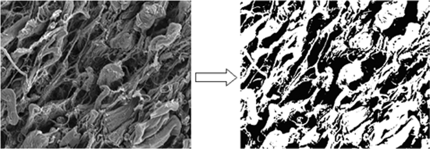
Illustration of the conversion of the scanning electron micrograph of a scaffold sample (left) to its corresponding binary image (right) with the Isodata algorithm (auto-thresholding) of Image J. The black pixels represent void areas (pores) and the white pixels represent the scaffold sample.
Assessment of pore size distribution
Because of the typically large range of pore size or pore area observed in an acellular biological scaffold, the variability or distribution of the pore size must be considered to better describe the dimensions of the pores. Based on their ECD as given in Equation (3), pores in each SEM micrograph can be divided into two groups (ranges) according to their dimensions: 1) ECD less than 10 μm and 2) ECD 10 μm or greater. Ten μm was considered to be the critical pore diameter, given that the characteristic diameter of mammalian cells is on the order of 10 μm. 23 For each image, the pore area in each group was normalized by dividing the total area of the pores in that group by the total area of all the pores shown in that micrograph, resulting in a “normalized pore area” given in percentages. The distribution of normalized pore area in the scaffold was measured from the same 45 SEM micrographs of five bovine acellular scaffolds as in the previous section on the measurement of pore size (ECD). Repeated-measures analysis of variance (ANOVA) was conducted to determine whether the distributions (proportions) of normalized pore area across the three regions (medial, intermediate, and lateral) were significantly different from one another in the five sample scaffolds for both pore size ranges (<10 μm and ≥10 μm). Also, paired Student t-tests were used to statistically compare the proportion of smaller pores (<10 μm) in each region with that of larger pores (≥10 μm) in the same region, for all three regions. The level of significance was set at 0.05.
Measurement of scaffold permeability
In material science, the term “permeability” is used to describe the conductivity of a porous medium with respect to permeation by a Newtonian fluid. 24 In the tissue-engineering literature, the term “porosity” has sometimes been used to refer to this concept of permeability,14,18,25 which is misleading. In the present study, the permeability of the bovine acellular scaffold was assessed as an indication of the overall porous nature of the material microstructure.3,5 Permeability was assessed by measuring the flow of deionized water through the scaffold as a barrier to hydrostatic pressure in a custom-built apparatus similar to that of Hiles et al. 25 Figure 3 shows the schematic design of the apparatus, in which a sample scaffold was subjected to near-constant hydrostatic pressure in a test chamber, with the flow across the sample measured by the rate of volume discharge in a measuring cylinder sampled at least five times over the duration of the experiment. A pressure range of 1.0 to 10.0 kPa was chosen, corresponding to the range of aerodynamic stress and impact stress values observed in human phonation (vocal fold vibration). 26 A water column, with effective heights of 10.2, 51, and 102 cm, created the hydrostatic pressures of 1.0, 5.0, and 10.0 kPa, respectively. To match with the typical dimensions of the bovine acellular scaffold, the orifice area in the test chamber from which the hydrostatic pressure was applied to the sample was 5.7256 mm2. At each pressure, five sample scaffolds of the reported dimensions obtained from five different animals were each mounted and rigidly secured in the test chamber as shown in Fig. 3, with no leakage of water observed. Also, to determine whether the permeability was dependent on the direction of flow, at the pressure of 10.0 kPa, five additional sample scaffolds were mounted with the medial surface facing the upper plate of the test chamber, whereas all other samples were mounted with the lateral surface facing the upper plate of the test chamber.

Schematic of the intrinsic permeability test apparatus. Details of the test chamber (
According to Darcy's law,
24
the coefficient of permeability (or hydraulic conductivity) K (in m/s) can be calculated for the bovine acellular scaffold:
Estimation of intrinsic permeability
Because the coefficient of permeability is dependent on the permeating fluid and the mechanism of permeation,
24
to characterize the hydraulic conductivity of a porous material independent of the properties of the fluid and the mechanisms of fluid flow, the “specific permeability” or “intrinsic permeability” can be derived from the coefficient of permeability, K. The intrinsic permeability, k, in m2 or in darcy, where 1 darcy ≈ 10−12 m2, is related to the coefficient of permeability as follows:
The effect of the direction of flow on the intrinsic permeability of the scaffold was assessed statistically using Student t-tests at all six time points from 30 min to 3 h. The level of significance was set at 0.05.
Results
Using the standard fluid replacement method with a pycnometer, the average porosity of the five bovine acellular scaffold samples was found to be 90.49%, with a standard deviation of 4.33% (n = 7). The porosity of acellular biological scaffolds has been reported to be approximately 63%, and upon chemical treatment (e.g., with acetic acid and collagenase), it could be improved to greater than 90%.16,17 With PBS solution as the replacement fluid, the porosity of the bovine acellular scaffold was found to be higher than that of other acellular scaffolds.
Three structural layers (regions) could be observed on the mid-coronal surfaces of the acellular scaffolds in SEM micrographs (Fig. 4). The pore architecture of these regions could correspond to that of the well-defined layered structure of the human vocal fold lamina propria, 27 with the higher density of matrix proteins in the medial region simulating the deep layer of the lamina propria. The medial region had an average thickness of 0.29 mm, the thinnest of the three regions. The average thickness of the intermediate region was 0.39 mm, and that of the lateral region was 0.44 mm.
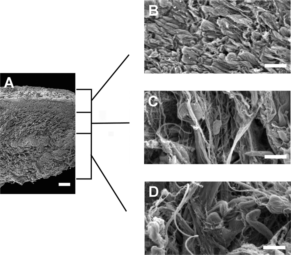
Scanning electron micrograph showing the mid-coronal cross-sectional surface of the bovine acellular scaffold at a total magnification of 40× (
Based on a total of approximately 80,289 pores in five bovine acellular scaffolds, the average pore diameter was 2.196 μm, with a large standard deviation of 9.311 μm. Because of the large variability in pore size or pore area in the scaffold, it was deemed that the dimensions of the pores may be better described according to the distribution of pore size. Thus, pores were divided into two ranges based on their dimensions (ECD < 10 μm and ECD ≥ 10 μm), with 10 μm chosen as the critical pore equivalent diameter. The largest pore equivalent diameter was measured as 98.9 μm. Figure 5 shows the percentage of pore area in both ranges of ECD across the three structural regions of the bovine acellular scaffold. It can be seen that, across all three regions, more than 60% of the total area of all pores was larger pores, with equivalent diameters of 10 μm or larger. On average, smaller pores (ECD < 10 μm) made up more of the pore area (39.17%) in the medial region than in the intermediate and the lateral regions, whereas larger pores (with ECD ≥10 μm) made up most of the pore area (73.58%) in the intermediate region. Results of repeated-measures ANOVA showed that the pore size distributions of the three structural regions (medial, intermediate, and lateral) were not significantly different in the five scaffold samples for both smaller pores and larger pores (F(2, 8) = 1.3355, p > 0.05) (Fig. 5). On the other hand, results of paired Student t-tests revealed that, for the medial region, there was no significant difference between the proportions of smaller pores (<10 μm) and larger pores (≥10 μm) (t = 1.5640, df = 4, p > 0.05), whereas larger pores made up significantly larger proportions of pore area than smaller pores in the intermediate region (t = 8.8196, df = 4, p < 0.01) and the lateral region (t = 3.4548, df = 4, p < 0.05) (Fig. 5).
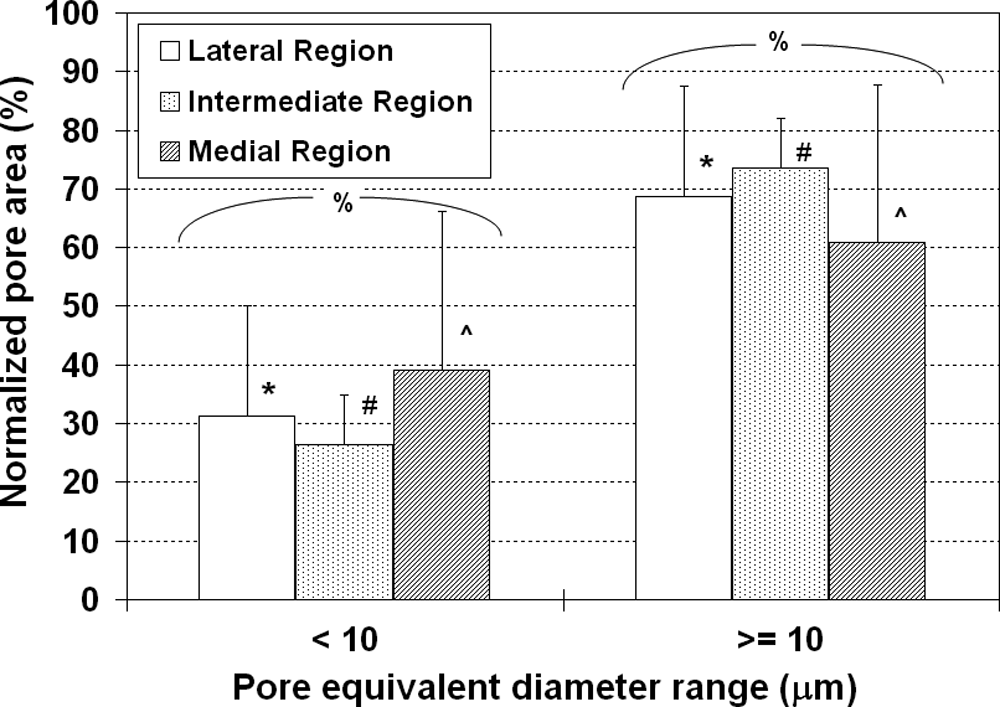
Distribution of normalized pore area (means ± standard deviations) in three regions of the mid-coronal surface of the bovine acellular scaffold, showing the percentage area of each group of pores relative to the total area of all pores. Pores are grouped based on their equivalent circle diameter (<10 μm and ≥10 μm) (n = 5). *p < 0.05 between the smaller pores and the larger pores in the lateral region; #p < 0.05 between the smaller pores and the larger pores in the intermediate region; ∧p > 0.05 between the smaller pores and the larger pores in the medial region; %p > 0.05 for the differences between the three regions for the smaller pores and the larger pores.
Figure 6 shows that the intrinsic permeability (k) of the bovine acellular scaffold increased linearly with time under three different levels of applied hydrostatic pressure. A linear increase of k with time was observed over a period of up to 15 h at a pressure of 1.0 kPa (Fig. 6A) and up to 3 h at pressures of 5.0 kPa (Fig. 6B) and 10.0 kPa (Fig. 6C). At the applied pressure of 10.0 kPa, although an apparently lower permeability was observed for the medial-lateral direction of flow at most time points, the variability was large, as shown by the error bars representing standard deviations. The differences between the opposite directions of flow were not statistically significant at all time points (e.g., at 3 h, Student t = 0.4573, df = 2, p > 0.05) (Fig. 6C). Figure 6D shows the dependence of k on the applied pressure. The mean values of k ranged from approximately 0.21 to 3.21 darcy (∼10−12 m2) (n = 5). According to the material science literature, 24 these values of intrinsic permeability can be considered semi-pervious or relatively porous, because they are also comparable with those of porous polymers such as poly(D,L-lactide)-co-glycolide, polyvinyl-alcohol, and poly-methyl methacrylate, with k values ranging from approximately 10−10 to 10−16 m2.28,29 It is also clear that k increased linearly with increasing applied pressure, consistent with previous results on other acellular biological scaffolds. 25
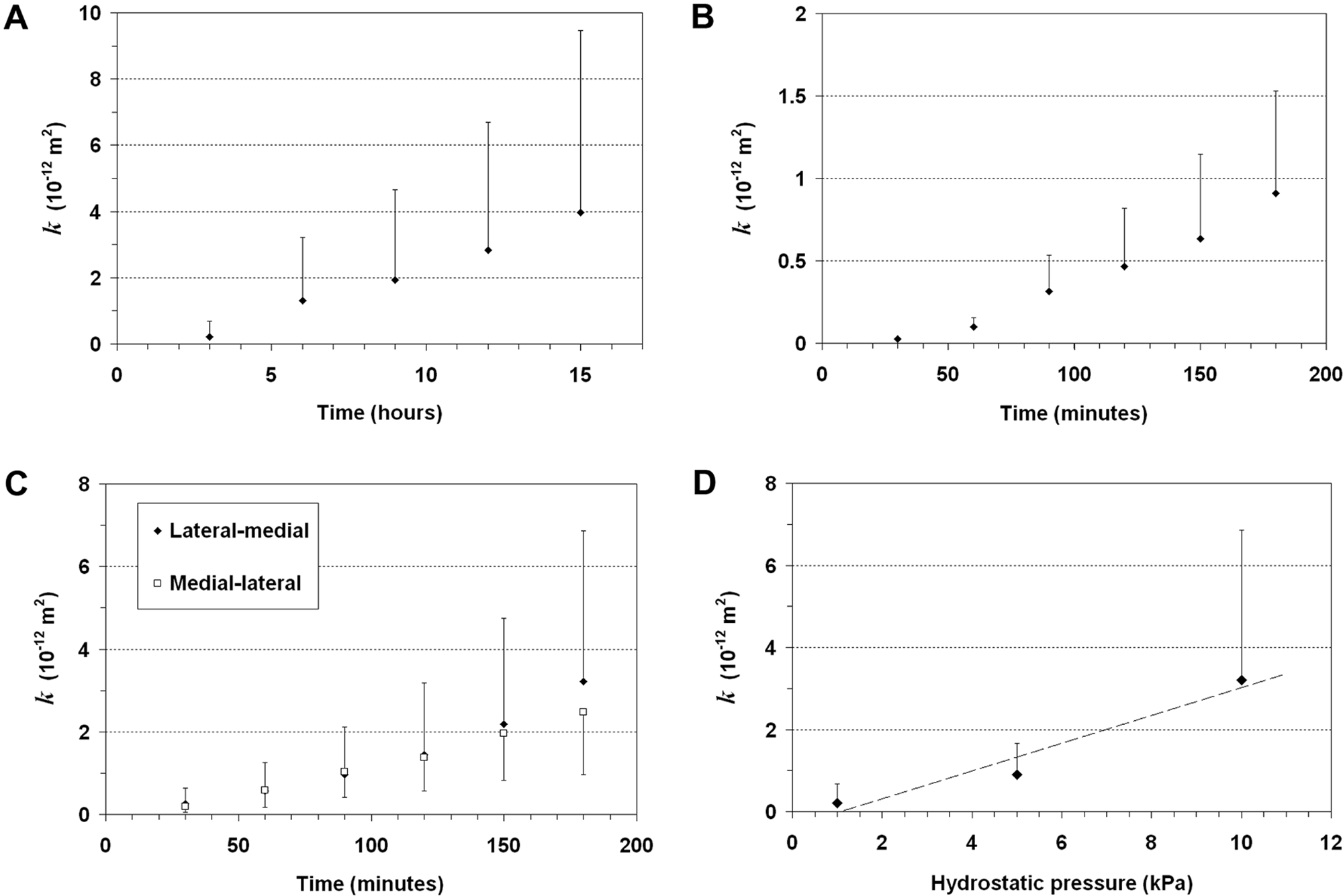
The intrinsic permeability (k) of the bovine acellular scaffold measured over (
Discussion
During phonation, the human vocal folds vibrate at high fundamental frequencies, ranging from approximately 100 to 300 Hz (for speech) or up to approximately 1000 Hz (for soprano singing), with an amplitude of approximately 1 to 3 mm and magnitudes of acceleration as high as 230 g. 30 The vocal fold lamina propria, primarily an ECM, is continuously subjected to a unique micromechanical environment.26,30,31 Thus, to facilitate the functional vibratory properties of the regenerated vocal fold, most of the attention in vocal fold tissue engineering has been on the development of implantable scaffolds with an optimal biomechanical effect. This is an appropriate focus, although an equally important aspect of tissue-engineering scaffolds, namely the pore architecture, has rarely been investigated for vocal fold scaffolds. The porous nature of the scaffold plays an important role in the transport of proteins, nutrients, growth factors, and other soluble factors, as well as in the promotion of cellular attachment, migration, and infiltration; the maintenance of tissue geometry and volume; and eventually the functional remodeling and regeneration of the ECM.1,32
Porosity can be expressed as the ratio of pore (void) volume to the apparent (total) volume. 4 It can be determined using several methods, including the buoyancy method, mercury intrusion, image analysis, and molecular probes. 21 In the present study, the fluid replacement method, also called the buoyancy method, was applied as one of the standard methods in materials science for the measurement of porosity.4,21,33,34 The fluid replacement method was straightforward, easy to conduct, safe, non-invasive, and reasonably accurate.21,33,34 One key advantage of this method is that the porosity of a sample in the wet condition can be measured, unlike with many other methods in which only the porosity of dried samples can be assessed, such as with helium pycnometry. 4 In the present study, PBS solution was used as the replacement fluid to minimize any interaction between the replacement fluid and the scaffold, such as dehydration and shrinkage of the collagen network associated with 100% ethanol. 35 PBS solution can also closely mimic the in vivo osmotic environment that the scaffold will be subjected to upon implantation into a host. It was found that the mean porosity of the bovine acellular vocal fold scaffold was 90.49%, with a standard deviation of 4.33% (n = 7). This result showed a much higher porosity than what was measured in our previous study using digital image analysis (47.03% ± 6.91%). 12 It is likely that this difference was due to the inherent limitations of the image analysis method for porosity measurement, because the process involved the estimation of three-dimensional (3-D) properties from 2-D images. The errors and uncertainties associated with image resolution and segmentation were difficult to quantify in image analysis. 21 Also, only a limited number of images was evaluated. On the other hand, the standard fluid replacement method was based on the penetration of fluid into the entire scaffold when it was fully saturated with the fluid, although dead-end pores might still have been missed. 4 The porosity of the bovine acellular scaffold was higher than that of other acellular biological scaffolds (e.g., ∼63% for bovine pericardia). 16
In vitro studies have shown that the topology of the scaffold, including that of the void space, affects the migration, attachment, proliferation, and cell–matrix interactions of the populating cells.32,36 To estimate the relationship between the average pore diameter of a matrix and its capacity for cellular attachment, Yannas 23 calculated the surface area available for cell binding per unit volume of a porous material with different pore sizes. It was found that the surface area per unit scaffold volume dropped dramatically with an increase in pore diameter. For example, using the model of Yannas, 23 the surface available for cell attachment per unit volume in a scaffold with a pore diameter of 300 μm is approximately 27 times lower than that of a scaffold with a pore diameter of 10 μm. This result suggested that, solely for the purpose of cellular attachment, the pore diameter should be kept small, although there is also a minimum limit on the pore diameter for the cells to approach the scaffold before attachment, which should equal to the characteristic diameter of mammalian cells, on the order of 10 μm. 23 The present study showed that, in the bovine acellular scaffold, large proportions of the void spaces consisted of large pores with equivalent diameters of 10 μm to 100 μm, such that unattached cells could easily approach the pores without compromising the availability of the scaffold surface for cellular attachment.
Unlike synthetic materials, acellular scaffolds derived from biological tissues consist of native ECM proteins (fibrous proteins and interstitial proteins). Hence, cellular interaction with an acellular scaffold should be similar to that with the native ECM, whereby host cells can attach to the matrix proteins of the acellular scaffold (e.g., collagens, elastin, fibronectin, laminins) using their cell surface adhesion molecules, such as integrins. 37 Host cell migration into the scaffold is key for wound healing and tissue remodeling. Generally, migration of stromal cells in the native ECM involves two processes. The first is related to the binding of integrins to provide signaling and traction for movement, and the second is the mechanical penetration or infiltration of cells into the ECM. The degradation of surrounding ECM by proteinases (e.g., MMPs), mechanical deformation of the ECM network, or simply the squeezing of cells through preexisting pores could facilitate the latter.38,39 Hence, pores provided by the acellular scaffolds are important for the infiltration and migration of host cells, especially when proteinase activities are limited. 38 Although the average pore size of the bovine acellular scaffold was small (2.196 μm), at least 60% of the total void space in the three different regions (medial, intermediate, and lateral) of the scaffold consisted of larger pores (ECD ≥ 10 μm), as shown in Fig. 5. Our previous study 12 on the acellular scaffold showed that, in SEM micrographs, pores occupied approximately 50% of the total surface area on the lateral, mid-coronal surface. Combining the findings of the present study with those of Xu et al. 12 suggested that more than 30% of the scaffold surface consisted of larger pores that would allow host cells to infiltrate the scaffold even when proteinase activities were limited.
Although the porosity and pore size reflect the volume and dimensions of the void space available in the scaffold, they provide no indication of the extent to which the pores are interconnected or how readily biofactors could move into and out of the scaffold. These properties are critical for the migration, nutritional support, and metabolism of cells populating the scaffold, allowing for the regeneration of viable tissue constructs. To describe the interconnectivity, the intrinsic permeability of the scaffold at three levels of applied hydrostatic pressure (1.0, 5.0, and 10.0 kPa) was measured in a custom-built test apparatus (Fig. 3). Results showed that the intrinsic permeability of the bovine acellular scaffold increased linearly with time, as well as with applied pressure (Fig. 6). These linear relationships were similar to those of the acellular porcine small-intestinal submucosa. 25 The intrinsic permeability of the scaffold was found not to depend on the direction of the flow (Fig. 6C).
According to Titze, 26 the typical magnitude of inertial stress in the vocal fold is approximately 1.0 kPa, which is also the typical aerodynamic driving pressure acting on the vocal fold surface during phonation at a comfortable pitch (fundamental frequency) and a comfortable loudness (subglottal pressure). The peak impact stress during vocal fold collision has been found to be approximately 5.0 kPa and could reach 10.0 kPa for phonation at high fundamental frequencies and high subglottal pressures. 26 Under the three levels of applied hydrostatic pressure (1.0, 5.0, and 10.0 kPa), the intrinsic permeability ranged from approximately 0.21 to 3.21 darcy, comparable with the permeability of porous synthetic polymer scaffolds. These data suggest that the capability of the scaffold to transport and deliver biofactors would probably remain intact even when it was subjected to typical magnitudes of mechanical stresses in phonation.
Our previous in vitro study demonstrated that primary human vocal fold fibroblasts seeded onto the lateral, cut surface of a bovine acellular scaffold were able to proliferate and infiltrate the scaffold to a depth of approximately 20 μm. 12 The vocal fold fibroblasts remained viable and active in protein synthesis, as shown by their morphological incorporation into the 3-D protein structure of the scaffold, as well as by results of biochemical assays on ECM proteins. 12 The depth of cellular infiltration was reasonable given the absence of any significant angiogenesis or the use of any perfusion system, because it is likely that only limited nutrients and oxygen supplies were available to any cells deep in the scaffold with a thickness of up to approximately 750 μm. 12 These previous results combined with the present findings provided preliminary support for the notion that the pores on the surface and in the 3-D protein network of the scaffold were capable of the transport of biofactors such as proteins, oxygen, nutrients, and other soluble factors.
Conclusion
Being porous is a crucial physical characteristic for tissue-engineered scaffolds. The pore architecture dictates the ease with which cells, proteins, nutrients, growth factors, and other biofactors can be transported through the scaffold, contributing to the functional processes of ECM remodeling and tissue reconstruction. Tissue-engineered scaffolds derived from biological tissues are known to have low porosity, but the results of this study showed that the bovine acellular scaffold developed in our laboratory 12 is reasonably porous (with a 90% porosity), has a proper pore size distribution (60% of pores with equivalent diameters ≥10 μm) for cellular attachment, migration, and infiltration; and has a high intrinsic permeability (0.21–3.21 darcy) to facilitate the transport of soluble factors (e.g., oxygen and nutrients) to the host cells once the acellular scaffold is implanted in vivo. Overall, these results supported the potential of the bovine acellular scaffold for the regeneration of the human vocal fold lamina propria conducive to functional tissue remodeling. Nevertheless, these observations must be considered preliminary, because the current findings were based on only a limited number of sample scaffolds. Since the variability of the data was large, further studies with a larger number of samples are required to corroborate the present results and to reveal any statistical significance. In addition, it is not yet established exactly what kind of pore structure might be the best for biological ECM scaffolds, particularly as it pertains to reconstruction of the vocal fold lamina propria. The optimal pore characteristics of acellular scaffolds for vocal fold regeneration and the decellularization processing methods for achieving such pore structure are currently unknown. Further studies along these important directions are warranted.
Footnotes
Acknowledgments
This work was supported by the National Institutes of Health, National Institute on Deafness and Other Communication Disorders Grant R01 DC006101. The authors would like to thank Dr. Jian Yang of the University of Texas at Arlington for his advice on the measurement of scaffold porosity.