
Abstract
Abstract
We describe a novel bioreactor system for tissue engineering of bone that enables cultivation of up to six tissue constructs simultaneously, with direct perfusion and imaging capability. The bioreactor was used to investigate the relative effects of initial seeding density and medium perfusion rate on the growth and osteogenic differentiation patterns of bone marrow–derived human mesenchymal stem cells (hMSCs) cultured on three-dimensional scaffolds. Fully decellularized bovine trabecular bone was used as a scaffold because it provided suitable “biomimetic” topography, biochemical composition, and mechanical properties for osteogenic differentiation of hMSCs. Trabecular bone plugs were completely denuded of cellular material using a serial treatment with hypotonic buffers and detergents, seeded with hMSCs, and cultured for 5 weeks. Increasing seeding density from 30 × 106 cells/mL to 60 × 106 cells/mL did not measurably influence the characteristics of tissue-engineered bone, in contrast to an increase in the perfusion rate from 100 μms−1 to 400 μms−1, which radically improved final cell numbers, cell distributions throughout the constructs, and the amounts of bone proteins and minerals. Taken together, these findings suggest that the rate of medium perfusion during cultivation has a significant effect on the characteristics of engineered bone.
Introduction
Cell-seeding density is known to influence the characteristics and functionality of engineered tissues. In prior studies, the effect of varying the density of cells seeded into constructs was investigated for bone, cartilage, and cardiac tissues.16–20 The effect of higher seeding densities was dependent on the tissue type as well as the culture conditions. For example, high seeding densities of chondrocytes in agarose gels yielded better mechanical properties, but these gels were less responsive to mechanical stimulation. 18 When the seeding density for tissue engineered bone was increased from 1 × 10⁶ cells/mL to 10 × 106 cells/mL, there was no enhancement in bone formation. 19 However, homogenous cell distribution throughout the constructs, which facilitates tissue development, was better at higher seeding densities. Hence, there is a need to better understand the effects of the initial cell density and medium flow on bone formation to support the required construct development and functionality.
In this study mineralized, fully decellularized bone (DCB) was used to generate scaffolds for tissue-engineering applications. Prior studies, including the seminal work done by Urist, 21 have demonstrated the utility of demineralized bone matrix as an osteo-inductive biomaterial and have led to the discovery of bone-derived growth factors, the primary osteoinductive components of bone matrix. 22 As a result, various forms of demineralized bone matrix, bone morphogenetic proteins, and bone graft substitutes (including allografts) are now commonly used in clinical applications for bone repair. 23 We hypothesized that the ability of DCB grafts to provide the osteoinductive growth factors, in addition to mechanical, biochemical, and architectural properties, without eliciting immunogenic responses makes them a suitable scaffold for bone tissue–engineering applications. DCB also has a potential advantage over the commonly used demineralized bone matrix in that its superior mechanical properties can help to expedite its functionality upon integration into defects in load-bearing sites. DCB scaffolds have already been employed for various tissue-engineering applications.24–27 However, here we describe the application of a rigorous technique for removing all of the cellular material from cores taken from the trabecular regions of bovine bone, with the aim of obtaining biological, osteoinductive, degradable scaffolds, which may be seeded with human mesenchymal stem cells (hMSCs) to tissue engineer bone grafts for the treatment of load-bearing, clinically relevant osseous non-unions.
To determine the relative contributions of perfusion rates and cell seeding density on the characteristics of tissue-engineered bone, a novel bioreactor system was developed, characterized, and used to investigate the effect of two different medium perfusion rates (superficial velocities through scaffold: 100 μms−1 and 400 μms−1) and two seeding densities (30 × 106 cells/mL and 60 × 106 cells/mL). The growth and tissue development of hMSCs seeded into the DCB scaffolds and cultured for 5 weeks were evaluated using biochemical, histological, and imaging techniques. It was found that perfusion rates had greater influence on cell growth and bone formation than did the initial seeding density.
Materials and Methods
Perfusion bioreactor
A novel, easy-to-use perfusion bioreactor system has been developed, which addresses several key design issues including the use of multiple scaffolds (4 to 10 mm in diameter and up to 4 mm high) and imaging capabilities. The bioreactor has the dimensions of a 10-cm glass Petri dish, and the glass cover of this dish serves as the removable bioreactor cover (Fig. 1A). This enables access to scaffolds as well as the use of traditional cell culture techniques for medium changes, thus eliminating any technical challenges of using an engineering system for cell culture. The bioreactor was machined from polycarbonate plastic with medical-grade (platinum cured) silicone gaskets separating the individual parts. Scaffolds were press-fit in place into a layer of polydimethylsiloxane (PDMS).

Bioreactor design. (
Scaffolds were placed into six wells arranged in a radial pattern. Medium entered the bioreactor through a central port from where it flowed to the center of the bioreactor and became evenly distributed into six channels leading to the scaffolds (Fig. 1B). The width and height of the inlet channel were 1.0 mm × 1.0 mm. The six channels leading to each scaffold were 1.0 mm wide and had a height that was tapered from 1.0 mm at the center to 0.5 mm below each scaffold. The channels were designed to provide relatively high flow resistance to the medium recirculation, as opposed to the resistance provided by the scaffolds themselves, and thereby maintained equal flow rates of culture medium through each scaffold. To verify this important feature of the bioreactor, we measured the flow rate through scaffolds with different densities at a constant pressure head. In addition, the flow rate through the bioreactor with and without scaffolds were compared at high and low pressure heads. This measurement was repeated at multiple time points. In each case, the flow rate was determined by measuring the total time taken for 20 mL of fluid to flow through, and this measurement was repeated at multiple time points.
The bioreactor was checked for the formation of bubbles every day to avoid obstruction of flow. Any gas bubbles that formed in the channels were removed using a syringe attached upstream of the inlet port using a three-way connector. During normal operation, the syringe is closed off from the flow of medium. Medium from the space above the scaffolds, which serves as a reservoir, flows through the scaffolds. During culture, the direction of flow is upwards through the scaffold into the reservoir, which enables the use of the reservoir as a bubble trap. The reservoir also serves as a gas exchanger for equilibrating oxygen and pH levels in culture medium with the incubator environment. Medium flows out of the reservoir through a port on the side of the bioreactor and is re-circulated by a low-flow multi-channel digital peristaltic Ismatec pump (Cole-Parmer, Vernon Hills, IL).
Decellularized bone scaffolds
Cylindrical, trabecular bone grafts (4 mm in diameter) were cored from the subchondral region of carpometacarpal joints of 2-week- to 4-month-old cows. The plugs were washed with a high-velocity stream of water to remove the marrow from the pore spaces and then for 1 hr in phosphate buffered saline (PBS) with 0.1% ethylenediaminetetraacetic acid (EDTA) at room temperature. This step was followed by sequential washes in hypotonic buffer (10 mM Tris, 0.1% EDTA, overnight at 4°C), detergent (10 mM Tris, 0.5% sodium dodecyl sulfate (SDS), for 24 h at room temperature) and enzymatic solution (50 U/mL DNAse, 1 U/mL RNAse, 10 mM Tris, for 3–6 h at 37°C) to remove any remaining cellular material. After each step, the scaffolds were rinsed with PBS for 1 h. After SDS treatment, several washes (≥7) were performed until no more bubbles indicating the presence of detergent were seen in the PBS during washing. At the end of the process, decellularized bone plugs were rinsed in PBS, freeze-dried, and cut to 4-mm-long cylinders. The weights and exact dimensions of the plugs were measured and used to calculate the scaffold density and porosity. Scaffolds were sterilized in 70% ethanol for 1 h and incubated in culture medium overnight before seeding cells.
In vivo studies of scaffolds
To test for any immunogenic properties, decellularized bone scaffolds were implanted subcutaneously into the ventral regions of Sprague-Dawley rats following an approved standard protocol. The samples were removed 2 weeks after implantation, fixed in 3.7% formaldehyde, demineralized, embedded in paraffin, and sectioned into 5-μm-thick slices. Slides were stained with hematoxylin and eosin.
Human mesenchymal stem cells
Bone marrow–derived hMSCs were isolated from bone marrow aspirates taken with donor consent during hip replacement surgeries, using previously described cell isolation methods. 28 Cells were culture expanded in low-glucose Dulbecco's Modified Eagle Medium (DMEM) supplemented with 10% fetal bovine serum and 1% penicillin-streptomycin (control medium) up to the fourth passage and were then used for seeding onto the scaffolds. Constructs were subsequently cultured with control medium supplemented with osteogenic supplements: 10 nM dexamethasone, 10 mM sodium-β-glycerophosphate, and 0.05 mM ascorbic acid-2-phosphate.
Construct seeding and culture
Cells at the fourth passage were resuspended at a low seeding density (LS) of 30 × 106 or a high seeding density (HS) of 60 × 106 cells/mL. A 50-μL aliquot of the cell suspension was pipetted to the top of blot-dried scaffolds and allowed to percolate through. After 15 min, the scaffolds were rotated 180°, and 10 μL of medium was added to prevent the cells from drying out. This process was repeated every 15 min for up to 2 h to facilitate uniform cell distribution. Constructs were then placed in the bioreactors, and 40 mL of medium was added. After 1 day, scaffolds were perfused at a superficial velocity of 10 μm/s for up to 1 week to provide local supply of oxygen to the cells while maintaining low hydrodynamic shear to promote cell attachment.
The medium flow rates were then set to provide the superficial velocity of medium through the scaffolds of 100 μm/s−1 (low-flow; LF) or 400 μms−1 (high flow; HF). This resulted in four experimental groups: unseeded scaffolds (US) placed in bioreactors and perfused at 400 μms−1, LS-LF, LS-HF, and HS-LF. During culture, 50% of the medium volume was replaced twice a week. This partial medium exchange was determined in previous studies to provide supply of fresh nutrients along with the maintenance of cell-secreted factors in the cell environment. After 4 weeks of culture, the constructs were harvested and cut in half and the wet weight of each half was determined. Four constructs were used to provide statistical evaluation of the DNA and protein contents and alkaline phosphatase activity. The two remaining constructs from each group were used for imaging (histology, scanning electron microscopy (SEM), and micro-computed tomography (μ-CT)). After fixation, samples were imaged using μ-CT, then half of each sample was used for histology, and the other half was used for SEM.
Analytical assays
DNA
Tissue constructs were washed in PBS and then placed in 100 μL of digestion buffer (10 mM Tris, 1 mM EDTA and 0.1% Triton X-100) with 0.1 mg/mL of proteinase K in micro-centrifuge tubes. Constructs were maintained in this solution overnight at 50°C. The supernatants were drawn off and diluted 10 ×. A standard curve was prepared from a solution of salmon testes DNA obtained from Molecular Probes (Eugene, OR). Picogreen dye (Molecular Probes) was added to the samples in duplicate in 96-well plates (100 μL dye:100 μL sample) and read in a fluorescent plate reader (Excitation 485 nm: emission 530 nm).
Protein and alkaline phosphatase
Tissue constructs were washed in PBS and then placed in 100 μL of lysate buffer (PBS, 1% Trition X-100, 0.5% sodium deoxycholate, 0.1% SDS, 0.1 mg/mL phenylmethylsulfonylfluoride, and 0.3% aprotinin). Samples were maintained on ice and vortexed every 30 min for up to 2 h to mechanically break down cell membranes. The extracts were removed and centrifuged and the supernatant stored at −20°C. For the protein assay, 10 μL of each sample was added to 200 μL of protein dye (Protein Assay, BioRad, Hercules, CA) and the absorbance read at 595 nm. The readings were compared with a standard curve made using bovine serum albumin. For the alkaline phosphatase assay, 50 μL of samples was added to 50 μL of alkaline buffer and 50 μL of nitrophenyl-phosphate substrate solution in micro-centrifuge tubes at 37°C for 15 min. The reaction was stopped using 0.5 N sodium hydroxide solution. The absorbance was read at 405 nm and compared with a standard curve obtained from p-nitrophenol solutions of known concentrations.
Histology and immunohistochemistry
Constructs were washed in PBS and fixed in 10% formalin for 1 to 2 days. Samples were then decalcified for 2 days with Immunocal solution (Decal Chemical Corp., Tallman, NY), dehydrated with graded ethanol washes, embedded in paraffin, sectioned into 5-μm slices, and mounted on glass slides. (All four groups were processed in the same batch, with two sections per experimental group on each slide.) Samples were stained using conventional hematoxylin and eosin. Immunohistochemistry staining for collagen I, bone sialoprotein (BSP), and osteopontin were also performed. Briefly, the sections were deparaffinized with Citrisolv (Fisher, Pittsburgh, PA) and rehydrated with a graded series of ethanol washes. The antigens were retrieved by incubating in 0.25% trypsin. Sections were blocked with normal serum, stained with primary antibodies (collagen 1: mouse monoclonal anti-collagen I from Abcam (Cambridge, MA) (ab6308), BSP: rabbit polyclonal anti-BSP II from Chemicon (Temecula, CA) (AB1854), and osteopontin: rabbit polyclonal anti-osteopontin from Chemicon (AB1870)) and then secondary antibody, and developed with a biotin/avidin system. The serum, secondary antibody, and developing reagents were obtained from Vector Laboratories (Burlingame, CA) and included in the Vector Elite ABC kit (universal) (PK6200) and DAB/Ni Substrate (SK-4100). For negative controls, the first antibody incubation step was omitted.
Scanning electron microscopy
Samples were washed in PBS and then fixed in 2% glutaraldehyde in sodium cacodylate buffer for 2 h. Constructs were washed in buffer and freeze-dried overnight in a lyophilizer. Before imaging, samples were coated with gold and palladium and used for SEM. One construct from each group was used to image the cut surface, and another was used to image the outer surface.
Micro-computerized tomography
μCT was performed using a protocol described in detail in Liu et al. 30 with the following settings: voltage 55 kV, current 0.109 mA, slice thickness 21 μm, and inter-slice spacing 22 μm. After fixation with glutaraldehyde, the samples were aligned along their axial direction and stabilized with wet gauze in a 15-mL centrifuge tube. The tube was clamped in the specimen holder of a vivaCT 40 system (SCANCO Medical AG, Basserdorf, Switzerland). The 4-mm length of the scaffold was scanned at 21 μm isotropic resolution. The bone volume was obtained using a global thresholding technique that was applied to binarize gray-scale μCT images, and the minimum between the peaks in the voxel gray values histogram was chosen as the threshold value. The bone volume fraction (BVF) was calculated by dividing the bone volume by the volume of the sample. Spatial resolution of this full-voxel model was considered sufficient for evaluating the micro-architecture of the samples, based on the previously established methods. 30
Statistical analysis
Pair-wise comparisons of results were carried out using multi-variate analysis of variance followed by Tukey's post hoc analysis using STATISTICA software (StatSoft Inc., Tusca, OK). P < 0.05 was considered significant.
Results
Medium flow and shear stress within tissue constructs
The flow channels in the bioreactor provided an even distribution of medium flow between the six scaffolds. When scaffolds (n = 9) with densities ranging from 0.30 to 0.49 mg/ml were individually placed in the bioreactor, the flow rate through each scaffold for a constant pressure head remained 0.85 ± 0.01 mL/s regardless of density. By comparing flow through the bioreactor with and without all six scaffolds in place, at high pressure drop, the flow rate in each of the six channels measured 0.161 ± 0.001 mL/s (scaffolds in place) and 0.161 ± 0.002 mL/s (empty wells). At low pressure drop, the values were 0.0695 ± 0.001 mL/s (scaffolds in place) and 0.0698 ± 0.001 ML/s (empty wells). All measurements were repeated five times.
Order-of-magnitude estimates for shear stresses associated with medium flow in tissue constructs were obtained by making several assumptions. The internal geometries of the scaffolds were simplified by representing the porous network as bundles of cylindrical tubes of a given diameter (Fig. 2A). The complex architecture of the bone was represented using three simplified geometric configurations, all of which were modeled for the void volume of 70%, and fully developed laminar flow, as follows: First, to obtain the absolute minimum of the shear stress, the bone was modeled as a single 3.4-mm-diameter channel (calculated as combined void volume of the bone sample) within an outer cylinder. Next, the scaffold was represented as a system with 10 channels, 1.1 mm in diameter. Finally, the scaffold was modeled for a system containing 125 channels 300 μm in diameter each (which is considerably smaller than the pore sizes in DCB because tortuosity of the system is being neglected). The density (ρ) and viscosity (μ) values for culture medium were obtained from the literature as 1.004 g/mL and 0.77 cP, respectively. 29 The values for wall shear stresses (τw) were calculated as a function of cylinder diameter (dc) and mean linear flow rate (u), using the equation τw = (8 μu)/dc derived from the Hagen-Poiseuille relation for laminar flow through a conduit. Reynolds numbers were calculated for each configuration to validate the assumption of laminar flow. The results of modeling in Figure 2B show the estimated shear stress as a function of the channel diameter and medium flow rate. For the conditions studied, the estimated levels of shear stress were between 0.7 and 10 mPa.
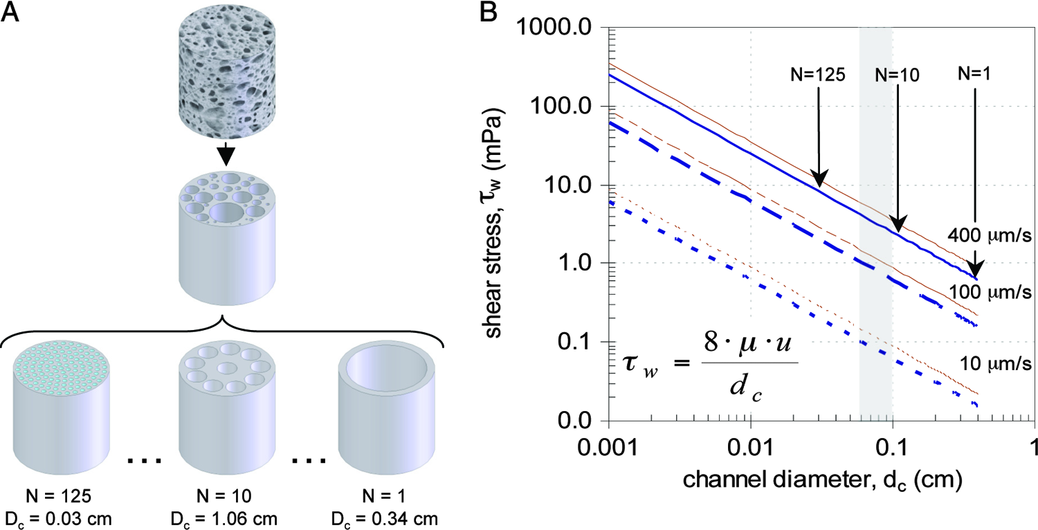
Estimates of shear stress in decellularized bone scaffolds. (
Scaffold properties
Native trabecular bovine bone was effectively decellularized by combining physical washing with detergent and enzymatic treatments. An average 6 × 10⁵ cells per scaffold was measured for native bone (Fig. 3A). This corresponds to a cell density of 1.2 × 10⁷ cells/mL of tissue, approximately ten times lower than typical values for native tissues, which may be due to high extracellular matrix (ECM) content in the bone marrow and the inability to fully digest the mineral encasing osteocytes (Fig. 3B). Also, the cellularity of the bone is heavily dependent on the age, the site of harvest, and the distance from the articular surface, resulting in large standard deviations of the DNA content of scaffolds. Picogreen assays indicated that washing the cored bone matrix removed roughly 80% of cells (Fig. 3A). Histological analysis indicated that osteocytes mostly remained trapped in the mineralized portion of the bone (Fig. 3C). Subsequent treatment methods removed an additional 98% of cells—including the osteocytes—resulting in virtually acellular scaffolds (Fig. 3D). Regardless of initial cell content, the decellularization procedure resulted in greater than 99.6% efficiency, with low standard deviations in the DNA content of processed bone scaffolds. The remaining components were primarily mineralized matrix and collagenous material and were found to have no apparent inflammatory or immunogenic responses after subcutaneous implantation into immuno-competent Sprague-Dawley rats. The decellularized scaffolds had approximately 70% to 80% void volume, with large pores approximately 600 to 1000 μm in diameter.
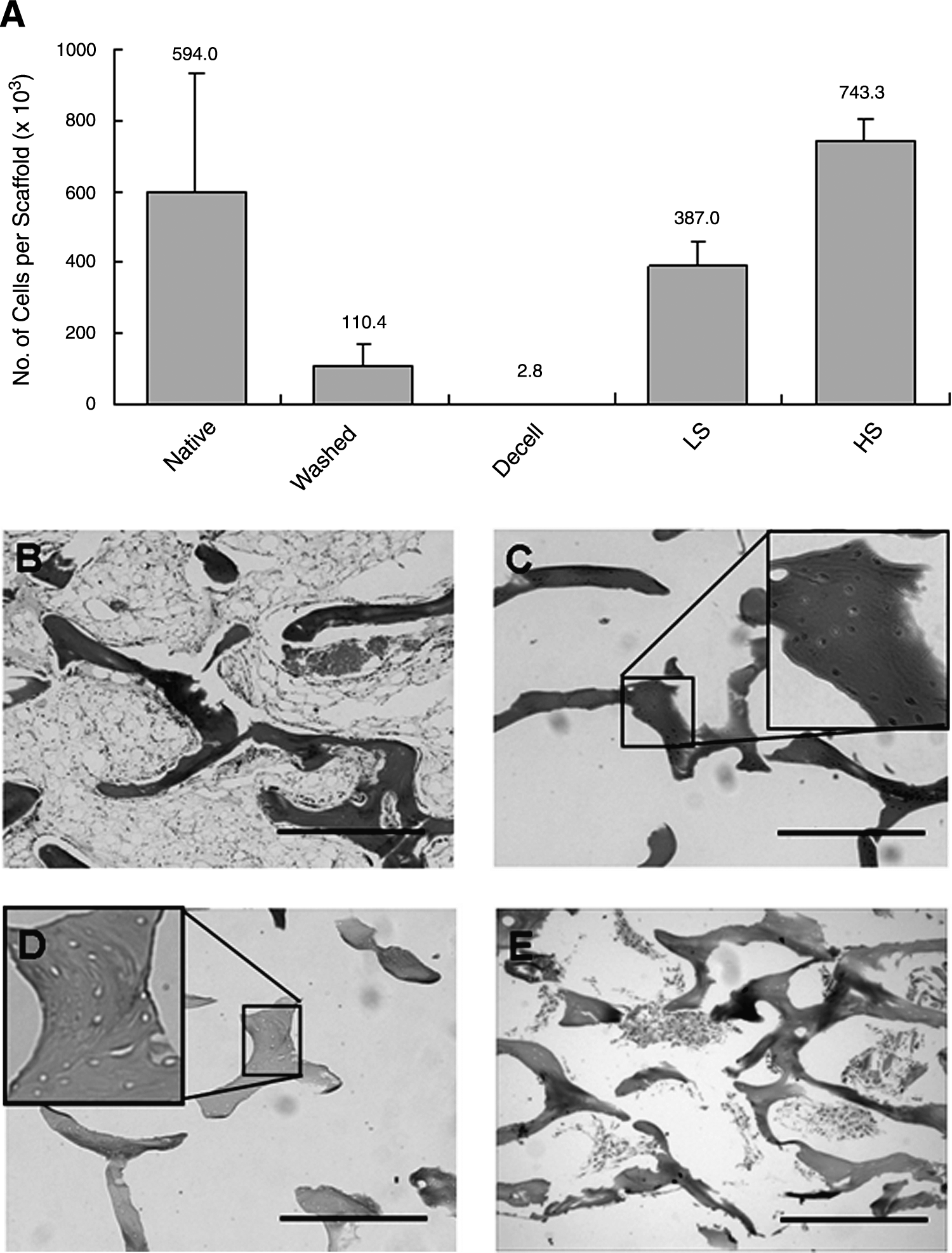
Decellularized bone scaffold. (
Cell distribution after seeding
For the LS-LF and LS-HF groups, a total of 1.5 × 10⁶ cells were seeded into the scaffolds. Immediately after seeding, 3.5 × 10⁵ cells per scaffold (28.1% of the total cells) were retained in the LS group (Fig. 3A), and 7.4 × 10⁵ cells per scaffold (24.7% of the total cells) were retained in the HS group (Fig. 3A). Seeding efficiency was thus independent of the initial density for the two cell densities used in the experiment. The cells that remained in the scaffolds after seeding were well distributed throughout the scaffold (Fig. 3E). The hMSCs appeared as clusters of cells at the edges and in the central regions, rather than being homogenously dispersed throughout the matrix. This contrasts with the distribution of cells at the end of the culture period.
Cell distribution after cultivation
Histological staining of the cell constructs in all of the seeded groups revealed acellular regions throughout the central parts of the grafts (Fig. 4A). A cross-section of the constructs in the axial direction exhibited a “shell” of cellular material at the upper and lower faces, as well as along the walls of the cylinders. The thickness of the cellular layer along the sides of the scaffolds appeared to be similar in all groups. However, cell distribution into the scaffold from the upper and lower ends and morphology throughout the scaffolds appeared to be dependent on the flow-rate and initial seeding density. The thicknesses of the cellular regions in the HS-LF group were estimated to be approximately 350 μm (Fig. 4A) and approximately 500 μm in LS-LF, whereas that of the high flow-rate (LS-HF) group was approximately 800 μm and approaching 1 mm from the edge in some places. At higher magnifications, however, another major difference was apparent in the two LF groups; the LS-LF group had thicker cell growth, but the cell density and ECM deposition were low, whereas the HS-LF group had a thin shell but densely packed cellular material (Fig. 4B). (The DNA values for the two groups are similar.) The cells in the HF groups exhibited dense packing as well as enhanced growth into the scaffolds.
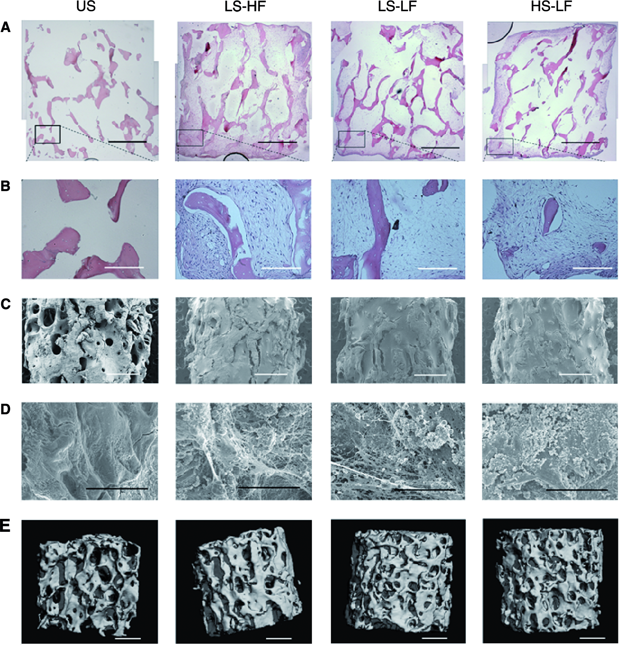
Tissue engineered bone: morphology. (
Cell morphology
Low-magnification (30 ×) images of the external surfaces of the seeded scaffolds indicated that hMSCs formed dense structures in the trabecular spaces (Fig. 4C). Examining the cut surfaces at higher magnifications (1000 ×) illustrated the formation of fibrillar ECM networks by the cells and demonstrated cells embedding themselves into the extracellular proteins (Fig. 4D). The large, empty pores at the beginning of culture would indicate that cells could adhere only to the walls of the pore spaces. However, by creating ECM networks and growing into the pore spaces, cells were able to provide themselves with a 3D context in localized regions.
Mineral deposition in engineered bone
Image analysis using μ-CT demonstrated that there were some differences in the microstructure of the bone grafts even after the cultivation period (Fig. 4E). All seeded groups exhibited greater bone volume fractions than the unseeded group. The BVF of the seeded groups was approximately 4% greater than that of the unseeded controls. The unseeded scaffolds had a BVF of 20%, whereas the volume fractions of the seeded groups ranged from 23.8% to 24.5%. Two other parameters were examined: connectivity density (a measure of the number of trabeculae that need to be cut in a given volume before the scaffolds break into two) and trabeculae number (the number of trabeculae per unit length). The average values for the unseeded scaffolds were 4.45 mm3 and 2.10 mm−1 respectively (Table 1). These values increased gradually: LS-LF (4.48 mm3, 2.21 mm−1), LS-HF (5.78 mm3, 2.28 mm−1), and HS-LF (6.34 mm3, 2.35 mm−1).
Biochemical composition of engineered bone
As expected, the seeded groups had statistically higher values for dry and wet weight (not shown), total protein, alkaline phosphatase, DNA, and BVF than unseeded scaffolds (Fig. 5A–D, respectively). However, the only statistical differences between the seeded groups were in the DNA values; the high-flow-rate (LS-HF) group facilitated higher cell numbers than either of the low-flow-rate groups. The DNA values (given as ngDNA/mg wet weight) corresponding to cell numbers in the LS-HF, LS-LF, and HS-LF groups that were 81.0%, 36.8%, and 23.2% of day 0 values, respectively.
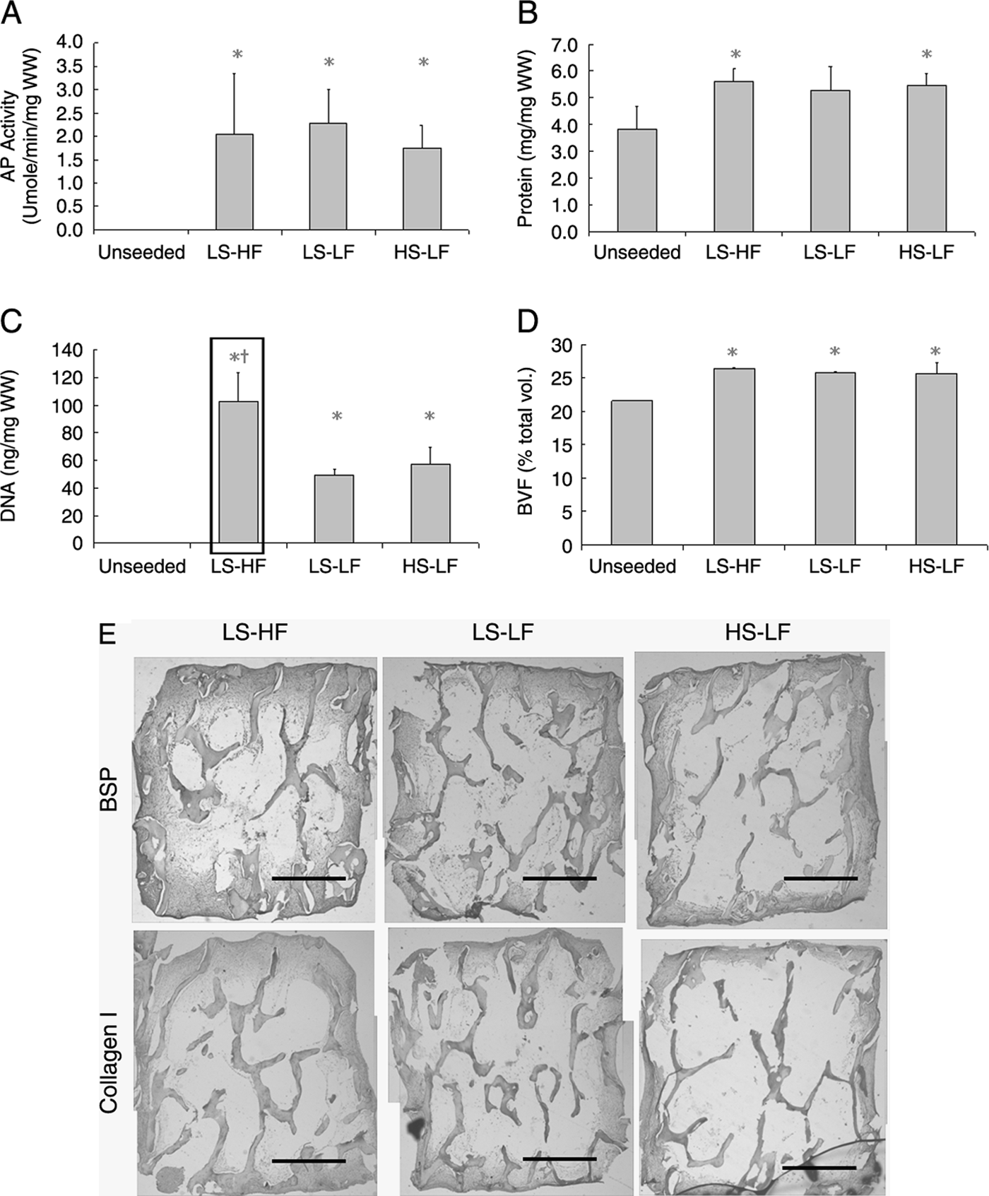
Tissue-engineered bone: marker expression.
Expression of bone markers
Cell constructs from each group were stained with antibodies to BSP, collagen I, and osteopontin that are all indicative of osteogenic differentiation (Fig. 5E). Unseeded constructs did not stain for BSP and osteopontin, but as expected, the mineralized regions of unseeded constructs stained positively for collagen I. The cellular regions of seeded constructs stained positively for collagen I, BSP, and osteopontin (not shown) content.
Discussion
Perfusion bioreactor system
We describe a novel perfusion system capable of culturing up to six tissue constructs simultaneously, thus enabling the collection of sufficient data for statistical analysis of each set of conditions (Fig. 1). The bioreactor design provided equal flow rates in the six individual scaffolds, for a wide range of flow rates (up to 1 mL/s of total flow) and scaffold densities (0.30–0.49 mg/mL). Even if the culture wells were empty (scaffold-free), the individual flow rates remained at the same level for a given pressure head. This feature is based on a higher flow resistance in the bioreactor channels than in the rest of the system and is important for preventing uneven distribution of flow due to the changes in scaffold density during cultivation. The system can also be modified in different ways. The scaffolds used in this study were 4 mm high × 4 mm in diameter. However, the bioreactor can be easily modified to culture cylindrical pieces of various diameters and thicknesses by having just one interchangeable layer with different well sizes. Another major advantage of the system is its ease of operation, acting, in essence, as a functionalized Petri dish. The system has the potential for online monitoring of bone growth and mineral deposition because scaffolds can be removed sterilely, imaged using non-invasive techniques such as nuclear magnetic resonance or μ-CT, and replaced. In these experiments, cell-seeded scaffolds were press-fit into the wells of the bioreactor with a surrounding gasket (made from PDMS), and flow experiments demonstrated that perfusion occurred through the interstices of the scaffolds rather than around its periphery.
Decellularized trabecular bone as a tissue-engineering scaffold
Functional bone grafts can be generated by seeding suitable biomaterial scaffolds with cells that have osteogenic potential and culturing them under conditions that optimize their bone-forming capabilities. Scaffolds should not exhibit immunogenic properties. For native tissues, this requires the effective removal of all cellular components and immunogenic proteins. In the present study, decellularization of bovine bone constructs resulted in the lack of invading leukocytes into scaffolds implanted subcutaneously into immuno-competent rats. From a tissue-engineering perspective, the topography of the scaffolds, as well as pore size, pore distribution, and biochemical content, should present hMSCs with a highly osteogenic microenvironment. Previous studies have shown that the presence of mineral in the constructs does not enhance or inhibit osteogenesis of hMSCs relative to demineralized bone constructs, 27 suggesting that growth factors trapped within the matrix remain accessible to hMSCs and that the mineral component does not enhance osteogenesis in hMSCs significantly better than in non-mineralized scaffolds.
Influence of seeding density and flow-rate
There were some, but not major, differences between the high-seeding-density and low-seeding-density groups (LS-LF and HS-LF) at the end of 5 weeks of culture with respect to the quantifiable parameters. Whereas the μ-CT analysis suggested differences in the microstructure of new bone formation (Fig. 5, Table 1), the histological analysis (Fig. 4, 5) indicated differences in cell distributions, cell morphologies, and ECM deposition. Hence, the initial seeding density may influence the cellular development in the resulting bone graft but does not appear to enhance bone formation or final cellular content. Comparisons with prior studies 19 suggest that the effect of increasing cell numbers is apparent only at lower seeding densities and may be widely negated in these experiments in which the seeding densities in the LS and HS groups were both high.
Conversely, flow rate appeared to play a more-significant role in the growth and tissue development of hMSCs within the scaffolds. The flow rates for the experiments were chosen to be within the ranges of superficial velocities used in other studies, in which considerable differences in mineral deposition were noted for variations in perfusion rates.5,10,31 The LS-HF group had twice as many cells as either of the low-flow-rate groups (Fig. 3A). This correlated with the findings in histology, which showed that the thickness and densities of the cellular regions were also considerably higher in LS-HF than in the other seeded groups (Fig. 4A, B). However, other measurable parameters, for example protein and BVF (Fig. 5B, D, respectively), were only slightly higher in the LS-HF group and not statistically different from in the low-flow groups. This suggests that the major advantage of high flow is better maintenance of cells rather than stimulation to form more mineralized deposits. To further support this, cell numbers at the end of the culture period were lower than at day 0 (Fig. 5C), suggesting that the differences were due to higher survival rates rather than enhanced proliferation of cells.
Oxygen becomes severely limiting for cells cultured statically within 3D matrices; 1 hence, medium perfusion is necessary to enhance cell survival.2,32 In this study, it appeared that increased nutrient transport from higher perfusion rates may be the primary factor contributing to differences observed between experimental groups. However, medium perfusion imparts fluid shear stresses to cells, and the shear levels within the system should be optimized for cell growth. A study done by Cartmell et al. 8 showed that low perfusion rates (0.01 mL/min) resulted in significantly greater osteoblast-like cell expansion than in static controls, whereas there was a decrease in cell viability within the constructs corresponding with increases in flow rates from 0.01 mL/min to 1.0 mL/min. In contrast, another group investigating the effects of perfusion rates on rat marrow-stromal osteoblasts found that greater flow rates (0.3, 1.0 and 3.0 mL/min) enhanced mineralization and calcium deposition in a dose-dependent manner. These inconsistencies may be explained in terms of differences in cell types, but differences in system geometries and internal architectures of the scaffolds such that similar volumetric flow rates result in highly dissimilar shear stresses imparted to cells also complicate direct comparisons. To aid in comparisons, flow rates in this study are reported as superficial linear velocities.
For the flow rates used, estimates of shear stresses within the constructs ranged from 0.0007 Pa (dc = 1100 μm) to 0.01 Pa (dc = 300 μm) (Fig. 2B). These values are similar to those reported in the literature for 3D scaffold systems.33,34 However, they are up to one thousand times lower than the shear levels required to elicit measurable responses from osteoblasts7,35 seeded on 2D surfaces or those predicted to act on osteocytes trapped in the lacuna system of native bone. 36 It is also roughly ten times lower than other in vitro studies designed specifically to elucidate the role of shear on bone formation in 3D scaffolds. 31 However, the approach used only gives order of magnitude estimates and does not account for the increases in shear with changes in internal architecture due to ECM deposition and mineralization. Hence, the role of shear in maintaining higher cell numbers and affecting osteogenesis in this system is not entirely clear, and further studies are ongoing to determine its role in tissue development.
Patterns of tissue development
In the engineering of a specific tissue phenotype, it is widely assumed that the eventual functionality is directly correlated with homogenous cell growth and matrix formation throughout the construct. One of the major motivations for using direct perfusion is to facilitate the uniform development of the tissue constructs. However, the growth patterns observed in these studies show cells developing preferentially on the edges of tissue constructs, as well as the inlet and outlet regions of the construct cross-section. This pattern is also observed in the mineralization data of perfusion studies performed by other groups. 8 One assumption is that, as cells grow into the pore spaces, they begin to occlude flow through the constructs and medium flows around the DCB scaffolds. However, the scaffolds are tightly press-fit into PDMS gaskets of a slightly smaller diameter, making it unlikely that fluid flows around the periphery rather than through the interstitial spaces, even at later stages of culture. The thicknesses of the cellular layers at the inlets and outlets correlate with perfusion rates, further suggesting that medium continues to perfuse through the interstices (Fig. 5E). The low-density central regions of constructs may therefore be indicative of insufficient nutrient delivery and may be solved by using even higher flow rates, the work now in progress.
Conclusion
This study describes the development and use of a versatile perfusion system in combination with decellularized bovine trabecular bone scaffolds to study the relative effects of greater seeding densities of hMSCs and higher medium perfusion rates. The results of the study indicate that, at the seeding densities evaluated, greater initial cell numbers do not necessarily result in increased bone formation. The flow rate of medium perfusion remains a significant parameter because the better nutrient transport afforded by higher perfusion rates enables more-uniform tissue development and supports higher cell densities within the constructs. Because the levels of shear stress are low in the range of flow rates studied, future studies need to investigate the ability of even higher perfusion rates to support cell growth throughout the 4-mm thickness of the scaffolds and determine at which point higher flow becomes detrimental to construct development.
Footnotes
Acknowledgments
The authors would like to thank Dr. Ed Guo for kindly sharing his μCT system and imaging expertise and Dr. Timothy Martens for help with animal surgery. The study was funded by the National Institutes of Health (R01 DE161525-01 and P41 EB02520-01A1, GV-N), Arthritis Foundation (PG Chao), and the Mandl Foundation (WG).