
Abstract
Abstract
Bridging of nerve gaps after injury is a major problem in peripheral nerve regeneration. Considering the potential application of a bio-artificial nerve guide material, polycaprolactone (PCL)/chitosan nanofibrous scaffolds was designed and evaluated in vitro using rat Schwann cells (RT4-D6P2T) for nerve tissue engineering. PCL, chitosan, and PCL/chitosan nanofibers with average fiber diameters of 630, 450, and 190 nm, respectively, were fabricated using an electrospinning process. The surface chemistry of the fabricated nanofibers was determined using Fourier transform infrared spectroscopy and X-ray photoelectron spectroscopy. Simple blending of PCL with chitosan proved an easy and efficient method for fabricating PCL/chitosan nanofibrous scaffolds, whose surface characteristics proved more hydrophilic than PCL nanofibers. Evaluation of mechanical properties showed that the Young's modulus and strain at break of the electrospun PCL/chitosan nanofibers were better than those of the chitosan nanofibers. Results of cell proliferation studies on nanofibrous scaffolds using 3-(4,5-dimethylthiazol-2-yl)-5-(3-carboxymethoxyphenyl)-2-(4-sulfophenyl)-2H-tetrazolium assay showed 48% more cell proliferation on PCL/chitosan scaffolds than on PCL scaffolds after 8 days of culture. PCL/chitosan scaffolds showed better cell proliferation than PCL scaffolds and maintained their characteristic cell morphology, with spreading bipolar elongations to the nanofibrous substrates. This electrospun nanofibrous matrix thus proved of specific interest in tissue engineering for peripheral nerve regeneration.
Introduction
Electrospinning is a well-established technique that has recently brought much attention to the fabrication of tissue-engineered scaffolds made of a non-woven, three-dimensional, porous, nanoscale fiber-based matrix. 11 In this process, a high-voltage electrical field is applied to a continuous strand of polymeric solution ejected from the open end of a needle spinneret to obtain nano- and micro fibers. The electrospun nanofiber matrix provides not only a high surface area–to-volume ratio, but also a structural morphology similar to the extracellular matrix (ECM), thereby serving as an effective tissue-engineered scaffold.12,13 A variety of polymeric biomaterials, including natural and synthetic polymers, have been investigated for their suitability in nerve tissue–engineering applications. Chitosan, a natural polymer, is an amino polysaccharide (poly-1,4-D-glucosamine) derived from chitin using de-acetylation. Because of its biocompatibility, biodegradability, and non-toxic and antimicrobial properties, chitosan has been widely studied for applications in biomedical, antimicrobial, and drug-delivery applications. 14 Fiber formation from chitosan has been met with challenges because of its limited solubility and poly-cationic nature in solution. Approaches chosen to overcome the limitations, including the weakness of chitosan, include graft polymerization and blending with other synthetic polymers, 15 although the cationic nature of chitosan provides an advantage in obtaining a suitable scaffold for cell adhesion in tissue-engineering applications. On the other hand, polycaprolactone (PCL) is an aliphatic, biodegradable, and biocompatible polyester with good tensile properties, although its poor hydrophilicity, slow degradation kinetics, and lack of natural cell recognition sites greatly limit its application in the biomedical field. Blending of chitosan with PCL provides an attractive option in improving its biological, mechanical, and degradation properties in comparison with individual components.
Schwann cells are the primary structural and functional cells that play an important role in peripheral nerve regeneration. Once a peripheral nerve is damaged, Schwann cells alter their morphology, behavior, and proliferation, being involved in Wallerian degeneration and Bungner bands. Schwann cells form myelin sheaths surrounding axons and guide and promote axonal growth to establish a precise innervation. 16 However, to our knowledge, there are few reports available on the in vitro responses of Schwann cells on electrospun biocomposite polymeric scaffolds. The present study explores the potential of blending PCL with chitosan and electrospinning to obtain PCL/chitosan nanofibrous scaffolds. Schwann cells were further cultured on this biocomposite nanofibrous scaffolds for evaluating biocompatibility and its application in nerve tissue engineering.
Materials and Methods
Materials
PCL (MW 80,000), low-molecular-weight chitosan (MW 120,000), 1,1,1,3,3,3-hexafluoro-2-propanol (HFIP), and fibronectin were obtained from Sigma-Aldrich (St. Louis, MO). Solvents such as methanol, chloroform, dichloromethane (DCM), and trifluoroacetic acid (TFA) were obtained from Merck (Hohenbrunn, Germany). The above-listed polymers and solvents were used as received without any further purification.
Fabrication of nanofibrous scaffolds
Polymeric solutions with suitable concentrations were prepared for electrospinning. PCL (15 wt%) was dissolved in chloroform/methanol (75:25 v/v) and used for electrospinning to obtain PCL nanofibers. Chitosan nanofibers were electrospun from 8 wt% of chitosan after dissolving in TFA/DCM (75:25 v/v). Chitosan scaffolds were prepared only for a comparison of surface morphology, chemical, and mechanical properties with PCL and PCL/chitosan scaffolds. For preparing PCL/chitosan nanofibrous scaffolds, PCL was first dissolved in HFIP and further blended with chitosan solution in TFA/DCM, to obtain a weight ratio of PCL/chitosan (75:25). Briefly, chitosan was dissolved in TFA/DCM, and the solution was stirred for 3 days for complete dissolution. PCL was dissolved in HFIP separately and stirred for a day. Further to this, chitosan solution was added to PCL solution to obtain a 75:25 weight ratio of PCL:chitosan, and this blend mixture was stirred for another day to obtain a homogenous solution before using for electrospinning. For electrospinning, the polymer solutions were individually fed into a 5-mL standard syringe attached to a 27G1/2 blunted stainless steel needle using a syringe pump (KD Scientific Inc., Holliston, MA) at a rate of 1 mL/h with an applied voltage of 12 to 20 kV (Gamma High Voltage Research, Ormond Beach, FL). A flat aluminum plate was kept 12 cm from the syringe needle tip for collecting the fibers. The polymer solution drawn from the syringe formed a pendant drop at the tip of the needle, and this positively charged jet travelled toward the collector. During this time, solvent evaporated, and fibers were deposited as a fibrous mat. Nanofibers were also collected on 15-mm-diameter cover slips for cell culture studies. The as-spun nanofibrous scaffolds were dried in a vacuum at room temperature before further investigation.
Structural morphology of nanofibrous scaffolds
The morphology of electrospun PCL, chitosan, and PCL/chitosan scaffolds were studied using field emission scanning electron microscopy (SEM; FEI-QUANTA 200F, Eindhoven, The Netherlands) at an accelerating voltage of 10 kV after sputter coating with gold (JEOL JFC-1600, Auto Fine Coater, Tokyo, Japan). Diameters of the electrospun fibers were analyzed from the obtained SEM images using image analysis software (Image J, National Institutes of Health, Bethesda, MD).
Characterization of nanofibrous scaffold
Attenuated total reflectance Fourier transform infrared (ATR-FTIR) spectroscopic analysis of electrospun PCL, chitosan, and PCL/chitosan nanofibrous scaffolds was performed on an Avatar 380 FTIR (Thermo Nicolet, Waltham, MA) over a range of 400 to 4000 cm−1 at a resolution of 2 cm−1.
X-ray photoelectron spectroscopy (XPS) is a widely used chemical analysis technique. XPS spectra of PCL, chitosan, and PCL/chitosan nanofibrous scaffolds were obtained from electrospun nanofibrous scaffolds. XPS analysis was conducted on a VG Escalab 2201-XL Base system (Thermo VG Scientific, West Sussex, England) with a take off angle of 90°. Binding energy was referenced to the C1S peak of saturated hydrocarbon at 285.0 eV.
Wettabilities of the electrospun nanofibrous scaffolds were measured using the sessile drop water contact angle measurement using a VCA Optima Surface Analysis system (AST Products, Billerica, MA). Distilled water was used as the testing liquid. Six samples were employed for each test, and the average value was calculated. The measured contact angle value reflects the hydrophilicity of the scaffolds. The lower the contact angle value, the higher the hydrophilicity of the scaffold is. 17
Tensile properties of electrospun nanofibrous scaffolds were determined at of 25°C and 74% humidity using an Instron 5845 Microtester (Norwood, MA) at a cross-head speed of 10 mm/min. Rectangular specimens 60 to 70 μm thick were cut from the as-spun membranes and used for mechanical testing studies. The ends of the rectangular specimens were mounted vertically on mechanical gripping units of the tensile tester. A minimum of six specimens of individual scaffolds were tested during this study.
Schwann cell culture
Schwann cell line (rat) RT4-D6P2T was obtained from ATCC (American Type Culture Collection, Manassas, VA) and cultured in Dulbecco's modified Eagle medium (DMEM) supplemented with 10% fetal bovine serum and a 1% antibiotic–antimycotic formulation (100×, containing 10,000 U/mL penicillin G sodium; 10,000 μg/mL streptomycin sulfate and 25 μg/mL amphotericin B in 0.85% saline; Invitrogen Corp., San Diego, CA). Cells were incubated at 37°C in a humidified atmosphere containing 5% carbon dioxide, and the culture medium was changed once every 2 days. Each of the nanofibrous scaffolds was placed on 15-mm cover slips in the wells of a 24-well plate and pressed with a stainless steel ring to ensure complete contact of the specimens and wells. The specimens were sterilized under an ultraviolet lamp, washed three times with phosphate buffered saline (PBS), and immersed in DMEM overnight before cell seeding. On confluency, cells grown in 75-cm2 cell culture flasks were detached using trypsinization with 0.25% trypsin containing 0.1% ethylenediaminetetra acetic acid (Invitrogen Corp.) and counted using trypan blue assay using a hemocytometer. Cells were further seeded at a density of 1.0 × 10⁴ and allowed to culture on tissue-cultured polystyrene plate (TCP as control), PCL and PCL/chitosan nanofibrous scaffolds. The viability of attached cells was determined by 3-(4,5-dimethylthiazol-2-yl)-5-(3-carboxymethoxyphenyl)-2-(4-sulfophenyl)-2H-tetrazolium (MTS) assay after 2, 4, 6, and 8 days.
Quantification of viable cells (MTS assay)
To study the cell adhesion and proliferation on different scaffolds, the number of viable cells were determined using the colorimetric MTS assay (CellTiter 96 AQueous One Solution, Promega, Madison, WI). The assay is based on the reduction of yellow tetrazolium salt in MTS to form purple formazan crystals by dehydrogenase enzymes secreted from the mitochondria of metabolically active cells. The amount of formazan crystals formed is directly proportional to the number of viable cells. MTS assay is an indirect measurement of cell proliferation that serves as an indicator of cell viability after seeding cells on different scaffolds. The metabolic activity of cells might also vary depending on the surface chemistry and composition of materials. Briefly, the samples were rinsed with PBS to remove unattached cells and further incubated with 20% MTS reagent in a serum-free medium for a period of 3 h. It was further aliquoted into 96-well plates, and absorbance was read at 490 nm using a spectrophotometric plate reader (FLUOstar OPTIMA, BMG Lab Technologies, Offenburg, Germany).
Morphology of cultured cells
Morphological observations of the in vitro culture of Schwann cells on PCL and PCL/ chitosan scaffolds were carried out. To provide a surface conducive to efficient cell attachment, scaffolds were pre-wetted with 10 μL of 0.1% fibronectin in PBS before cell seeding. After 6 days of cell proliferation, the cell-cultured scaffolds were rinsed twice with PBS and fixed with 3% glutaraldehyde solution for 3 h at 37°C. Then the scaffolds were washed with deionized water and dehydrated with gradient concentrations of ethanol (50%, 70% 95%, 100%) for 15 min each. After final washings with 100% ethanol, specimens were added with hexamethyldisilazane (HMDS, Sigma, Singapore). Finally, HMDS was air-dried, and scaffolds were coated with gold, mounted on a SEM stub, and observed under SEM. For comparison, the morphology of cells seeded on TCP was investigated. SEM observation was also carried out on cells grown on PCL/chitosan nanofibrous scaffolds without prior fibronectin coating.
Statistical analysis
Data were presented as means ± standard deviations from triplicate analysis. Single-factor analysis of variance was carried out to compare the means of different data sets, and a value of p ≤ 0.05 was considered statistically significant.
Results
Morphology and characterization of PCL, chitosan, and PCL/chitosan nanofibers
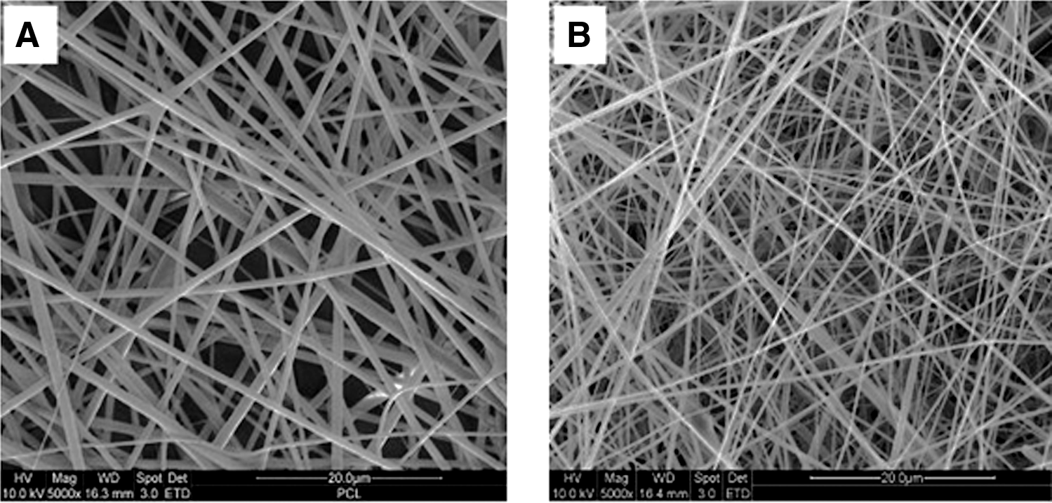
Electrospinning parameters were optimized during this study to obtain bead-free and relatively uniform nanofibers of PCL, chitosan, and PCL/chitosan. Figure 1 reveals the SEM micrographs showing the morphology of electrospun PCL, chitosan, and PCL/chitosan nanofibrous scaffolds. PCL nanofibers with average diameters of 630 ± 40 nm were obtained, whereas PCL/chitosan composite nanofibers were 190 ± 26 nm. Blending of PCL with chitosan had the advantage of producing thin fibers with diameters less than 200 nm.
Scanning electron micrography micrographs of electrospun (
The presence of amine groups on the surface of PCL/chitosan nanofibrous scaffolds was characterized using ATR-FTIR spectroscopy studies. Figure 2 shows the ATR-FTIR spectra of three different electrospun nanofibrous scaffolds produced during this study, extracted within a range of 1000 to 1800 cm−1. The ester stretching of PCL at 1724 cm−1 was observed as a major peak on PCL scaffolds. The FTIR spectra of chitosan scaffold exhibited the resonance bands at 1087 to 1203 cm−1 characteristic of its saccharide structure and those at 3400, 1680, and 1544 cm−1 commonly known for the N-H stretching of the primary amino groups, the carbonyl stretching (amide I), and N-H bending (amide II), respectively. The transmission peaks at 1680 and 1544 cm−1 corresponding to amide I and II were present on PCL/chitosan nanofibrous scaffolds, along with the major ester stretching of PCL matrix at 1724 cm−1 (Fig. 2).

Attenuated total-reflectance Fourier transform infrared spectra of electrospun polycaprolactone (PCL), chitosan, and PCL/chitosan nanofibers. In situ figure shows the major N-H stretching vibration of chitosan at 3400 cm−1.
Surface chemistry of the blended nanofibers was further verified using XPS studies. Table 1 shows the atomic ratios of carbon, nitrogen, and oxygen on the surface of PCL/chitosan nanofibers, together with those of PCL and chitosan nanofibers. Nitrogen atoms were not present within the chemical composition of PCL, whereas they were present in chitosan. The presence of N1S peak in the survey scan of XPS spectra of chitosan nanofibers confirmed this (Fig. 3), whereas no nitrogen peak was observed in the XPS spectra of PCL nanofibers. The presence of N1S peak observed in Figure 3 and indicated as the N atomic ratio in Table 1 also confirmed the presence of chitosan molecules on the electrospun PCL/chitosan nanofibrous surface. However, less nitrogen was found on the surface of PCL/chitosan scaffolds than on chitosan nanofibers.
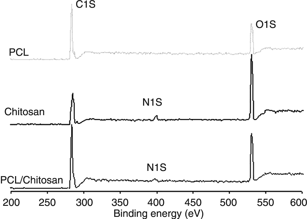
Survey scan X-ray photoelectron spectroscopy of electrospun polycaprolactone (PCL), chitosan, and PCL/chitosan nanofibers. N1S peak verified the presence of chitosan on electrospun PCL/chitosan nanofibrous scaffolds.
PCL, polycaprolactone.
Cells attached and spread more easily and effectively on surfaces with proper hydrophilicity than on hydrophobic surfaces.18,19 The effectiveness of blending PCL with chitosan in increasing the wettability of PCL/chitosan scaffolds was also checked using water contact angle measurement. The results are summarized along with the fiber diameters in Table 2. The contact angle of PCL scaffolds was 118°, indicating that water does not spread on this scaffold surface because of its hydrophobic characteristics. Apparently, chitosan showed zero water contact angle. Blending of PCL with chitosan increased the wettability of nanofibrous PCL/chitosan scaffolds, as observed from their water contact angle measurement results, as shown in Table 2.
PCL, polycaprolactone.
Mechanical properties of PCL, chitosan, and PCL/chitosan nanofibers
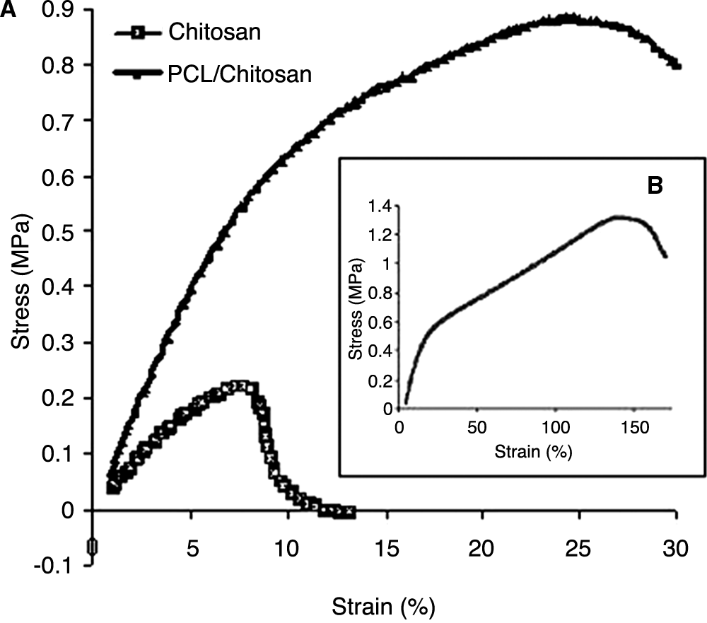
Figure 4 shows the typical stress-strain curve of PCL, chitosan, and PCL/chitosan nanofibrous scaffolds under tensile loading. For PCL scaffolds, the average tensile strength was 1.31 ± 0.25 Mpa, with an ultimate strain of 145 ± 35%. Experimental results show that the mechanical strength of PCL nanofibers was higher than that of other nanofibrous scaffolds (Table 3). Chitosan nanofibers have a low tensile strength of 0.22 Mpa, with a strain at break of 7.1%. However, PCL/chitosan composite nanofibers had superior mechanical properties to those of chitosan nanofibers. Table 3 summarizes the mechanical integrity in terms of tensile strength, Young's modulus, and elongation at break of the electrospun nanofibrous scaffolds. The Young's modulus of different scaffolds in tension was calculated from the slope of the stress-strain curve in the linear region.
Stress-strain curve of electrospun (
PCL, polycaprolactone.
Proliferation of Schwann cells on PCL/chitosan nanofibers
The proliferation of Schwann cells on PCL, PCL/chitosan nanofibrous scaffolds, and TCP cultured on days 2, 4, 6, and 8 was determined using MTS assay (Fig. 5). The Schwann cell proliferation rate was significantly (p ≤ 0.05) greater in PCL/chitosan scaffolds than PCL scaffolds. Although the PCL nanofibers showed a slow increase in cell proliferation throughout the designated time intervals, it was significantly lower than in other groups. Results of this study showed that PCL nanofibers supported the growth of Schwann cells, but the introduction of amine groups via chitosan in PCL/chitosan enhanced the proliferation rate of Schwann cells.

Schwann cell proliferation on polycaprolactone (PCL), PCL/chitosan nanofibers, and tissue-cultured polystyrene plate, as determined using 3-(4,5-dimethylthiazol-2-yl)-5-(3-carboxymethoxyphenyl)-2-(4-sulfophenyl)-2H-tetrazolium assay.
Morphology of Schwann cells on PCL/chitosan nanofibers
Figure 6 shows the SEM micrographs of Schwann cell morphology on TCP, PCL, and PCL/chitosan scaffolds. A positive effect on the physiology of Schwann cells is an essential requirement of a guidance material, for example, nanofibers used during this study. After 5 h of cell seeding, cells appeared to have a round shape, but they stretched into spindle shape after 24 h of cell seeding. Schwann cells interacted and integrated well with the surrounding fibers in PCL and PCL/chitosan nanofibrous scaffolds (Fig. 6), but the cells had a typical oval-shaped cell body with long extensions only on PCL/chitosan nanofibers, giving an overall spindle shape that was typical with normal cell morphology on TCP. The morphology of cells on PCL/chitosan scaffolds without fibronectin coating was also similar to those on PCL/chitosan scaffolds coated with fibronectin (Fig. 7). Moreover, the proliferation rate was lower on PCL scaffolds than PCL/chitosan nanofibrous scaffolds. Fiber architecture appeared to guide cell growth, giving rise to a three-dimensional, multi-cellular network guided by the architecture of the fibrous scaffold.

Comparison of Schwann cell morphology under low magnification and high magnification on (

Schwann cell morphology without fibronectin coating on (
Discussion
A wide variety of materials and approaches are being explored in an effort to identify a material to facilitate the regeneration of severed nerves. Synthetic polymers, including PCL, have been used widely in a variety of biomedical applications because of their physicochemical and mechanical characteristics, but they are bio-inert and do not possess biological functions. The introduction of functional groups onto their surfaces for an enhanced cellular response has previously been achieved using aminolysis, 20 plasma treatment, 21 or a simple adsorption process. 22 Santiago and co-workers 23 introduced amine groups onto the surface of PCL films through treatment with a diamine followed by attachment of RGD peptide using carbodiimide, whereas other researchers used epoxy-amine chemistry. 24 However, such modification processes involving severe chemical reactions might destroy the polymer surface or result in an exchange or removal of the bioactive groups during in vitro culture or in vivo implantation. Blends of PCL with polysaccharides prepared recently by Ciardelli et al. and hollow fibers were fabricated for tissue-engineering applications using PCL and poly(3-hydroxybutyrate-co-3-hydroxyvalerate).25,26
Collagen and chitosan are two of the most commonly used naturally derived polymers in tissue engineering, but material availability, cost, and poor immunogenicity have hindered the widespread application of collagen. Chitosan fibers supported the adhesion, migration, and proliferation of Schwann cells, 27 but a major drawback of using chitosan alone for tissue-engineering applications is its enhanced dissolution due to its hydrophilicity along with swelling characteristics. 28 Cao et al. used cross-linking agents such as hexa-methylene diisocyanate, epichlorohydrin, and glutaraldehyde to improve the swelling and degradation properties of chitosan films. 29 Hexa-methylene diisocyanate—cross-linked chitosan films were found to enhance the proliferation of Schwann cells and proved a promising method to modify the chitosan conduits for nerve repair. Enhancing chitosan nerve cell affinity was also attempted by blending chitosan with protein or peptide 30 or by using hydroxy-apatite–coated chitosan tubes impregnated with laminin or laminin peptides. 31 In another study, composite materials of chitosan/poly(L-lysine) were used for enhanced nerve cell affinity. 32 Ciardelli et al. coated melt-extruded PCL/chitosan guides with diameters ranging from 500 to 800 μm with gelatin, and neuroblasts were found to adhere better on these guides. 33
Chitosan has a molecular structure similar to that of glycosaminoglycan (GAG) of mammalian connective tissues. GAGs have specific interactions with growth factors and adhesive proteins, which suggests that chitosan also has related bioactivities. Upon seeding cells onto a polymeric biomaterial, cell adhesion is the first cellular event to take place, and cell proliferation and differentiation follow only after the cells are securely attached. 34 Fibers 250 μm in diameter or less are found to guide the axons along their length and hence are attractive guidance substrates for nerve regeneration. 35 Moreover, Schwann cells secrete a variety of cell-adhesion molecules, neurotrophic factors, and associated factors essential for the development of, maintenance of, and response to injury of the peripheral nervous system. We investigated the in vitro biocompatibility of PCL/chitosan nanofibers as a potential growth substrate for Schwann cells.
Electrospinning produces nano-sized, nonwoven fibrous structures suitable for nerve tissue engineering. In this study, blending of PCL with chitosan was carried out to prepare PCL/chitosan nanofibers. Direct electrospinning of a blended mixture of PCL and chitosan resulted in PCL/chitosan nanofibers (190 ± 26 nm) with fiber diameters smaller than PCL (630 ± 40 nm) and chitosan (450 ± 48 nm) nanofibers. The surface characterization studies showed the presence of chitosan molecules on the surface of PCL/chitosan scaffolds. The ATR-FTIR studies of PCL/chitosan scaffolds showed intense transmission peaks characteristic of PCL with less intensity of amide I and amide II peaks, because of the higher percentage composition of PCL than for chitosan in the biocomposite nanofibrous scaffold. Results of XPS measurements also showed a similar trend, with the N atomic ratio of PCL/chitosan nanofibers lesser than for chitosan nanofibers. The presence of chitosan on the surface of PCL/chitosan scaffolds resulted in better hydrophilicity than with PCL scaffolds. PCL/chitosan scaffolds became more wettable, showing a water contact angle of 30°, which shows that the surface wettability of PCL/chitosan nanofibers was significantly better than with the PCL nanofibrous scaffolds (p ≤ 0.05).
Mechanical strength of nanofibrous scaffolds is critical for tissue-engineering applications, but blended nanofibers have the advantage of combining the mechanical properties of synthetic and natural materials. This might provide PCL/chitosan nanofibers with better mechanical strength than pure chitosan nanofibers. The tensile strength of PCL/chitosan nanofibers was 0.87 MPa, with a strain at break of 26.98%. Blending of PCL with chitosan resulted in greater tensile strength and percentage strain at break for PCL/chitosan nanofibers than for chitosan nanofibers. The present study proved that a simple blending of PCL with chitosan is a fast and efficient method of tailoring the mechanical properties of nanofibrous scaffolds, as observed with PCL/chitosan scaffolds.
The growth and morphology of Schwann cells along was studied after seeding the cells on PCL and PCL/chitosan scaffolds. Procuring adult Schwann cells from peripheral nerves is a difficult task; therefore, we utilized rat Schwann cells as a model system for evaluating the effect of newly fabricated PCL/chitosan composite scaffolds for Schwann cell proliferation, which might eventually lead to an enhanced nerve regeneration process. Schwann cells attached and spread on PCL and PCL/chitosan scaffolds, but their proliferation rates were different. Results of the present investigation showed that the proliferation rate of Schwann cells on PCL/chitosan nanofibers was significantly faster than the rate of cells grown on PCL nanofibers. The growth rate of Schwann cells on PCL/chitosan nanofibers was 51% higher than cell growth on PCL nanofibers after 6 days. A similar trend (48%) was also observed after 8 days of experiment, mainly because the amino group on chitosan imparts more hydrophilic sites on PCL/chitosan scaffolds, resulting in greater attachment and proliferation of Schwann cells. Human nerve exhibited many of the same characteristics as rat sciatic nerve, albeit with a slightly slower time course. 36 Therefore, the Schwann cells (RT4-D6P2T) used in this study might have shown good proliferation, which might not be the case with Schwann cells from adults. Suwantong and co-workers, 37 who studied the growth of RT4-D6P2T cells on poly (3-hydroxybutyrate) and poly (3-hydroxybutyrate-co-3-hydroxyvalerate) films, reported similar results. They found that Schwann cells preferred to attach on a hydrophilic surface (polyhydroxybutyrate) than a hydrophobic surface (polyhydroxybutyrate-valerate). Blending of PCL with chitosan caused greater surface hydrophilicity, whereby the water contact angle of PCL/chitosan scaffolds was reduced from 118° to 30°. The cell proliferations were also greater to a higher extent although still small than with TCP. With the inclusion of chitosan, other researcher's also observed greater adhesion and proliferation of S5Y5 neuroblastoma cells after 7 days of culture on melt-extruded PCL/chitosan guides. 33 It was concluded that a simple blending of PCL with chitosan introduced NH2 groups and had a positive effect on the biocompatibility effect of composite nanofibrous scaffold.
Howling and co-workers found that the low-molecular-weight chitosan displayed higher cell proliferation than the high-molecular-weight chitosan. 38 Hydrophilicity and biocompatibility of blended films might be enhanced by increasing the degree of de-acetylation of chitosan. It was suspected that the appropriated charge between negatively charged cells and positively charged surfaces of material was one of the important factors responsible for this effect. However, our investigations were only carried out on low-molecular-weight chitosan with a degree of acetylation of greater than 85%. Moreover, we did not probe the effect of molecular weight of chitosan and PCL on cell proliferation.
The morphology of Schwann cells was more-highly retained on PCL/chitosan nanofibers than on PCL nanofibers. On PCL nanofibers, Schwann cells appeared flatter, and their original morphology seemed to be lost after 6 days of culture. The SEM observations at higher magnification showed that the ECM components produced by Schwann cells were expressed on their surfaces as well as deposited on different locations for PCL/chitosan scaffolds cultured with Schwann cells. However, such ECM secretions were not observed on PCL scaffolds after cell culture. Lengthy elongations of Schwann cells were also found frequently on PCL/chitosan nanofibers after seeding Schwann cells for 6 days, whereas this was not found on PCL nanofibers. Hydrophobic PCL blended with hydrophilic chitosan resulted in greater hydrophilicity of PCL/chitosan nanofibers with the presence of NH2 groups on their surfaces. This might have resulted in better viability and supportive proliferation behaviour and normal morphology of Schwann cells on PCL/chitosan nanofibrous scaffolds. The PCL/chitosan composite nanofibrous scaffold developed in this study is considered to be a potential substrate for the adhesion and proliferation of Schwann cells for nerve tissue regeneration.
Conclusions
Nanofibrous scaffolds of PCL, chitosan, and PCL/chitosan were fabricated with average fiber diameters of 630, 450, and 190 nm, respectively. Results of FTIR and XPS measurements of PCL/chitosan scaffold showed the presence of chitosan on their surface, whereas the water contact angle measurement confirmed greater hydrophilicity. PCL/chitosan scaffolds showed better tensile property than chitosan scaffolds, indicating that the blend material had taken advantage of the superior mechanical properties of PCL through this blending process. Better cell attachment and proliferation of Schwann cells on PCL/chitosan scaffolds was observed during this study, confirming them as a potential biocomposite material for effective nerve regeneration. Furthermore, the cell adhesion and spreading behavior of Schwann cells on electrospun nanofibrous scaffolds were studied using SEM. PCL/chitosan nanofibers not only promoted the adhesion of Schwann cells, but also maintained their characteristic cell morphology and cell phenotype and may serve as a potential substrate for neural tissue engineering.
Footnotes
Acknowledgments
This research was supported by a research grant from the National Medical Research Council, Singapore.