
Abstract
We developed a novel pH- and thermo-sensitive hydrogel as a scaffold for autologous bone tissue engineering. We synthesized this polymer by adding pH-sensitive sulfamethazine oligomers (SMOs) to both ends of a thermo-sensitive poly(ɛ-caprolactone-co-lactide)–poly(ethylene glycol)–poly(ɛ-caprolactone-co-lactide) (PCLA-PEG-PCLA) block copolymer, yielding a pH/thermo-sensitive SMO-PCLA-PEG-PCLA-SMO block copolymer. The synthesized block copolymer solution rapidly formed a stable gel under physiological conditions (pH 7.4 and 37°C), whereas it formed a sol at pH 8.0 and 37°C, making it injectable. This pH/thermo-sensitive hydrogel exhibited high biocompatibility in a Dulbecco's modified Eagle's medium extract test. Under physiological conditions, the hydrogel easily encapsulated human mesenchymal stem cells (hMSCs) and recombinant human bone morphogenetic protein-2 (rhBMP-2), with encapsulating efficiencies of about 90% and 85%, respectively. To assay for ectopic bone formation in vivo, we subcutaneously injected a polymer solution containing hMSCs and rhBMP-2 into the back of mice, after which we could observe hMSC differentiation for up to 7 weeks. Histological studies revealed mineralized tissue formation and high levels of alkaline phosphatase activity in the mineralized tissue. Therefore, this pH/thermo-sensitive SMO-PCLA-PEG-PCLA-SMO block copolymer demonstrated potential as an injectable scaffold for bone tissue engineering, with in situ formation capabilities.
Introduction
Members of the osteoinductive bone morphogenic protein (BMP) family are often used as stimulators to communicate with MSCs. BMPs initiate a signaling pathway by binding to a transmembrane serine-threonine kinase receptor on the MSC surface. 11 The resulting BMP signaling cascade induces commitment and terminal differentiation of MSCs, which ultimately leads to new bone formation.12–14 In particular, several bone tissue engineering studies have co-delivered BMP-2 and bone marrow MSCs.11,15,16
Various synthetic or natural scaffolds for MSCs and/or BMPs delivery have been described, including peptides,17,18 collagen, 19 poly(fumarate),20,21 methyl cellulose, 22 and poly(lactic-co-glycolic acid).23,24 Hydrolysis of some biodegradable scaffolds can produce acidic side products that cause inflammation. 25 Since this acidic environment accelerates scaffold degradation, these scaffolds may also not retain the mechanical properties required for new bone formation. In addition, traditional scaffolds are pre-formed prior to grafting, whereas bone defects are often irregular in shape, requiring a free-forming scaffold that can be applied in a minimally invasive manner. In this regard, injectable scaffolds are now replacing traditional preformed solid scaffolds in bone tissue engineering.26–28 Ideally, these scaffolds are liquid upon injection, foregoing an open surgical procedure, but they undergo gelation in situ, allowing them to fill in a bone defect of any shape. In addition, therapeutic agents such as growth factors and cytokines are more easily incorporated into an aqueous solution without the need for organic solvents, thus better preserving the biological activities of the molecules. Accordingly, an in situ–forming injectable hydrogel scaffold with the ability to respond well to physiological stimuli meets all of these requirements. The material used for such a scaffold must also satisfy the traditional requirements of a scaffold, including tailored biodegradability, appropriate mechanical stability, and biocompatibility.
With these criteria in mind, we synthesized a novel synthetic injectable pH/thermo-sensitive biodegradable block copolymer, consisting of sulfamethazine oligomer–poly(ɛ-caprolactone-co-lactide)–poly(ethylene glycol)–poly(ɛ-caprolactone-co-lactide)–sulfamethazine oligomers (SMO-PCLA-PEG-PCLA-SMO). Conjugation of pH-sensitive SMOs to the thermosensitive biodegradable block copolymer, poly(ɛ-caprolactone-co-lactide)–PEG–poly(ɛ-caprolactone-co-lactide) (PCLA-PEG-PCLA) imparted dual responsiveness to both pH and temperature stimuli.29–31 The SMO-PCLA-PEG-PCLA-SMO block copolymer solution is a stable gel under physiological conditions (pH 7.4 and 37°C), but it is a sol at pH 8.0, making it possible to inject the material as a scaffold. In addition, conjugation of the SMO groups impeded the in vivo degradation rate and reduced the resulting inflammation from acidic by-products, due to the buffering effects afforded by the sulfonamide moieties. Herein, we present a detailed study characterizing this material as an in situ–forming injectable scaffold used to deliver human mesenchymal stem cells (hMSCs) and recombinant human bone morphogenetic protein-2 (rhBMP-2) for bone tissue engineering.
Materials and Methods
Materials
PEG (Mn = 1750 Da; ID Biochem, Seoul, Korea) was dried for 4 h at 110°C in vacuo. D,L-lactide (LA), ɛ-caprolactone (CL), sulfamethazine, methacryloyl chloride, stannous 2-ethyl-hexanoate (Sn(Oct)2), 3-mercaptopropionic acid, dicyclohexyl carbodiimide, 4-(dimethyl amino) pyridine, N,N-dimethyl formamide, and methylene chloride were all purchased from Sigma-Aldrich (St. Louis, MO) and used as received. The chemical 2,2′-azobisiobutyronitrile (AIBN; Junsei, Tokyo, Japan) was purified three times by recrystallization from methanol. All other chemicals and reagents were of analytical grade and were used as received.
Synthesis of the pH/thermo-sensitive block copolymer
The pH/thermo-sensitive SMO-PCLA-PEG-PCLA-SMO block copolymer was synthesized by chemically grafting pH-sensitive SMOs to the thermo-sensitive PCLA-PEG-PCLA block copolymer, according to a previously described method. 31 Briefly, the pH-sensitive SMOs were prepared by polymerizing methacryloyl chloride–modified sulfamethazine in the presence of AIBN and 3-mercaptopropionic acid in dimethylformamide at 85°C for 2 days. The SMOs (Mn =1140 Da, pKa = 7.0 at 37°C) were chemically coupled to the –OH groups at both ends of the thermosensitive PCLA-PEG-PCLA copolymer (Mn = 4840 Da), which were made available through the ring-opening polymerization of CL and LA, using PEG as a macroinitiator in the presence of Sn(Oct)2. Based on NMR and GPC analyses, 1.5 molar equivalents of SMO oligomers were chemically conjugated to the PCLA-PEG-PCLA block copolymer.
Sol–gel transition characteristics of the pH/thermo-sensitive block copolymer
The SMO-PCLA-PEG-PCLA-SMO block copolymer was dissolved in phosphate-buffered saline (PBS), left overnight in an ice bath, and adjusted to various pH values by adding 5 M HCl or 5 M NaOH. The resulting block copolymer solutions were vortexed for 1 h and centrifuged to remove air bubbles. The block copolymer solutions (1 mL, 20% [w/v]) were placed in 5 mL tubes and tightly capped with rubber septa. The solutions were maintained at 4°C for 1 h prior to the sol–gel transition studies. A simple test was employed to determine whether a sol–gel transition had occurred. 32 Tubes were inverted, and the sol phase was defined as a free-flowing liquid, whereas the gel phase was nonflowing. The sol–gel transitions of block copolymers at different pH values (pH 7.4 and pH 8.0) were examined several times over a 10-min period at 37°C.
Cytotoxicity tests on pH/thermo-sensitive block copolymers
To measure the in vitro cytotoxicity of the pH/thermo-sensitive block copolymer, the ISO/EN 10993 Part 5 Guidelines were followed 33 ; these guidelines prescribe the use of the Dulbecco's modified Eagle's medium (DMEM) extraction test, aimed at establishing possible toxic effects of components released from medical polymers during extraction. Various amounts (50–400 mg/mL) of the pH/thermo-sensitive SMO-PCLA-PEG-PCLA-SMO block copolymer were extracted for 24 h at 37°C, using DMEM culture medium as the extraction fluid. After incubation for 24 h, the extracts were filtered (0.2 μm pore size; Advantec MFS, Dublin, CA) and 1 mL of each extract was added to L929 fibroblast cells that had been seeded in 24-well plates. Fresh DMEM was used as a negative control. After 36 and 72 h of incubation, the degrees of confluence, cell morphologies, cell death levels, and numbers of floating cells were evaluated. After 72 h, cell viability and proliferation were determined with an MTT assay, as follows: The tetrazolium compound 3-(4,5-dimehtylthiazol-2-yl)-2,5-diphenyltetrazolium (20 μL) bromide (MTT) was added to 100 μL of fresh DMEM, and the L929 fibroblast cells were incubated in the solution for 4 h at 37°C. The absorbance of the solutions at 570 nm was directly proportional to the number of living cells.
Human mesenchymal stem cell cultures
Third-passage hMSCs were a kind gift from Kangnam St. Mary's Hospital (Seoul, Republic of Korea), and passed the Institute on Religion and Democracy (IRD). The hMSCs were cultured in osteogenic DMEM (supplemented with 10% [v/v] fetal bovine serum, 1% [w/v] antibiotic–antimycotic mixture, 50 μg/mL ascorbic acid, 10 mM β-glycerolphosphate, and 10−9 M dexamethasone) in a humidified atmosphere with 5% CO2 at 37°C. The osteogenic medium was changed after 24 h to remove nonadherent cells. Subsequently, the hMSCs used for implantation were grown under osteogenic conditions for 7 days. To track the hMSCs in vivo, they were labeled with the fluorescent dye PKH26 (MINI26, Sigma-Aldrich, St. Louis, MO). The labeled hMSCs were rinsed twice with PBS solution and resuspended in PKH26 dye/diluent C solution at a dye concentration of 4 μM. After incubation for 5 min at room temperature, the cells were pelleted by centrifugation at 400 rpm for 5 min, washed with PBS for 1 min, and diluted again with complete DMEM medium. The dye PKH26-labeled osteogenic cells were again concentrated by centrifugation, rinsed, resuspended in DMEM medium, and seeded into the pH/thermo-sensitive block copolymer solution, with or without rhBMP-2.
Implantation of pH/thermo-sensitive block copolymer solution with hMSCs and rhBMP-2
All animal tests were conducted with approval from the Institutional Animal Care and Use Committee, Kangnam St. Mary's Hospital. The pH/thermo-sensitive SMO-PCLA-PEG-PCLA-SMO copolymer solution [250 μL, 20% (w/v)] was mixed with fluorescently labeled hMSCs (1 × 10⁵) and rhBMP2 (0.5 μg; Peprotech, Rocky Hill, NJ) at 4°C. Each pH/thermo-sensitive block copolymer solution was subcutaneously injected into BALB/c nude mice (6-week-old, male) using syringes fitted with 18-gauge needles. Four experimental groups (n = 5 per group) were used. Group 1 received 20% pH/thermo-sensitive block copolymer (hydrogel), group 2 received 20% pH/thermo-sensitive block copolymer and rhBMP-2 (hydrogel/rhBMP-2), group 3 received 20% pH/thermo-sensitive block copolymer and hMSCs (hydrogel/hMSCs), and group 4 received 20% pH/thermo-sensitive block copolymer with hMSCs and rhBMP-2 (hydrogel/hMSCs/rhBMP-2).
After 2, 5, or 7 weeks postimplantation, the hydrogels and surrounding tissues were harvested, fixed in 10% (v/v) neutral buffered formalin, and cryosectioned to slices 6 μm in thickness at −20°C. The specimens were then stained with von Kossa stain or Alizarin Red O to highlight mineralized bone and calcium deposits, respectively. Alkaline phosphatase (ALP) activities were quantified by measuring the hydrolysis of p-nitrophenyl-2-phosphate (pNPP) into p-nitrophenol (pNP) using an ALP assay kit (Wako Pure Chemical Industries, Osaka, Japan). The samples obtained from the three groups were frozen at −20°C and homogenized in 1 mL of lysis buffer (0.2%[v/v] IGEPAL CA-630, 10 mM Tris-HCl, 1 mM MgCl2, pH 7.4). The lysates were centrifuged twice at 13,000 rpm for 15 min at 4°C, and the supernatants (300 μL) were homogenized in 700 μL of a mixed solution of 5 mM MgCl2 and 5 mM pNPP (pH 9.0) in 1 M Tris-HCl buffer, followed by incubation at 37°C for 30 min. Then, 1 N NaOH was added to stop the reactions. After standing at 4°C for 10 min, absorbance was measured at 405 nm, with a reference scan at 620 nm, using a spectrophotometer.
Statistical analysis
Statistical significance was analyzed using Student's t-tests and defined as a p-value of ≤0.05. Data are presented as means and standard deviations.
Results
Physicochemical properties of the pH/thermo-sensitive block copolymer
The synthetic pathway for the pH/thermo-sensitive block copolymer (SMO-PCLA-PEG-PCLA-SMO) is shown in Figure 1a, in which we introduced pH-sensitive moieties, SMOs, to both ends of the thermo-sensitive PCLA-PEG-PCLA copolymer. The 1H-NMR spectrum revealed proton signals corresponding to LA (5.2 ppm), CL (2.4 ppm), PEG (3.6 ppm), and SMO (imidazole ring 2.2 and 6.8 ppm; aromatic ring 7.6–8.0 ppm), which we used in calculations to determine the detailed composition of the pH/thermo-sensitive block copolymer. Based on GPC measurements, the average molecular weight of the SMO-PCLA-PEG-PCLA-SMO block copolymer was 6550 Da, indicating that 1.5 molar equivalents of SMO were chemically conjugated to the PCLA-PEG-PCLA. Detailed characteristics of the chemical structure of this pH/thermo-sensitive block copolymer are summarized in Table 1.

(
Measured by GPC, relative to PEG standards.
Determined by 1H-NMR on the basis of the Mn of PEG (Mn = 1750) provided by ID Biochem.
The SMO-PCLA-PEG-PCLA-SMO block copolymer exhibited a distinctive sol–gel transition that varied with both pH and temperature, as described in detail in previous works.29,30 Briefly, at pH values less than 8.0, a 20% (w/v) solution began to form a gel as the temperature increased to 37°C (Fig. 1b). This property arises from the SMO-PCLA segments, which are more strongly hydrophobic under physiological conditions (pH 7.4 and 37°C). The block copolymer solution did not form a gel at high pH values (pH > 7.9), even after the temperature increased to 37°C.
Cytotoxicity of the pH/thermo-sensitive block copolymer
We evaluated the cytotoxicity of the pH/thermo-sensitive block copolymer with the DMEM extraction test recommended by the ISO/EN 10993 Part 5 Guidelines. 33 This test is designed to reveal possible toxic effects of materials extracted from biomaterials implanted in the body. To obtain the extracts, we incubated different amounts of the block copolymer in DMEM for 24 h at 37°C. We then exposed L919 fibroblast cells to the extracts for 72 h and evaluated the cytotoxic effects of the extracts with a score of 0–8 (from lowest to highest cytotoxicity, as recommended by the Guidelines). Figure 2a shows the morphologies and viabilities of the fibroblasts exposed to the extracts. Unmodified DMEM, the negative control, did not affect cell proliferation or morphology, and the cells spread into a typical monolayer. When the extract concentration in the DMEM was less than 200 mg/mL, most of the cells maintained a normal cellular morphology, and viability was greater than 90% of that seen with unmodified DMEM (Fig. 2b). These results warranted “pass” cytotoxicity scores; they demonstrated no cytotoxic effects. A 400 mg/mL polymer extract, however, noticeably altered cellular morphology and inhibited cell proliferation, indicating cytotoxicity. Thus, the cytotoxicity of the pH/thermo-sensitive SMO-PCLA-PEG-PCLA-SMO block copolymer depended on polymer concentration, and the polymer would be expected to be biocompatible in vivo at lower concentrations (<200 mg/mL).

In vitro cytotoxicity of the pH/thermo-sensitive block copolymer toward L929 fibroblasts. DMEM extracts (24 h at 37°C) of different amounts of the SMO-PCLA-PEG-PCLA-SMO block copolymer (50–400 mg/mL) were assayed for cytotoxicity. (
Implantation of pH/thermo-sensitive block copolymer/hMSCs/rhBMP-2 into nude mice
To implant the pH/thermo-sensitive block copolymer containing hMSCs and rhBMP-2 into nude mice, we simply mixed fluorescently labeled hMSCs (1 × 10⁵) and rhBMP-2 (0.5 μg) with a 20% (w/v) block copolymer solution (250 μL in PBS, pH 8.0) at 4°C, and we subcutaneously injected the solution into the mice. Control groups received block copolymer solutions without/with rhBMP-2 or block copolymer solutions with only hMSCs. The injected solutions formed gross gels at the injection sites within just 10 min, due to the decreasing pH and increasing temperature in vivo (Fig. 3a). To determine the encapsulating efficiencies of hMSCs and rhBMP-2, we sacrificed the mice within 10 min and carefully isolated the injectable hydrogels with tweezers. The isolated hydrogels showed a higher encapsulating efficiency for hMSCs than for rhBMP-2 (90% and 85%, respectively). As the time following implantation increased to 2, 5, or 7 weeks, the sizes of gross gels decreased, indicating that the hydrogels were degraded and absorbed in vivo.

(
In particular, after 7 weeks postimplantation, all the hydrogels had firmly localized in vivo and showed clean interfaces without reactive tissue (Fig. 3b). In all four mouse groups, the hydrogel nodules exhibited semi-hard, bone-like admixtures and were resistant to compression (Fig. 3c). The margins of the nodules were well defined upon dissection from subcutaneous tissues. Throughout the experimental period (7 weeks), the hydrogels maintained their gel-like shapes, but their size became smaller over time, due to gel degradation resulting from hydrolysis of the polyester blocks. These results indicated that the pH/thermo-sensitive hydrogel of SMO-PCLA-PEG-PCLA-SMO is biocompatible in vivo and is thus suitable for use in tissue engineering.
Histological and immunohistochemical measurements of osteogenesis
To evaluate whether the PKH26-labeled hMSCs co-localized to sites of mineralization, we harvested the hydrogels and the surrounding tissues 7 weeks after implantation, and we fixed and cryosectioned each specimen into 6-inch thick slices. We serially assayed each specimen with fluorescence microscopy following von Kossa staining, Alizarin Red O staining, and ALP activity assay. First, we monitored the fluorescently labeled osteogenic hMSCs within the hydrogels in vivo in the absence or presence of rhBMP-2, to determine their viability and continuous proliferation. After isolating each hydrogel, we took images of cross-sectioned tissues using fluorescence microscopy. Control images taken from both cell-free hydrogels did not exhibit any fluorescence over the course of 7 weeks (Fig. 4). As expected, images of hydrogels containing hMSCs, with or without rhBMP-2, showed large numbers of fluorescently labeled osteogenic cells that were uniformly distributed throughout the implanted hydrogels. This demonstrated that the hMSCs were viable and proliferated during the 7-week experiment. Importantly, the hMSCs were more confluent when rhBMP-2 was also present in the hydrogel, suggesting that the rhBMP-2 was released in a sustained manner and stimulated the proliferation of hMSCs. The data suggest that the injectable in situ–forming hydrogels provided a useful scaffold that delivered hMSCs and rhBMP-2 for bone tissue engineering.
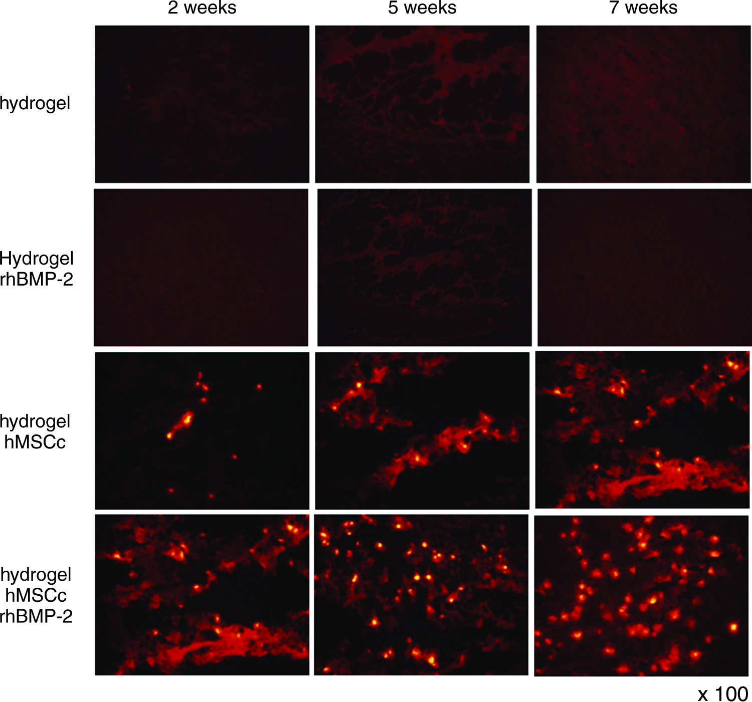
Fluorescence microscopy images showing the fluorescent dye PKH26 in labeled osteogenic hMSCs. Hydrogels with rhBMP-2, with hMSCs, or with hMSCs and rhBMP-2 were implanted into nude mice, and the images shown were obtained after 7 weeks (magnification ×100). Color images available online at www.liebertonline.com/ten.
Second, we examined mineralization, as a measure of new bone formation, using von Kossa stains and Alizarin Red O stains to reveal calcium deposits. When each specimen was stained with von Kossa, we observed mineralization at the hydrogels containing hMSCs, indicative of hMSC differentiation into bone-forming cells, after 7 weeks (Fig. 5a). The areas staining black with von Kossa stain resulted from deposition of biological minerals and reduction of organic components. As expected, the empty hydrogel (no cells) did not exhibit any mineralization, but importantly, the positive control consisting of rhBMP-2–encapsulated hydrogel without hMSCs did not exhibit noticeable positive von Kossa staining. However, the black-stained areas within the hydrogels containing hMSCs and rhBMP-2 increased more significantly over time than the areas within the hydrogels containing only hMSCs. These results indicated that mineralization and ectopic bone formation matured with time and that rhBMP-2 enhanced these activities. We also observed calcium deposits after Alizarin Red O staining of the samples (Fig. 5b). In agreement with the von Kossa results, the empty hydrogel encapsulated with rhBMP-2 exhibited no mineral or calcium deposition, but we observed calcium-positive orange-red areas in the hydrogels containing hMSCs, either alone or together with rhBMP-2. Moreover, in the case of the hydrogels with hMSCs and rhBMP-2, the areas colored orange-red increased in a time-dependent manner, indicating that calcium deposition increased over time in the presence of rhBMP-2. These results suggested that the hydrogels with hMSCs and rhBMP-2 provided a structural bridge, wherein the hMSCs proliferated and differentiated into osteoblasts.
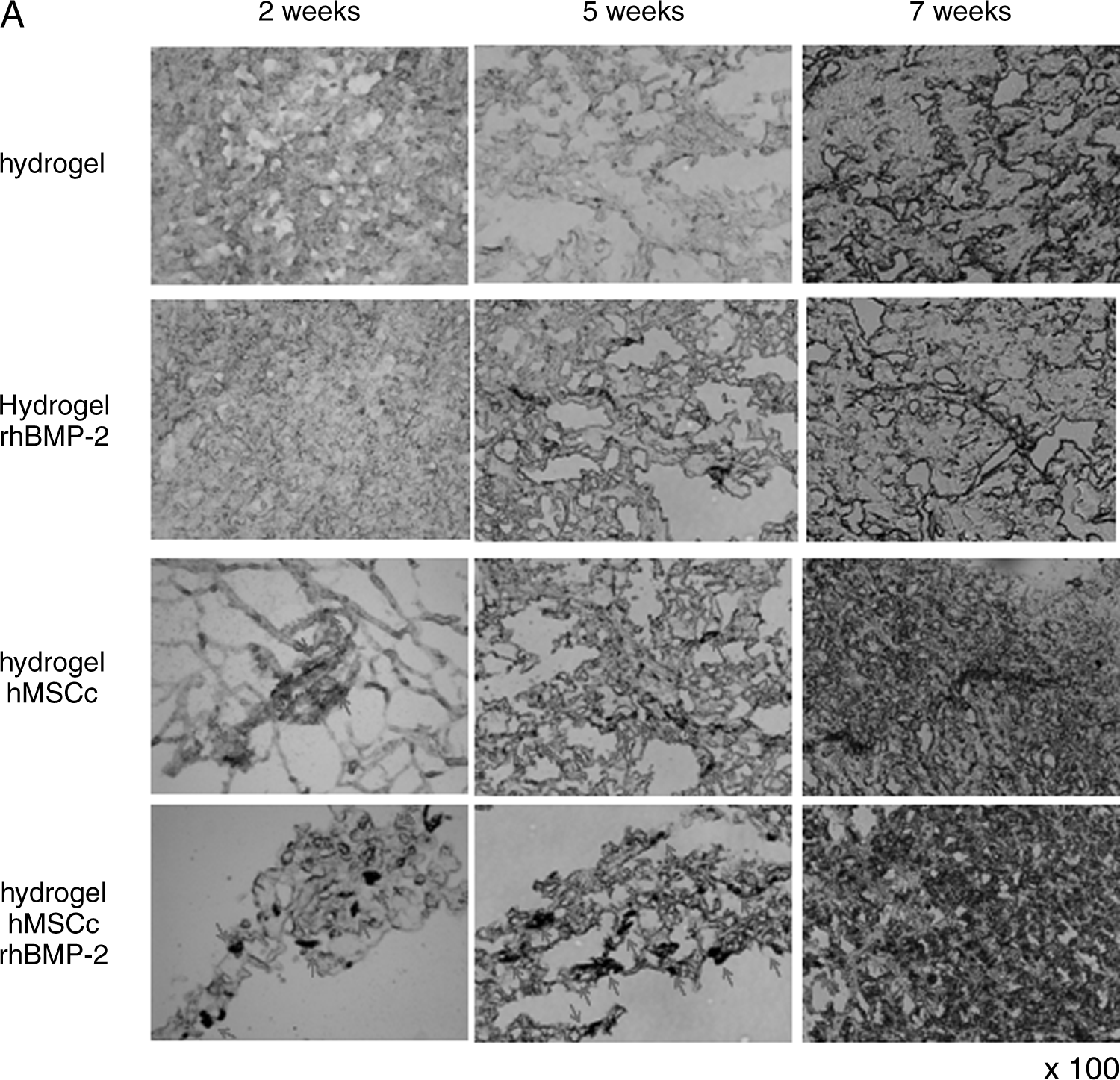
Microphotographs (magnification ×100) of histological sections stained with (
To estimate the osteogenic differentiation of hMSCs, we analyzed ALP activity 2, 5, and 7 weeks after implantation, as shown in Figure 6. The control groups harboring empty hydrogel or a cell-free hydrogel with BMP-2 did not exhibit any ALP activity throughout the 7-week period. This result is inferred that ingrowing host cells did not affect on the ALP activity in vivo. In contrast, at 2 weeks postinjection, we detected low ALP activity levels from the hMSCs in the hydrogel, indicating that the hMSCs had started to differentiate into an osteoblast-like cell lineage. The levels of ALP activities from the hydrogel loaded with hMSCs and rhBMP-2 quickly increased in a time-dependent manner compared to those in the control hydrogels or the hydrogels with hMSCs alone, over the course of 7 weeks postimplantation.
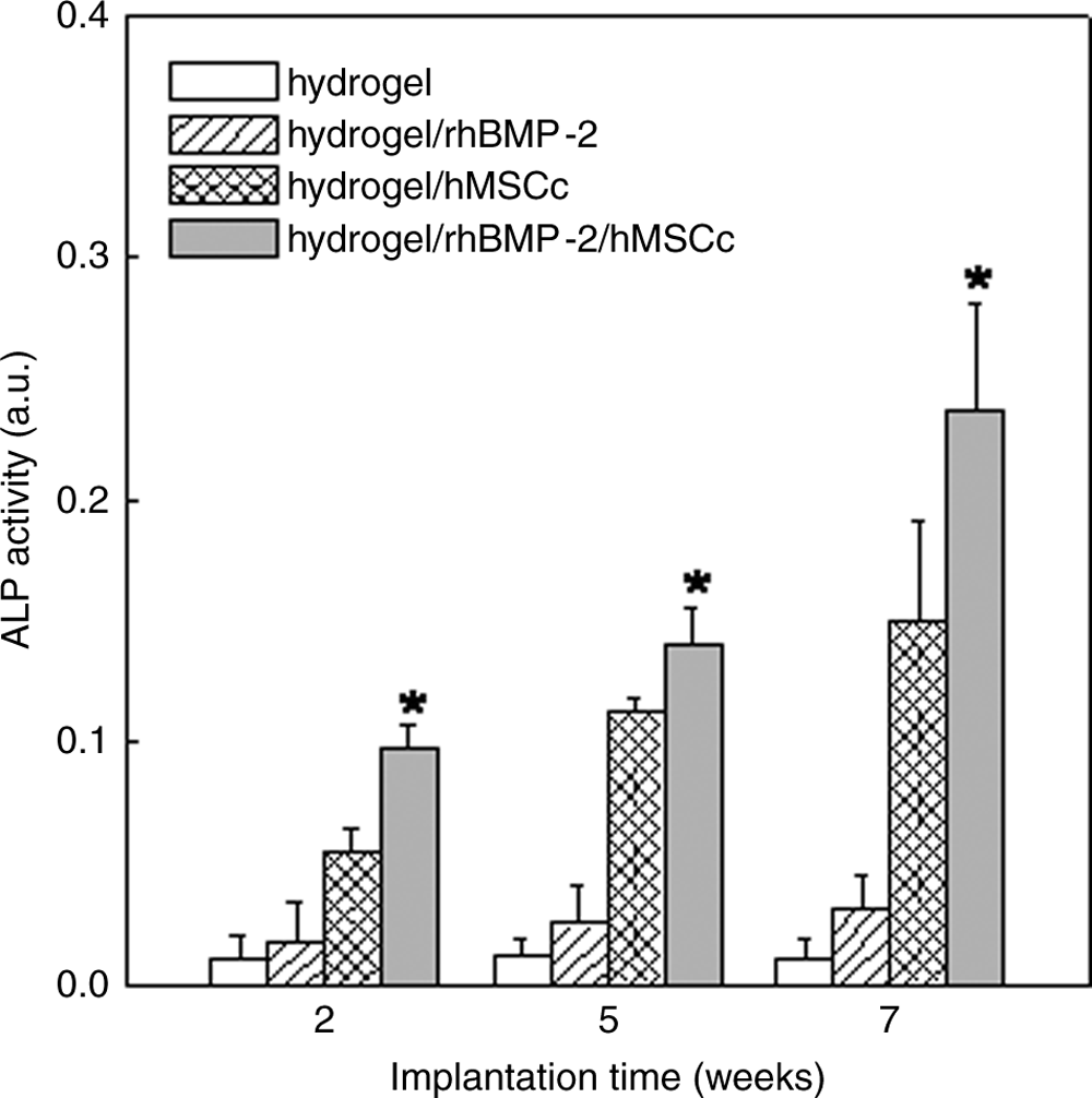
Time-dependent ALP activities of the hydrogels following implantation into nude mice. The ALP enzyme activities were measured and expressed as nanomoles of p-nitrophenol produced per min per mg of protein. Data are presented as means ± S.D. *p < 0.05.
Discussion
The objective of this study was to examine whether our novel synthetic pH/thermo-sensitive hydrogel provided a bridge for delivering hMSC and rhBMP-2 in an in vivo bone tissue engineering application. Specifically, we evaluated the hydrogel's biocompatibility and its ability to encapsulate stem cells and growth factors with a high encapsulating efficacy. We also histologically assessed the in vivo response to the hydrogel following implantation in nude mice.
To synthesize this novel protein and stem cell delivery system, we added pH-sensitive SMOs to the ends of thermo-sensitive block copolymer of PCLA-PEG-PCLA. The resulting pH- and thermo-sensitive SMO-PCLA-PEG-PCLA-SMO block copolymer solution underwent a reversible sol–gel transition, induced by a narrow pH change, from pH 8.0 to pH 7.2, at body temperature (37°C). This property stems from the hydrophobic interactions between the SMO-PCLA segments, which are perturbed by the ionized sulfonamide groups in the block copolymer. 30 Notably, this process is reversible over a narrow pH range. These pH/thermo-sensitive gelling characteristics of the sulfonamide-modified block copolymer are some of its most crucial injectable scaffolding properties, in that they prevent premature gelation of the hydrogel during injection into anatomical sites deep within the body. After injection of the sol–sate polymer solution into the body, we anticipate that our hydrogel yields a three-dimensional environment under physiological conditions, in which stem cells are encapsulated.
In addition, the pH/thermo-sensitive SMO-PCLA-PEG-PCLA-SMO block copolymer exhibited a very low degradation rate over a 1-month period at physiological conditions, wherein the pH remained at around pH 5.5 due to the buffering effect of the sulfonamide moieties on the acidic PCLA monomers (data not shown). 29 However, an aliphatic copolymer, PCLA-PEG-PCLA, degraded rapidly within 1 week under physiological conditions (pH 7.4 and 37°C), and the pH value dramatically decreased from 7.4 to 2.3 as a result of hydrolytic degradation. It is well known that block copolymers composed of aliphatic PLA or PLGA polyesters tend to undergo rapid degradation, producing acidic monomers such as lactic or glycolic acid. 34 This reaction creates an acidic environment around the hydrogel that is deleterious to bioactive proteins and cells. In a previous report, we confirmed that the sulfonamide groups of SMO-PCLA-PEG-PCLA-SMO act as buffers, slowing the rapid hydrolytic degradation of aliphatic block copolymers under physiological conditions. 29 Presumably, the buffering sulfonamide moieties within the hydrogel decreased the production of cytotoxic acidic monomers in cell culture. Indeed, the DMEM extraction test recommended by the ISO/EN 10993 Part 5 Guidelines revealed that when the extract concentration was less than 200 mg/mL, the hydrogel did not exhibit noticeable cytotoxicity. We would therefore expect the copolymers to be biocompatible at lower concentrations (200 mg/mL), and we anticipate that the enhanced stability of SMO-PCLA-PEG-PCLA will be useful in sustained drug delivery systems or long-term cell therapy.
To evaluate the potential of the hydrogel as a vector for stem cells or therapeutic proteins in bone tissue engineering, we added rhBMP-2 and/or hMSCs to the pH/thermo-sensitive polymer solution, and we subcutaneously injected the solution into nude mice with a syringe needle. Ten minutes after injection, we could isolate gross gels formed at the injection sites, indicating that the block copolymer solution gelled rapidly in the lower pH and higher temperature of the tissue fluid surrounding the injection site. Importantly, the isolated hydrogel showed remarkable encapsulating efficiencies for hMSCs and rhBMP-2 (90% and 85%, respectively), possibly because the pH/thermo-sensitive block copolymer containing hMSCs and rhBMP-2 formed a gross gel so rapidly, within just 10 min, under in vivo conditions (pH 7.4 and 37°C). Therefore, bioactive stem cells and growth factors could easily be suspended into an aqueous solution of the hydrogel and encapsulated with high efficiency upon gellation, sidestepping the need for undesirable elements and processes such as organic solvents and drying/separation steps. The implanted hydrogel maintained its gel shape over the entire 7-week study, indicating that it might provide a long-lived three-dimensional environment to permit proliferation and differentiation of hMSCs at implanted sites. Moreover, the implanted hydrogels maintained their firmness for up to 7 weeks postinjection, suggesting that the SMO groups on the hydrogels prevented rapid in vivo hydrolysis of the aliphatic caprolactone and lactide block copolymers. This remarkable stability indicated that conjugation of the SMO groups solved one of the major drawbacks associated with aliphatic PLA- or PLGA-based hydrogels (PLA and PLGA): rapid degradation of the block copolymer and the associated deleterious acidic monomers. 34 We had previously confirmed that the buffering effect of the sulfonamide moieties within the hydrogels reduced the rapid gel degradation and its acidic by-products, and thus it may have largely prevented chronic inflammation in SD rats.29,30 Importantly, in the rat model, the implanted hydrogel did not produce noticeable chronic inflammation in rat that might have resulted from infiltration of polymorphonucleocytes, macrophages, fibroblasts, and lymphocytes; in addition, we did not observe thick fibrous encapsulation of the hydrogel. Also, infections or severe fistulae were not observed. These results indicate that the pH/thermo-sensitive hydrogel of SMO-PCLA-PEG-PCLA-SMO is biocompatible in vivo and is thus suitable for delivering hMSCs and rhBMP-2 in bone tissue engineering.
To evaluate the viability of hMSCs in hydrogels, we monitored the PKH26-labeled hMSCs in the hydrogel, with or without rhBMP-2, for up to 7 weeks using fluorescence microscopy. The result suggested that the hMSCs were viable and proliferated over the course of the experiment. Importantly, the hMSCs were more confluent when rhBMP-2 was also present in the hydrogel, presumably because the hydrogel permitted a sustained release of rhBMP-2 that stimulated the proliferation of the hMSCs. The data suggest that these injectable in situ–forming hydrogels provided a useful scaffold, in which hMSCs and rhBMP-2 could successfully be delivered for bone tissue engineering.
Mineralization and new bone formation within the different hydrogels closely correlated with the in vivo viability of hMSCs. Upon dissection, the control hydrogel (no cells), with or without BMP-2, did not exhibit detectable von Kossa–positive or Alizarin Red O–positive compartments, indicating that host mouse cells had not infiltrated into the hydrogel or differentiated into bone-forming osteoblasts. Reportedly, subcutaneous implantation is less favorable for rhBMP-2–induced bone formation than intra- or intermuscular implantation.35,36 Herein, however, the hydrogel containing hMSCs and rhBMP-2 exhibited much more mineralization and calcium deposition after 7 weeks postinjection than did the hydrogel containing hMSCs alone. These results demonstrated that the implanted hydrogel provided a structural bridge wherein the hMSCs proliferated and differentiated into osteoblasts. In addition, the enhanced differentiation and mineralization of hMSCs in the hydrogel indicated that rhBMP-2 within the gel matrix underwent sustained released in an active form, stimulating hMSC differentiation. Thus, in our in vivo model, the pH/thermo-sensitive block copolymer functioned effectively as a growth factor delivery carrier for bone tissue engineering. In the ALP activity test, which is one of the most commonly used osteogenesis tests, only the hydrogel containing hMSCs and rhBMP-2 displayed significant increases in ALP activity at 7 weeks postimplantation (p < 0.05), indicating that including osteoinductive rhBMP-2 within the hydrogel strongly enhanced hMSC differentiation. The ALP activity tests suggested that the hydrogel-encapsulated growth factor maintained its biological activity and might have been continuously released for a long period in vivo.
In summary, we assessed the potential of a pH/thermo-sensitive SMO-PCLA-PEG-PCLA-SMO block copolymer as an in situ–forming injectable hydrogel to induce autologous ectopic bone formation. The pH/thermo-sensitive hydrogel showed a sol–gel transition that rendered it suitable for in situ use, in that it formed a gel under physiological conditions (pH 7.4 and 37°C). In addition, this hydrogel was biocompatible in vivo; it functioned as a hMSC bridge that permitted continuous differentiation and proliferation, and it did not produce signs of severe inflammation within 7 weeks. Finally, this new synthetic pH/thermo-sensitive hydrogel, carrying MSCs and a recombinant human bone morphogenetic protein, permitted continuous stem cell differentiation, enhanced mineralization, and elevation of ALP levels, for prolonged periods in vivo. Future studies will evaluate the in vivo osteogenesis capabilities of our pH/thermo-sensitive in situ–forming hydrogel, using bone-fracture models and quantitative bone formation assays, such as calculation of bone surface areas in histological sections and determination of calcium content.
Footnotes
Acknowledgments
This research was financially supported by the Basic Research Program of the Korean Science & Engineering Foundation (grant no. R01-2006-000-10629-0), by the Ministry of Science and Technology (F104AA010003-06A0101-00310) in Korea, and by the Real-Time Imaging Project of the KIST Intramural Research Program.