
Abstract
All engineered bioartificial structures developed for tissue regeneration require oxygen and nutrients to establish proper physiological functions. Aiming to improve vascularization during dermal regeneration, we combined the use of a bioartificial collagen scaffold and a defined human mesenchymal cell (MC) line. This cell line, termed V54/2, exhibits typical morphologic and immunohistochemical characteristics of MC. V54/2 cells seeded in the scaffold were able to survive, proliferate, and secrete significant amounts of vascular endothelial growth factor (VEGF) and basic fibroblast growth factor (bFGF) during 2 weeks in vitro. To induce dermal regeneration, scaffolds with or without cells were transplanted in a nude mice full skin defect model. After 2 weeks of transplantation, scaffolds seeded with V54/2 cells showed more vascularization during the dermal regeneration process than controls, and the presence of human cells in the regenerating tissue was detected by immunohistochemistry. To confirm if local presence of angiogenic growth factors is sufficient to induce neovascularization, scaffolds were loaded with VEGF and bFGF and used to induce dermal regeneration in vivo. Results showed that scaffolds supplemented with growth factors were significantly more vascularized than control scaffolds (scaffolds without growth factors). The present work suggests that combined use of MC and bioartificial scaffolds induces therapeutic angiogenesis during the scaffold-based dermal regeneration process.
Introduction
A tissue-engineered skin substitute that promotes vascularization would be a valuable tool for plastic and reconstructive surgery. Vascular endothelial growth factor (VEGF) and basic fibroblast growth factor (bFGF) are among the best-described growth factors involved in angiogenesis during wound repair. 10 Previously, we as well as others have shown that the addition of angiogenic growth factors as recombinant proteins or preseeding with genetically modified cells improves vascular regeneration in vivo in an SDR-based process.8,15,16 Certain well-defined cell populations could act as delivery vehicles by continuously releasing growth factors thus enhancing vascularization in vivo. In vitro bioactivation of SDR by preseeding of cells may be an ideal method to promote angiogenesis in vivo.
Similarly to other fields of tissue engineering, the combined use of bioartificial scaffolds and cells is a rational way to create skin substitutes for clinical application. Moreover, incorporation of cells into SDR may also serve to improve our knowledge about the physiological mechanisms involved in dermal vascular regeneration process. Here, we investigated whether the in vitro incorporation of MCs could improve vascularization in vivo. The major aim of this study was to analyze the behavior in vitro and in vivo of a novel MC line seeded in SDR, focusing on their proangiogenic potential in vitro and in vivo. The experiments were performed to answer the following questions: (a) Are MCs viable in SDR, bioactivating the dermal scaffold? (b) Can bioactivated SDR improve vascular dermal regeneration in vivo? and (c) Is the presence of angiogenic proteins secreted from cell-bioactivated SDR sufficient to improve vascular dermal regeneration?
Materials and Methods
Cell line
V54/4 cell line was generated as previously described. 17 Briefly, CD34− cells derived from granulocyte colony-stimulating factor mobilized donors were immortalized by SV40 Large-T antigen expression. Clone V54/2 was selected and used for the cell line establishment. V54/2 cells were cultured in Dulbecco's modified Eagle's medium (DMEM) supplemented with 10% or 2% fetal calf serum (FCS) (both supplied by Gibco, Karlsruhe, Germany) depending on the experimental setting described below.
Fluorescent activated cell sorting (FACS) analysis
V54/2 cells were detached by 5-min incubation with trypsin–EDTA solution and washed once with phosphate-buffered saline (PBS). Next, fresh samples were incubated for 1 h with fluorophore-conjugated antibodies raised against the following cell markers: CD11a, CD11c, CD14, CD29, CD34, CD38, CD41, CD44, CD45, CD49d, CD49e, CD73, CDw90, CD105, CD117, CDw123, CD133, CD146, CD166, and CD235a (all diluted 1:100). As isotype controls, IgG associated with fluorescent isothiocyanate (IgG-FITC) or phycoerythrin (IgG-PE) were used (all antibodies from BD Biosciences, San Jose, CA). Samples were examined with an FACScalibur flow cytometer (BD Biosciences).
VEGF proliferation assay
V54/2 cells were seeded in 24-well plates (1 × 10⁴ cells per each well) and cultured in DMEM 2% FCS for 24 h (Gibco). Next, cells were cultured for 4 days in medium daily supplemented with varying concentrations of recombinant VEGF (from 0 up to 100 μg/mL; R&D System, Minneapolis, MN). For cell quantification, a 3-(4,5-dimethylthiazol-2-yl)-2,5-diphenyltetrazolium bromide (MTT) assay was performed as described below. Results were expressed as folds compared to control (cells without exogenous VEGF).
Scaffold for dermal regeneration
Integra matrix (IM) is a scaffold based on bovine collagen fibers cross-linked with glycosaminoglycans that forms a porous biodegradable structure of 1.9-mm hydrated thickness (Integra LifeScience, Plainsboro, NJ). On top, the scaffold is covered with a removable silicon layer, which acts as a temporal epidermis.
Cell seeding in the scaffold
Pieces of IM (15-mm diameter) were dried with sterile gauze and placed in a 24-well plate, and 300 μL of medium containing 2.5 × 10⁵ cells was dropped over the scaffold and it was quickly absorbed. After 30 min of incubation, 1 mL of DMEM 10% FCS was added into each well. Cell seeding efficiency was evaluated by removing the scaffold from the well and counting cells adhered to culture dish.
Quantification of metabolic activity in the scaffold
About 2.5 × 10⁵ cells were seeded into each scaffold (1.5 mm of diameter) in 1 mL DMEM 10% FCS. At days 1, 8, and 15 after seeding, medium was removed and scaffolds were incubated for 3 h in fresh medium containing 5 ng/mL MTT (Sigma-Aldrich, Turkirchen, Germany). Next, medium was removed and replaced by 300 μL dimethyl sulfoxide (DMSO; Sigma-Aldrich). To quantify metabolic activity, absorbance at 570 nm was measured in the DMSO containing soluble formazan blue. Scaffolds without cells were used as negative control.
Cell visualization in the scaffold
At different time points, scaffolds containing cells were fixed in 3% paraformaldehyde (PFA) (Sigma-Aldrich) for 1 h and embedded in paraffin. Fifteen-micrometer sections were deparaffinized, stained for 20 min with 4′,6-diamidino-2-phenylindole (DAPI; Sigma-Aldrich), and washed twice with PBS. Finally, samples were mounted and analyzed by an Axioskop 2 fluorescence microscope (Zeiss, Jena, Germany) at 400×. For the three-dimensional reconstruction of the cell-containing scaffold, cells were seeded for 72 h and then fixed and stained as described in this section. Tetramethyl rhodamine iso-thiocyanate conjugated phalloidin (TRITC-phalloidin; Sigma-Aldrich) was used according to manufacturer's instructions. Pictures were taken with an LSM (Zeiss LSM 510), and reconstruction was performed with Volocity 3D imaging software.
Growth factor secreted from scaffolds containing cells
About 2.5 × 10⁵ cells were seeded and cultured in scaffolds (15-mm diameter) during 2 weeks in standard conditions. After 48 h, medium was replaced by DMEM 2% FCS. Every 48 h, medium was removed and replaced by fresh medium. VEGF (all isoforms) and bFGF concentrations were measured by ELISA according to manufacturer's instructions (quantikine ELISA kits; R&D Systems). Scaffolds without cells were used as negative controls. Measurements were performed over five seeded scaffolds.
Transplantation of cell-seeded scaffolds
Cells were seeded as described in this section, and after overnight culture in standard conditions, scaffolds containing 2.5 × 10⁵ V54/2 cells each or empty scaffolds were transplanted into an in vivo model. Six- to 8-week-old athymic nude mice (Takomi, Copenhagen, Denmark) were anesthetized with a mix of ketamine (10 mg/kg of body weight) and xylazin (2.4 mg/kg body weight) via intraperitoneal injection. Under general anesthesia, a bilateral full skin defect was created (15 mm of diameter) in the back of the animals, and scaffolds were implanted to induce dermal regeneration in six animals per group (12 scaffolds per group). Scaffolds were fixed to the wound by using nonabsorbable sutures, and wounds were bandaged (Varihesive®; Convatec, Deeside, UK). All procedures in vivo were approved by the corresponding local ethical committees.
Blood vessel quantification in the scaffolds
Quantification of the vascularization levels was performed as we previously described. 18 Briefly, after 2 weeks, animals were sacrificed, and whole skin from the back, including scaffolds, was removed. For vessel visualization, tissues were quickly placed on a transilluminator (LP 5000K; Hama, Monheim, Germany) and digital pictures acquired (Olympus camera; C-5060). For vessel quantification, digital pictures were analyzed with a software developed to perform digital segmentations. Vascularization levels were calculated as percentage of white pixels from the total number of pixels in the analyzed area. Normal skin of the back of the same animal was used for normalization, and results were expressed as a percentage of vascularized area in the scaffold related to normal skin. After visualization of the vessels, scaffolds were fixed for further immunohistological analysis. The vessel segmentation program can be downloaded free of charge from www.isip.uni-luebeck.de.
Scaffold modification by direct incorporation of growth factors
Scaffolds were incubated overnight in 500 μL PBS containing 1 μg of recombinant VEGF and 0.5 μg bFGF (both from BD Science, Minneapolis, MN) and washed once for 5 min with PBS. Then, eight scaffolds (four animals) per group were used to induce dermal regeneration in vivo. As controls, scaffolds were preincubated overnight in PBS without growth factors.
Histological analysis
After 2 weeks of transplantation, animals were sacrificed by overdoses of anesthesia, and then scaffolds were harvested, fixed with 3% PFA for 1 h, dehydrated, and embedded in paraffin. Five-micrometer-thick paraffin sections were pretreated for 45 min in PBS (pH 9.5) and incubated with an anti-HLA-class 1 polyclonal goat antibody (A-18; dilution 1:50; Santa Cruz Biotechnology, Santa Cruz, CA), followed by incubation with the secondary antibody (biotin-SP–conjugated mouse anti-goat IgG; dilution 1:50; Dianova, Hamburg, Germany). Next, the slides were incubated for 15 min with horse-radish peroxidase (Dako, Glostrup, Denmark). After incubation for 10 min with DAB (Dako), the slides were counterstained with hematoxylin (Merck, Darmstadt, Germany).
Statistical analysis
All assays were repeated at least in three independent experiments. ANOVA test was performed to compare group of samples using GraphPad software. p < 0.05 was considered as significant. In the graphs, error bars represent SEM.
Results
Cell characterization
V54/2 is a novel cell line, which has not been fully characterized. 17 Under normal culture conditions, V54/2 cells were strongly adherent to polycarbonate, exhibiting a homogeneous fibroblast-like morphology (Fig. 1A). Immunophenotypical characterization of the cells by FACS showed a typical mesenchymal expression profile, being strongly positive for the markers CD73, CDw90, CD105, CD146, and CD166 and negative for the hematopoietic markers CD11a, CD11c, CD14, CD34, CD38, CD41, CD45, CD117, CDw123, CD133, and CD235a (Fig. 1B). To evaluate the responsive potential of V54/2 cells to external stimuli, cells were cultivated under increasing concentrations of recombinant human VEGF (0, 25, 50, and 100 ng/mL) for 4 days. In the presence of VEGF, the cell number was significantly increased after 4 days, compared to controls (p < 0.05) (Fig. 1C).

Characterization of V54/2 cell line. (
Cell viability and proliferation in scaffold
Next, we evaluated the potential of V54/2 to be cultured in scaffolds. As shown in Figure 2, IM confers enormous surfaces for cell adhesion, enabling a presumably different growth condition as compared to two-dimensional culture plates. To test cell–scaffold interaction, cell morphology of seeded cells was evaluated after 72 h of culture. As shown in Figure 2A, cells (red/blue) adhere to the scaffold (green) by direct interaction with its structure. Moreover, V54/2 cells express several adhesion molecules such as CD29, CD44, CD49d, and CD49e, which could interact with extracellular matrix (ECM) elements.

Interaction, viability, and proliferation of the V54/2 cells in scaffolds. Cell–scaffold interaction was analyzed by confocal microscopy (
To evaluate long-term viability of cells in the scaffolds, samples were incubated with MTT. In the presence of MTT, scaffolds containing cells turn to dark blue color in contrast to control scaffolds where no color appeared. Microscopically, cells were clearly visualized as blue structures in the scaffold (Fig. 2C, upper panel). After 8 and 15 days of V54/2 cell culturing in the scaffolds, formazan blue formation was significantly increased (3.14 ± 0.44- and 4.46 ± 0.05-fold, respectively) as compared to 1 day after cell seeding (Fig. 2C, lower panel). Cell visualization by nuclear staining with DAPI shows that cells were homogeneously distributed in the scaffold (Fig. 2B). Moreover, increased number of cells after 8 and 15 days of cultivation in the scaffold confirms the results obtained by MTT assays (Fig. 2C).
Growth factor secretion from scaffolds containing cells
After having confirmed viability of V54/2 in the scaffold, we evaluated their potential to bioactivate IM by secreting proangiogenic growth factors to the environment. Medium from scaffolds containing cells was replaced every 48 h during 2 weeks. Results showed a continuous secretion of VEGF and bFGF. Concentration of VEGF in conditioned media was stable during 2 weeks with values ranging from 1.5 to 2.1 ng/mL. After 48 h, the concentration of bFGF was close to 0, rising up to 0.35 ng/mL with a stabilization of values around 0.2 ng/mL (Fig. 3).

Growth factor secretion from cell-seeded scaffold. Media from five scaffolds containing cells were collected every 48 h during 2 weeks and analyzed by ELISA to quantify the levels of VEGF (squares, upper curve) and bFGF (rhombus, lower curve).
In vivo dermal regeneration model
Under sterile condition, scaffolds were transplanted into a bilateral full skin defect nude mice model (Fig. 4) and fixed with nonabsorbable 5/0 sutures (Ethicon, Norderstedt, Germany). No major complications were observed during the operation procedure or postoperatively. After 2 weeks, no macroscopic signs of infection or foreign body reaction were observed in the wound area. The outer silicone layer of the SDR had prevented shrinkage of the neighboring skin, avoiding wound contraction. To evaluate the contribution of V54/2 cells to the neovascularization process, scaffolds were analyzed as described in Materials and Methods section. Results showed that the presence of V54/2 cells significantly contributed to improve neovascular regeneration during the scaffold-based dermal regeneration process in vivo (p < 0.001) as well as incorporation of VEGF and bFGF (p < 0.05) (Fig. 5). Histological analysis of tissue sections (four scaffolds per group) showed high levels of cellularization in both groups. Presence of human cells (HLA-class 1) was detected only in the group of animals transplanted with scaffolds containing V54/2 cells (Fig. 6).
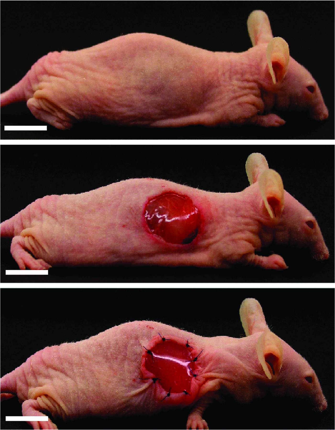
Nude-mouse full skin defect model for dermal regeneration. Fifteen-millimeter-diameter full skin was surgically and symmetrically removed from each side of the animal and was replaced by a scaffold to create a bilateral full skin defect model for dermal regeneration. Scale bars represent 1 cm. Color images available online at www.liebertonline.com/ten.
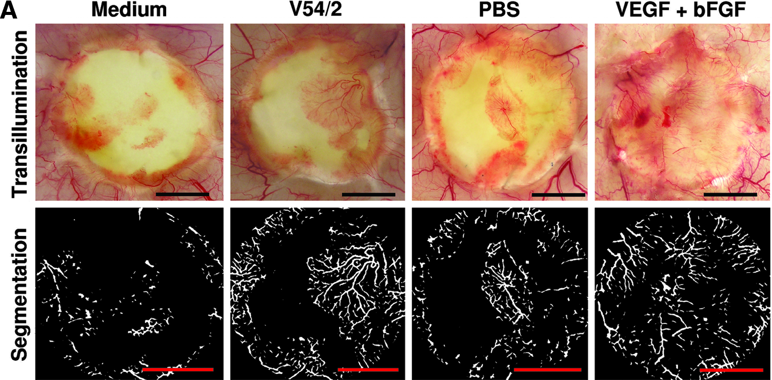
Blood vessel visualization and quantification. After 2 weeks of dermal regeneration, animals were sacrificed and skin was removed to analyze scaffold neovascularization. A representative picture of tissue transillumination and digital segmentation of one scaffold per group is shown (n = 12 for medium and V54/2 cells, and 8 for PBS and VEGF+bFGF) (
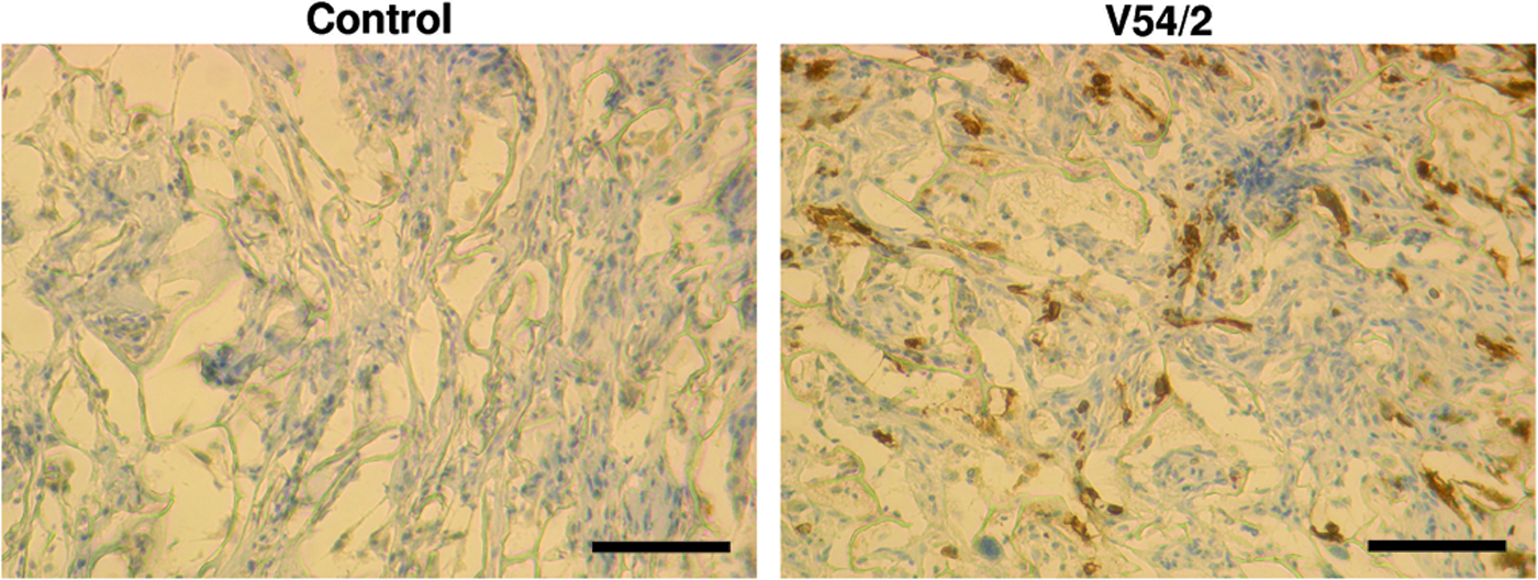
Histological analysis. Tissues were analyzed after 2 weeks of dermal regeneration. In both conditions, high cellularization levels were observed by eosin staining. Presence of V54/2 cells was visualized by HLA staining (brown). Scale bars represent 100 μm. Color images available online at www.liebertonline.com/ten.
Discussion
Inflammation, proliferation, and remodeling are the three different overlapped steps that mediate tissue regeneration. In normal conditions, skin cannot regenerate and can only be repaired by scar tissue formation, which, in general, is mainly composed by fibroblast and ECM. The scars restore the integrity of the tissue but not its functionality. This fact is critical in massive (e.g., burns) or specific (e.g., neck, hands, and elbows) skin injuries where a functional tissue is required. In this context, efforts in tissue engineering are focused to reach skin regeneration by mimicking the normal physiological wound regeneration processes. For that purpose, SDR have been used as template where regeneration could take place. Although the idea is good, clinical success has been poorer than expected.
Here, we investigated dermal vascular regeneration by cell-mediated scaffold bioactivation in vitro, enhancing angiogenesis in vivo. Angiogenesis is defined as the sprouting of preexisting vessels and is a process mainly regulated by growth factors, such as VEGF, angiopoietins, and bFGF,19–21 which are released to tissues from different cell types or by degradation of ECM. Such ECM, same as IM, is mainly composed of collagen that can directly interact with VEGF and bFGF. 22 VEGF plays a key role in angiogenesis during wound repair since its vasopermeability actions increase hydraulic conductivity and fenestration. 23 Moreover, VEGF is a potent mitogen for endothelial cells and induces endothelial cell migration, sprouting, and survival. Expression of VEGF inhibitor has been shown to be reduced in the dermal layer of acute wounds compared to chronic wounds or normal skin. 24 Similarly, bFGF promotes endothelial cell proliferation, differentiation, and migration into the wound area 22 and has been previously used in combination with SDR to improve angiogenesis during matrix-based dermal regeneration in a diabetic full skin defect model. 25 V54/2 cells seeded in IM secrete VEGF and bFGF (Fig. 3). As shown in Figure 1C, VEGF induces cell proliferation in these cells, suggesting that it enhances the V54/2 cell pool by autocrine signaling and promotes angiogenesis during skin regeneration. Scaffold bioactivation by cell seeding or by direct incorporation of recombinant growth factors could enhance angiogenesis by local growth factor release and by blocking the growth factor interaction sites present in the scaffold, which could inhibit angiogenesis by sequestration of the growth factor released by the tissue during wound healing. Expression, in vitro and in vivo, of 15 different growth factors in a scaffold seeded with keratinocytes and fibroblasts has been previously shown. 26 Those growth factors are known to promote wound healing, which could modulate the scaffold-dependent skin regeneration process.
To enhance angiogenesis, here we selected a cellular technology based on the intrinsic capacity of certain cell lines to secret proangiogenic growth factors. This approach allows seeding a large number of cells in the scaffold within a short time, thus constantly releasing growth factors in the wound area. Since no additional genetic manipulation is required, this approach could present advantages compared to others, where clinical translation could be difficult due to technical and legal problems associated with the use of gene vectors.
In our study, V54/2 cell line was characterized (Fig. 1) and seeded in IM. After seeding, cells were able to interact with the scaffold (Fig. 2A). Cell attachment could be mediated by α2β1 and Arg-Gly-Asp binding integrins, as was previously demonstrated seeding different cell types in IM. 27 Previous evidence showed that keratinocytes and fibroblast can survive and proliferate when they are seeded in IM. 28 Similarly, here we show cell survival and proliferation of V54/2 cells seeded in IM in vitro (Fig. 2B, C). Presence of human cells in the neodermal mouse tissue was confirmed in vivo by immunohistology (Fig. 6). Survival in vitro (Fig. 2), presence of the cells in vivo (Fig. 6), and constant growth factor secretion in vitro (Fig. 3) suggest that scaffolds containing such cells were constantly bioactivated.
VEGF and bFGF levels secreted from bioactivated scaffolds were rather stable in time and probably represent only a part of the total amount of growth factors secreted. Another important fraction could be interacting with the scaffold and inside the cells. Moreover, due to the lack of vascularization in vivo, scaffolds containing cells were primarily under hypoxic conditions that could enhance the production of proangiogenic growth factors during the dermal regeneration process.29–31 Continuous growth factor secretion from these cells in the matrix (Fig. 3) ensures prolonged bioactivity of the molecules in the targeted area as compared to the addition of recombinant proteins.
After 2 weeks of transplantation, in a bilateral full skin defect model (Fig. 4), bioactivated scaffolds were able to improve vascularization levels (Fig. 5). Those levels were evaluated with a novel method that presents advantages compared to traditional techniques. Using this technology, a broad target area can be analyzed at once, allowing visualization and quantification of the whole vascular network during the scaffold-based dermal regeneration process. Similarly, incorporation of recombinant VEGF and bFGF in the scaffold also improves vascularization during matrix-based dermal regeneration (Fig. 5).
Although our results suggest a possible angiogenic contribution of V54/2 cells, we cannot exclude the possibility of vascularization enhancement mediated by vasculogenesis, where V54/2 cells could act as endothelial progenitor cells (EPC), differentiating into endothelial cells or facilitating recruitment of those cells in the scaffold by releasing chemoattractant molecules such as SDF-1α. 32 However, experiments performed in vitro showed that V54/2 cells do not express endothelial features under previously described differentiation condition 33 and do not release SDF-1α (data not shown). Here we showed that cells were able to secret proangiogenic growth factors (Fig. 3) and that local presence of them improves vascularization (Fig. 5). Although this data support the hypothesis of a paracrine contribution of V54/2 cells to the vascular regeneration process, the ideal way to prove that is by the constant application of fresh conditioned media in the matrix in vivo. Unfortunately, this experiment is technically impossible to perform among others, due to the presence of a silicon layer on top of the scaffold that acts as physical barrier between the scaffold and the external environment.
Cells precultured in scaffolds to improve regeneration have been used in several fields of tissue engineering, including bone, cartilage, and nerve regeneration, being successfully applied in experimental models and clinical trials.34–36 In skin tissue engineering, clinical reports have shown that good results can be obtained in patients with different skin-related problems when they are treated with a combination of autologous cells and SDR.37,38
In the present study, cells were immortalized by transfection with the SV40 large antigen. Expression of this protein allows improving cell viability and proliferation in vivo. SV40 large antigen binds to heat-shock chaperone 70, the retinoblastoma family of tumor suppressors, and the transcription factor p53. All those interactions result in alterations in several signaling pathways that trigger, among others, unlimited cell division capacity and apoptotic resistance. 39 Although the role of SV40 in tumorigenesis is controversial, 40 the use of immortalized cell lines is a powerful tool in biomedical research and has been previously suggested to develop new therapies in regenerative medicine.41,42 Moreover, the new development of conditional SV40 cell lines is promising for their clinical translation. 43 Although in this work we present a xenotransplantation model, similar cell lines could be created from each patient to develop autologous therapies.
In this work, we characterize a new cell line showing its potential to activate a bioartificial scaffold. Here we also provide evidence supporting the idea that growth factors released from cell-seeded scaffolds could enhance angiogenesis during dermal regeneration. The data presented here suggest that combined use of SDR and certain MC populations may be useful to improve angiogenesis in skin tissue engineering technologies. However, further studies should be performed to cover several aspects nonincluded in this work such as the optimum density for cell seeding in vitro, and the comparison of results between different types of primary and immortalized cells. Also larger and nonimmunocompromised animal models (e.g., pigs) with major skin defects and long-term analysis experiments should be performed. Finally, further analysis focused on relevant clinical aspects such as quality of the regenerated dermis or infection rates would be extremely important.
Footnotes
Acknowledgments
The authors would like to thank Dr. Ralf Werner for his critical discussion during the experimental design and Ignacio Bazán for the critical review of the manuscript. This work was supported by grants to H.-G.M., provided by the University of Luebeck, and FONDAP 15010006 to S.L.
Disclosure Statement
No competing financial interests exist.