
Abstract
The goal of this study was to investigate the effect of cyclic mechanical stimulation on mesenchymal stem cells (MSCs) seeded within human umbilical veins (HUVs), and to determine the potential of the engineered constructs to function as tendon tissue replacement models. Decellularized HUVs were seeded with MSCs embedded in type I collagen hydrogel. A mechanical stimulator for tissue engineering applications was specifically designed to cyclically tension the constructs for durations up to 2 weeks, where controls were left untensioned. This HUV model system seeded with a cellular collagen gel, coupled with mechanical stimulation, resulted in improved mechanical properties compared to other tendon tissue engineered constructs composed of cellular collagen gel alone, without any additional supporting scaffold. After 2 weeks of culture an increase in cell number was measured for both tensioned and untensioned constructs; however, the increase was at least eightfold higher for stimulated samples. Microscopically, cyclically tensioned samples showed parallel orientation of collagen fibers and spindle-shaped cell nuclei mimicking the morphology of native tendons. Moreover, mechanostimulation resulted in significantly stronger (156%) and stiffer (109%) constructs compared to untensioned samples. This engineered tendon model had an ultimate tensile strength value only one order of magnitude lower than human tendons and strain values in the range of human tendons. The results documented are promising and can be further improved by optimizing potentially critical culture parameters such as seeding density, loading regimes, and mechanostimulation durations.
Introduction
Because immobilization severely lowers the ultimate strength of tendons, 8 current research has focused on the effect of stretching cell-seeded constructs to enhance mechanical properties.9–12 In a recent investigation two-dimensional tissue sheets secreted by human fibroblasts were subjected to continuous (noncyclic) mechanical loading; results showed that cells and the extracellular matrix became aligned and increased both the tensile strength and modulus of the tissue. 13 Mechanical stimulation of collagenous constructs seeded with mesenchymal stem cells (MSCs) has also been shown to increase the expression of ligament and tendon markers,14,15 indicating the potential of MSCs to differentiate into tenocytes under the sole action of mechanical stimulation. However, poor mechanical properties of cultured collagen gel constructs prevent their use as tendon replacements. 16
Biological scaffolds, such as small intestinal submucosa,17,18 collagen fibers, 19 and human umbilical veins (HUVs),20,21 have been targeted for tissue engineering applications because they are biocompatible, evade the immune response, and have initial ultimate stress and stiffness values significantly higher than collagen gels. It has been shown that HUV provides a suitable environment for musculoskeletal tissue engineering applications. 21 In this study we investigate the effect of cyclic mechanical stimulation on HUVs seeded with MSCs. To accomplish our goals a bioreactor was designed to cyclically tension the seeded scaffolds for periods of 1 and 2 weeks. We hypothesize that cyclic mechanical tensioning would increase MSCs proliferation rates and enhance the mechanical and morphometric properties of seeded HUVs, giving them the potential to act as tendon replacement models.
Materials and Methods
Bioreactor design
A mechanical stimulator for tissue engineering applications (MSTE) was designed specifically for this project. The main goals behind constructing the MSTE were (1) to culture multiple tissue constructs under a sterile environment and (2) to apply controlled mechanical stimuli at different frequencies, intensities, and waveforms to cell-seeded scaffolds. The MSTE is capable of stimulating triplicate constructs simultaneously (Fig. 1a). Three samples cultured in separate vessels can be tensioned via a linear actuator controlled by a signal converter (Wavetec model 185, San Diego, CA). The applied signal is amplified before being transferred to the actuator (Fig. 1b). The specifications of the actuator are as follows: linear travel zone = 5 inches (12.7 cm), peak force sustained for 10 s = 49 lb (22.22 kg), and maximum force sustained continuously = 14 lb (6.35 kg). Using the signal converter box (Fig. 1c), it is possible to apply several waveform signals with variable frequencies and amplitudes.
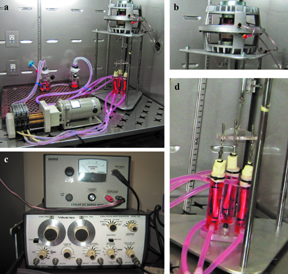
(
The cylindrical reactor vessels were made of glass (diameter = 2.5 cm, length = 10 cm) with an individual capacity of 49 mL. The total volume of medium circulated through the system is 240 mL. Medium is flowed around the constructs continuously using a multichannel pump at the rate of 1 mL/min. Two medium reservoirs are used to recirculate the medium around the MSTE perfusion circuit. The use of the two-reservoir system facilitates the procedure of changing the medium without having to move the system out of the incubator. Keeping the bioreactor in place in a closed environment minimizes contamination events. A filter on the medium container and gas-permeable silicone tubing allow medium oxygenation. The whole system is kept in a humidified incubator at 37°C, 5% CO2, and 95% air.
The load is transferred from the actuator to the samples via a piston that connects to a triangular plate, which in turn is hooked to the three constructs (Fig. 1d). The samples are attached to the triangular plexiglass plate via stainless steel adapters that can be screwed up or down to adjust the height of each sample separately accounting for individual plastic deformations upon stretching. To seal the top of each reactor vessel, surgical gloves were used. The three middle finger portions of the surgical gloves were cut, and then attached to the top of the vessel from one side and to the stainless steel adaptors from the other side using zip-ties (Fig. 1d). All of the materials used to assemble the bioreactor can be sterilized either by ethylene oxide or by steam (stainless steel adapters, glass bottles, plexiglass bases, glass reactor vessels, zip ties, and surgical gloves).
Preparation of scaffolds
Scaffolds were prepared as previously documented. 21 Briefly, fresh human umbilical cords were obtained from Norman Regional Hospital in Norman, Oklahoma. They were then thoroughly washed with distilled water and sliced into 8.5-cm-long sections. For each segment the HUV was mounted on a steel mandrel (McMaster-Car, Atlanta, GA), zip tied on both ends, and gradually frozen in a Styrofoam container to −80°C. Using a lathe (Central Machinery, Mod 33647, Harbor Freight Tools, Camarillo, CA) distributed by the HUVs were dissected out of the frozen umbilical cord sections to a uniform wall thickness of 0.75 mm and outer diameter of 6.75 ± 0.25 mm. The HUVs were thawed in phosphate buffered saline (PBS; Atlanta Biologicals, Lawrenceville, GA), sectioned to a length of 6.5 cm by discarding 1 cm from each end where the zip ties compressed the scaffold, and inverted such that the wall of the vein is on the outside. Cords were decellularized in 1% weight/volume sodium dodecyl sulfate (Baker, Phillipsburg, NJ) on an orbital shaker for 24 h. HUVs were then thoroughly washed in distilled water, and placed in 75% ethanol for 24 h. Finally, the scaffolds were sterilized in 0.2% peracetic acid (Sigma-Aldrich, St. Louis, MO), washed thoroughly in sterile water, and placed in PBS, where their PH was balanced between 7.2 and 7.4. HUVs were stored no more than 2 weeks in PBS at 4°C before being used.
Extraction of MSCs
Rat bone marrow MSCs were extracted from male Wistar rats (Harland Laboratories, Indianapolis, IN) using well-documented procedures. 22 Briefly, the femur and tibia of male rats (41–50 days old; 150–200 g) were dissected and suspended in alpha minimum essential medium (α-MEM; Invitrogen, Carlsbad, CA) supplemented with 10% fetal bovine serum (FBS; Atlanta Biologicals), 1000 U/mL penicillin G (Invitrogen), 1 mg/mL streptomycin sulfate (Invitrogen), and 2.5 μg/mL amphotericin B (Invitrogen). Four sterile syringes were each filled with 5 mL of medium (α-MEM supplemented with 10% FBS, 100 U/mL penicillin G, 100 μg/mL streptomycin sulfate, and 0.25 μg/mL amphotericin B). The bone marrow was flushed out of four bone aspirates into a 50 mL falcon tube using the sterile syringes. Large conglomerates of bone marrow were separated and homogenized using a sterile pipette. The cells were plated on four 75 cm2 culture plates and incubated in a humidifier at 37°C and 5% CO2. Nonadherent cells were removed 3 days later after the first medium change. Thereafter, the medium was changed every 2–3 days. When the cells became 75% confluent, they were split into three new flasks indicating a new passage. Cells used for seeding in the HUV were between passages 3 and 5.
Seeding of constructs
MSCs were washed with 5 mL of PBS and detached using 2 mL of 0.25% trypsin-EDTA (Invitrogen) for 5 min. The cell suspension was centrifuged at 2200 rpm for 5 min, and the supernatant discarded. The cell pellet was dissolved in medium and mixed with type I collagen (Angiotech Biomaterials, Vancouver BC, Canada) to yield a final cell density of 1 million cells/mL and collagen concentration of 2 mg/mL. Using 1 mL pipettes, 0.6 mL of cellular collagen solutions was inserted in each HUV. Cell attachment and polymerization of collagen were allowed for a full hour after which the seeded constructs were divided into three groups as follows: (group 1) seeded HUVs that were placed in 100-mm-diameter well plates; (group 2) seeded HUVs that were cultured in the MSTE without any mechanical stimulation; (group 3) seeded HUVs that were cultured in the MSTE and subjected to mechanical stimulation of 2% strain for 1 h/day at a frequency of 0.0167 Hz. A fourth group (group 4) consisted of acellular unseeded HUVs that were mechanically stimulated in the MSTE. Constructs were cultured for durations of 1 and 2 weeks and then tested for (1) cell density, (2) gene expression, (3) morphology, and (4) mechanical properties.
Cell density per construct
At the end of the culture duration, cords that to be tested for cell number were immediately processed as documented herein. Three 5-mm-long ringlets were dissected from the center and 1 cm away from each end of the cord. Ringlets were then sliced into fragments ≤ 0.25 mm2 and incubated with 2 mL of collagenase type I (MP Biomedicals, Solon, OH) overnight at 37°C. To expedite the release of cells from the matrix, samples were sonicated for 1 min. Finally, the cellular solution was subjected to two freeze/thaw cycles to enhance cells lyses and thus the exudation of all DNA into the solution. Using a PicoGreen DNA quantification kit (Invitrogen), it was possible to measure the cell density in each of the three cellular solutions and compute an average cell density per construct. A Synergy HT plate reader (Bio-Tek, Winooski, VT) was used to measure fluorescence at a wavelength of 550 nm. The DNA concentration in μg/mL per cellular solution was computed using a standard curve and converted to cell density, knowing that the DNA content per MSC was 3 pg/cell. The PicoGreen DNA assay gives an indirect indication of cell viability by showing an increase in DNA concentration, and thus an increase in cell number per construct, with time.
Ribonucleic acid extraction and real-time polymerase chain reaction
Tissue samples (≤25 mg) dedicated for gene expression analysis were stored in 1.5 mL of RNAlater® (Ambion, Austin, TX) at −20°C to preserve RNA until further analysis. RNA was extracted from the tissue using an RNAqueous®-4PCR Kit (Ambion) following manufacturer's directions. cDNA was synthesized from RNA using TaqMan® Reverse Transcription Reagents (Applied Biosystems, Austin, TX) in the presence of multiScribe reverse transcriptase (50 U/μL), RNase inhibitor (20 U/L), deoxyNTPs mixture (2.5 mM), random hexamers (50 μM), 10× buffer, and MgCl2 solution (25 mM). Gene expression levels of collagens type I and type III, and glyceraldehyde 3-phosphate dehydrogenase (GAPDH) were quantified using a Power SYBR® Green PCR Master Mix kit (Applied Biosystems). Primers used were as follows: collagen type I forward 5′-GGAGAGTACTGGATCGACCCTAAC-3′, backward 5′-CTGACCTGTCTCCATGTTGCA-3′; 23 collagen type III forward 5′-CAGCTGGCCTTCCTCAGACTT-3′, backward 5′-GCTGTTTTTGCAGTGGTATGTA ATGT-3′; 24 and GAPDH forward 5′-AACTCCCTCAAGATTGTCAGCAA-3′, backward 5′-GTGGTCATGAGCCCTTCCA-3′. 24 Quantitative real-time polymerase chain reaction (qRT-PCR) was conducted according to the following steps: 95°C for 10 min, 45 cycles of 95°C for 15 min, and 60°C for 1 min. At the end of RT-PCR, melting curves were generated for each amplified product. The threshold cycle (CT) for collagens type I and III was normalized to the housekeeping gene GAPDH, and the relative expression was computed following the ΔΔCT method. 25
Light and fluorescent microscopy
Supplies for conducting histology, including neutral buffered formalin, paraffin, clear-Rite, hematoxylin and eosin (H&E), and mounting medium, were purchased from Richard-Allan Scientific (Kalamazoo, MI). Cords dedicated for histological evaluation were cut in two different directions: (1) cross sections corresponding to 5-mm-thick ringlets and (2) longitudinal sections corresponding to 5-mm-thick portions cut parallel to the axial direction of the HUV. Tissue samples were fixed in 10% neutral buffered formalin overnight at room temperature and stored in 70% ethanol no more than 2 weeks before embedding. Fixed samples were dehydrated at 45°C in a series of increasing alcohol concentrations, cleared with clear-Rite, infiltrated with melted paraffin at 60°C under vacuum, and embedded in paraffin blocks. Seven-μm-thick sections were sliced using a manual microtome, mounted on Histobond slides (VWR), and baked overnight at 45°C. Use of the Histobond slides minimized tissue loss during H&E staining. Tissue samples on slides were deparaffinized in clear-Rite, and then rehydrated in a series of decreasing alcohol concentrations. Slides for light microscopy were stained progressively in hematoxylin, cleared in acid alcohol, counterstained in eosin, and finally dehydrated and secured by cover slips. Slides for fluorescent microscopy were incubated for 30 min in the dark at 37°C with 100 μL of dye solution. The dye solution was composed of 5 μL of lipophilic stain DID (Invitrogen) and 1 mL of medium. Tissue samples were finally washed three times in medium (10 min per wash).
All slides were studied under a Nikon E800 microscope, and images were captured by a Nikon camera and analyzed using the MetaMorph software V6.2. For fluorescent microscopy an omega XF110 optical filter (Omega Optical, Brattleboro, VT) was employed to detect the fluorescence of the DID dye with an excitation wavelength of 644 nm and an emission wavelength of 665 nm. Using high excitation and emission wavelengths was necessary to avoid background autofluorescence from the tissue.
Analysis of histological slides
Three main criteria were followed to analyze histological slides. For each criterion the analysis was done on at least three slides, and four different locations per slide were studied.
Unoccupied luminal space
The unoccupied luminal space in the central portion of the HUV was measured and reported as a percentage of the ratio of the central area that is devoid of cells and matrix to the total cross-sectional area of the HUV. This parameter was monitored to evaluate the formation of new extracellular matrix in the central portion of the HUV beyond the collagen hydrogel that was originally inserted. For that reason, values of this parameter in decellularized HUV were compared with those of seeded HUV at different time points.
Fiber alignment
To analyze fiber alignment the axial direction of the HUV, which is parallel to the direction of mechanical stimulation, was considered the reference axis. The angular deviation of fibers from the axial direction was measured, and the mean with the standard deviation was reported.
Shape of cells
Using the Metamorph program, it was possible to count the cells that appear in a longitudinal slide and compute their dimensions (length, width, and area). The shape factor is a measure of the ratio of the shorter dimension (width) to the longer dimension (length). This number is smaller than 1. The closer it is to 1, the more round the shape of the cell is.
Mechanical testing
Samples were preconditioned for five cycles before being stretched to failure at a strain rate of 1%/s using a uniaxial tensile testing frame (United Testing Systems, model SSTM-2K, Flint, MI). To account for end effects, force and extension data were collected only from samples that failed in the region away from the clamps (a minimum of 10% of the gauge length). Maximum tensile stress was computed by dividing the maximum force by the cross-sectional area. Similarly, the modulus of elasticity was computed by dividing the stiffness by the cross-sectional area and multiplying the results by the gauge length. The gauge length and cross-sectional dimensions were accurately measured for every sample using digital calipers.
Statistical analysis
For each culturing duration, at least three samples were dedicated to cell density computations and histological evaluations (n ≥ 3), and six samples for mechanical analysis (n ≥ 6). Thus, for each group and each culturing duration, at least nine constructs were prepared corresponding to a total of 72 scaffolds or 36 human umbilical cords. All results were expressed as mean ± standard deviation. Statistical analysis to compare results among groups was performed using the ANOVA method. A significant difference corresponds to p < 0.05 (confidence level >95%).
Results
Cell proliferation and RT-PCR
The culturing conditions of the four different groups are summarized in Table 1. For all seeded groups and all culturing durations, a statistically significant increase in cell number was measured (Fig. 2). After 1 week of culturing, group 1 (constructs cultured statically in a Petri dish) had the lowest cell density. The total cell number increased from 600,000 MSCs to 1.08 ± 0.03 million cells for group 1, 2.16 ± 0.36 million cells for group 2 (constructs cultured in the MSTE without tensioning), and 1.82 ± 0.19 million cells for group 3 (constructs tensioned in the MSTE). However, after 2 weeks of culture, group 3 had more than 29 million cells compared to 1.57 ± 0.20 million cells in group 1 and 3.56 ± 2.04 million cells in group 2.
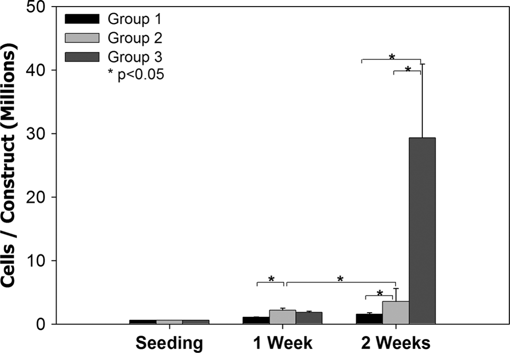
Proliferation of cells after 1 and 2 weeks of culture.
HUVs, human umbilical veins; MSTE, mechanical stimulator for tissue engineering.
qRT-PCR conducted after 2 weeks of culture showed an upregulation in the expression of both collagen I (fourfold) and collagen III (threefold) in stretched constructs of group 3 compared to groups 1 and 2.
Histology
Figure 3a and 3b shows cross sections of the HUV before and after decellularization, respectively, fluorescently tagged by the DID dye. Numerous cells appear in Figure 3a, reflecting the Wharton's Jelly and HUV cells originally present in the umbilical cord before decellularization. However, no intact cells were apparent in Figure 3b after decellularizing the HUV.

Cross-sectional view of the HUV stained with DID lipophilic dye (
Cross-sectional histological images of constructs cultured statically in the MSTE (Fig. 4a) showed large unoccupied spaces in the central portion of the HUV compared to dynamically cultured samples (Fig. 4b) in which the central area was almost totally occupied with proliferating cells. The decellularized HUV had a cylindrical structure with a central unoccupied luminal space of 14.4 ± 4.3%. After 2 weeks of culture the unoccupied luminal space significantly decreased to 5.7 ± 2.0% for group 3 (Fig. 5). No statistically significant decrease was measured for group 2. For all cultures, cells were able to integrate with the HUV and migrate through the scaffold. For group 2, cells only reached the first quarter of the scaffold (Fig. 6a), whereas cells seeded in group 3 (constructs stimulated in the MSTE) migrated deep into the scaffold almost reaching the outer end of the HUV (Fig. 6b).
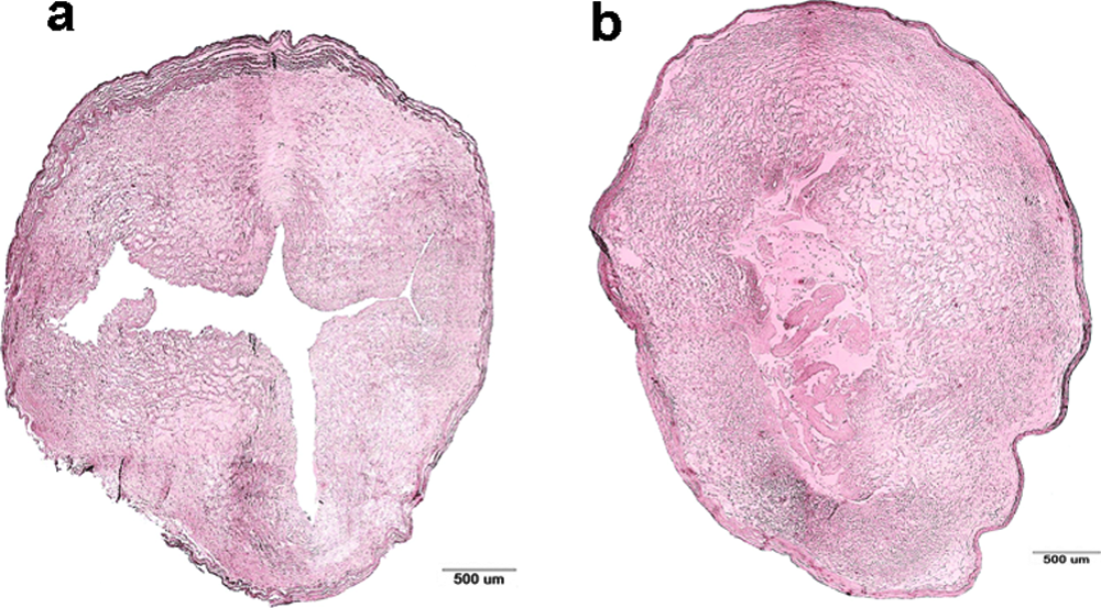
Cross section of constructs cultured for 2 weeks in the MSTE under (

Percent of unoccupied luminal space in the seeded constructs cultured in the MSTE.

Histological cross sections of seeded HUV after 2 weeks of culture in the MSTE: (
Longitudinal histological sections show extracellular matrix fiber alignment for static and dynamic cultures (Fig. 7). For static cultures, extracellular matrix fibers had random orientations 1 (Fig. 7a) and 2 weeks (Fig. 7b) postculture. Contrarily, mechanically stimulated constructs showed parallel alignment of fibers for both culture periods (Fig. 7c, d). After 2 weeks of culture, the average fiber deviation from the axial direction was measured to be 1.69° ± 17.6° for mechanically stimulated samples (Fig. 7f), while this value was 34.57° ± 48.16° for static cultures (Fig. 7e). A closer view at the longitudinal sections showed rounded nuclei for cells in the static cultures (Fig. 7g) with a shape factor of 0.9 compared to spindle-shaped nuclei for the dynamic cultures with a shape factor of 0.7 (Fig. 7h).
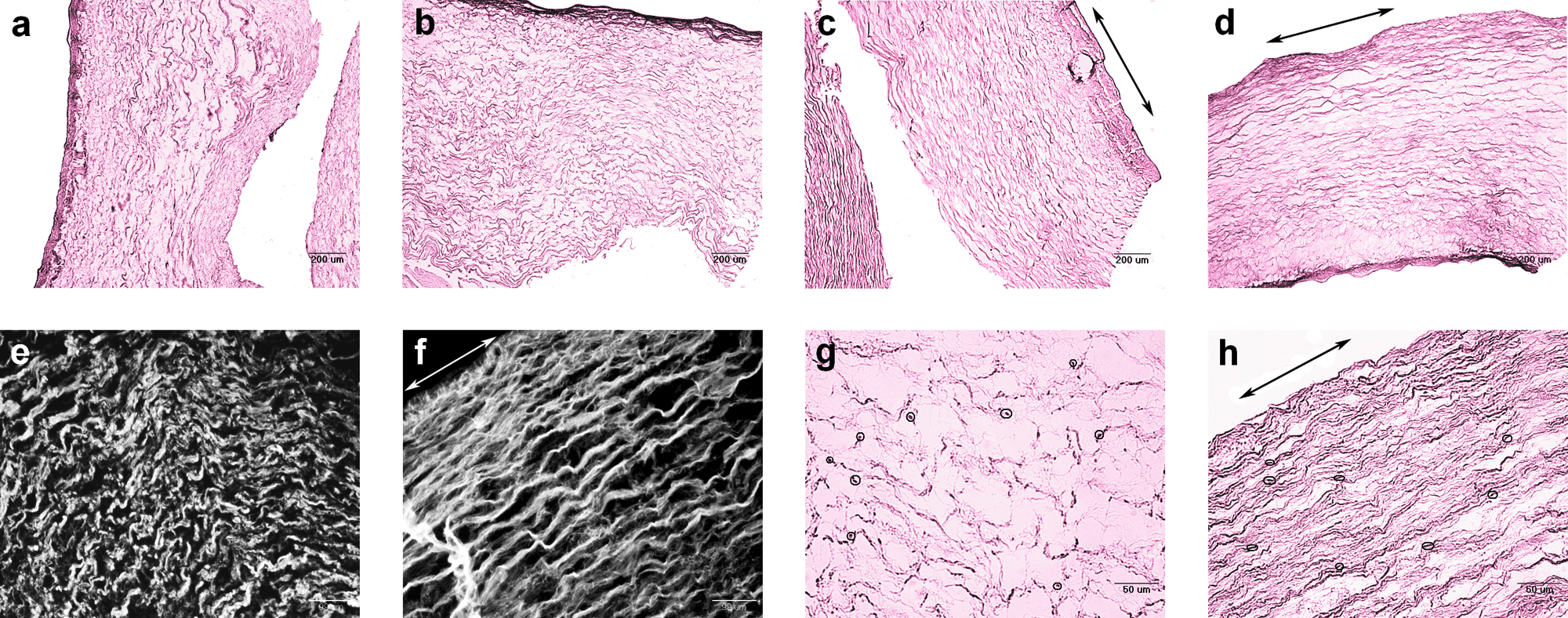
Longitudinal sections of HUVs cultured in the MSTE taken at 4× magnification. (
Mechanical analysis
Mechanical properties were assessed by stretching HUV constructs to failure while recording deformation versus load data. After 1 week of culture a statistically significant increase in the ultimate tensile stress values was recorded for all cellular groups (Fig. 8a). Group 3 had the highest ultimate stress value 2 weeks postculture (2.63 ± 0.65 MPa). No significant increase in ultimate tensile stress was measured for acellular dynamic samples (Fig. 8b).
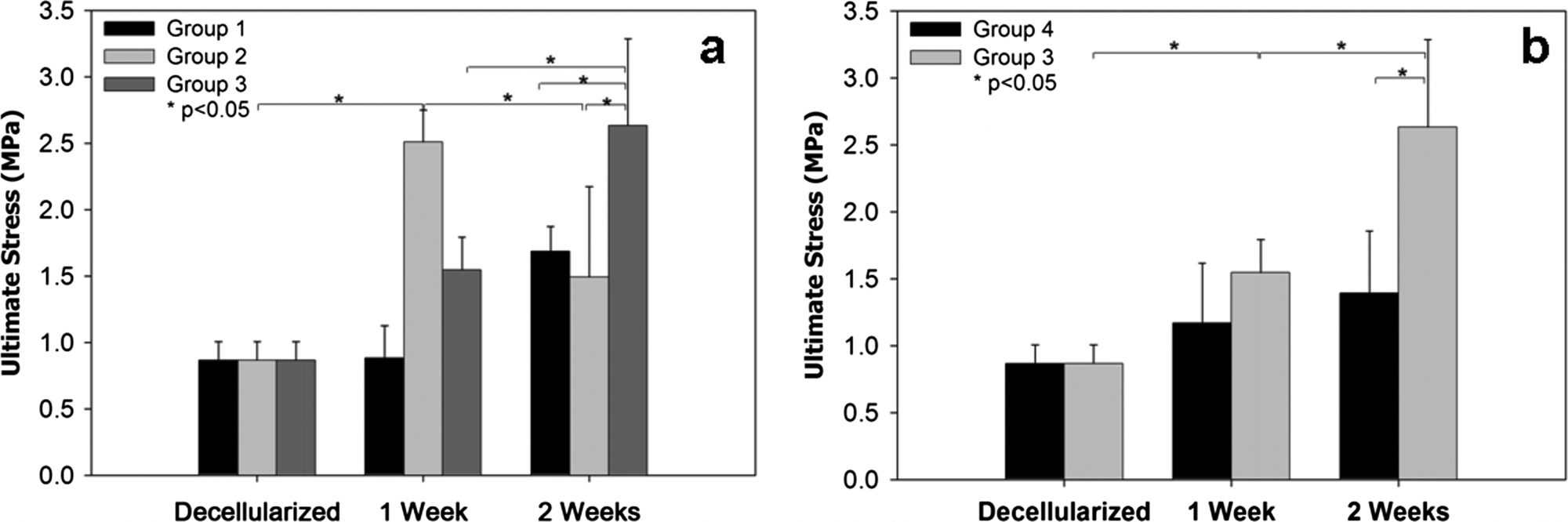
Ultimate tensile stress of (
A similar trend was observed for modulus of elasticity (E) values (Fig. 9). Decellularized scaffolds had a modulus of elasticity E = 3.31 ± 0.64 MPa. After 2 weeks of culture, group 1 had E = 9.04 ± 1.85 MPa, group 2 had E = 7.32 ± 2.37 MPa, and group 3 had the highest modulus value of 9.91 ± 2.22 MPa. Acellular samples of group 4 had inferior modulus of elasticity values (Fig. 9b) compared to other groups. One week after culture the modulus decreased from 3.31 ± 0.64 MPa to 2.93 ± 1.35 MPa. This value further decreased to 2.40 ± 0.52 by the end of the second week of culture.

Elastic modulus of (
Discussion
The goal of this study was to investigate the effect of cyclic mechanical stimulation on MSC-seeded HUVs and to assess the construct's potential as an engineered tendon replacement. We hypothesized that cyclic mechanical tensioning would (1) increase MSCs' proliferation rates; (2) modify the morphometric characteristics of seeded HUVs, giving them a tendon-like appearance; and (3) enhance the mechanical properties of the construct.
For all cellular groups a statistically significant increase in the cell number was measured, indicating that MSCs were able to proliferate within the scaffold (Fig. 2). Mechanical stimulation significantly increased cell proliferation over the 2-week culture period by at least eightfold. Under similar loading conditions, canine tendons deprived from stretching showed a decreased cell density compared to mechanically stimulated tendons. 8 In several other studies it was reported that mechanical stimulation increased proliferation rates of MSCs.11,15 Stretching the constructs may have a direct or an indirect effect on the cells, where applying a shearing load may affect surface receptors and/or stretch activated ion channels 26 leading to an increase in proliferation rates. Another explanation could be that cyclic loading enhances mass transport through the HUV, thus mitigating potentially existing mass transport limitations of nutrients. Contrary to our findings, Androjna et al. reported no significant effect of mechanical stimulation on proliferation rates of primary tenocytes seeded on small intestinal submucosa. 17 The disagreement between the two studies could be due to differences in cell and scaffold type, and mechanical stimulation scenarios. Interestingly, after 1 week of culture, group 2 (constructs cultured in MSTE without stimulation) had the highest cell number among all three groups. Initial stretching of the constructs after seeding may have caused some cells to detach from the matrix, resulting in lower cell numbers in group 3 than in group 2 after the first week of culture.
For all time points group 1 (constructs cultured in a Petri dish) had the lowest cell number compared to the other two groups that were cultured in the MSTE statically and dynamically. Constructs in group 1 were lying horizontal with their bottom part touching the Petri dish and stagnant medium covering the rest of the constructs. Groups 2 and 3, on the contrary, were cultured in the MSTE with medium continuously flowing around the samples on all sides. The lower cellularity in group 1 could imply the presence of external mass transport limitations to the HUV.
The significant increase in cell number after 2 weeks of culture for dynamic samples over static controls was also reflected in histological analysis. The unoccupied luminal space in the middle portion of the HUV remained almost unchanged for the static cultures of group 2 (Fig. 5) compared to a 50% decrease for mechanically stimulated constructs in which proliferating cells within collagen matrix filled up the central portion of the scaffold (Fig. 4b). The increase in extracellular matrix deposition was also reflected in RT-PCR results where collagen types I and III were upregulated four- and threefold, respectively, in tensioned samples compared to the static cultures. Cells within mechanically stimulated constructs penetrated almost 75% of the thickness of the HUV unlike the static controls, where this penetration was limited to the first quarter of the scaffold (Fig. 6). Clearly, mechanical stimulation enhanced the migratory capacity of MSCs into the tortuous porous network of the HUV extracellular matrix. Mechanical stimulation had also an effect on fiber alignment and cell shape within the scaffold (Fig. 7). Stretching the samples aligned collagen fibers parallel to each other and gave the cells a spindle shape (shape factor of 0.7), mimicking the morphology of native tendons, 27 while fibers of static controls had a random orientation, and the cells remained round typically like MSCs. 28 Stretching of the nuclei of the MSCs could be an indication of their differentiation into a specific lineage—namely, teknocytes. 15 Several cell types have been widely found to respond to mechanical stimuli.5,29 It has been suggested that the shape of the cell and its nucleus are governed by cytoskeletal tension.26,30 Nuclei of native tendons that were deprived for 2 weeks from stretching transformed from elongated to rounded. 8 Upon the application of longitudinal force to tissue constructs, cells aligned in the direction of stretching,13,31,32 giving the construct a tendon-like appearance. 10 Human fibroblasts plated on RGD-coated silicone dishes were mechanically stimulated with varying loading values (0%, 4%, 8%, and 12% strain) at 1 Hz for 24 h. Straining substrates at a higher percent lengthened cells and resulted in an increase in their alignment. 32
All cellular groups had improved mechanical properties 1 and 2 weeks postculture, showing that the presence of cells in the scaffold contributed to the increase in the strength and stiffness of the seeded HUVs. De novo secreted extracellular matrix and generated contractile forces through adhesion proteins are two ways in which cells could contribute to the increase in mechanical properties of the constructs. 17 Stretching significantly improved the mechanical properties of cellular constructs of group 3 compared to groups 1 and 2. The ultimate tensile stress and modulus of elasticity values for tensioned constructs were at least onefold higher than untensioned constructs after 2 weeks of culture (Figs. 8a and 9a). The significant increase in mechanical properties of the stimulated samples compared to all the controls was due to the combined effect of stretching and the presence of cells within the constructs. Stretching collagenous tissues or polymeric scaffolds has previously been found to align the in vitro synthesized or already existing fibers in a parallel manner improving the mechanical properties of the biotissue.33–35 Mechanically stimulating MSC-seeded collagen gels for 2 weeks increased their linear modulus fourfold compared to the untensioned controls. 34 Similar results were reported when rat knee patellar tendons, transplanted into the subcutaneous tissue, were cyclically tensioned for 4 weeks. 33
To determine whether the presence of cells played a role in improving mechanical properties of tensioned constructs, group 4 was introduced. Acellular scaffolds were dynamically cultured in the MSTE in the same manner and under the same conditions as group 3. If the increase in mechanical properties (ultimate tensile stress and modulus of elasticity) was solely due to stretching, then group 4 should have similar mechanical properties as group 3. However, after 2 weeks of culture group 4 had an ultimate tensile stress of 1.39 ± 0.46 MPa, which is significantly lower than group 3 (2.63 ± 0.65 MPa). Similarly, the modulus of elasticity for group 4 was fourfold lower than that of group 3. Our results are in agreement with previous studies that reported that the presence of cells significantly increased the mechanical properties of collagen scaffolds. 19
After 2 weeks of stimulation, the resulting constructs in this study had ultimate tensile strength values only one order of magnitude lower than human tendons (50–125 MPa)36,37 and within the range of ultimate strain of human tendons (9%–35%) 36 (results not included). Moreover, the HUV scaffold had a characteristic toe region in the stress–strain curve 21 reflecting crimp, which is an extremely important property of tendons and ligaments because it allows the soft tissue to briefly stretch before it acquires significant force, thus protecting the muscle and the joints. 38 The results obtained are promising and could lead to bioartificial tendon tissue cultured in vitro with the careful optimization of potentially critical culture parameters such as seeding density, loading regimes, and stretching durations.
A limitation of the current design is the lack of incorporating a transitional region that allows fixation of the construct to the bone. The major function of tendons is to transmit loads from the soft tissue to the bone; thus, failure to fix the engineered construct to the bone might render the bioartificial tendon dysfunctional. 38 In an attempt to reproduce and better understand the bone to soft tissue attachment region, a number of studies were conducted in which osteoblasts, fibroblasts,39,40 and chondrocytes 41 were cocultured. Several fixation methods of soft tissue to bone were investigated, including staples, sutures, screws,42,43 and soft tissue plates. 43 Screws were reported to give superior results over the rest of the choices. After attaching soft tissue engineered constructs into bone via insertion through a drilled bone tunnel, bone growth into the construct was observed.44,45 Therefore, it is possible that upon implanting the HUV tendon model in vivo, the segment that is enveloped by the bone tunnel would calcify due to signaling effects from cells at the insertion point, giving the construct enough strength at the enthesis. Further research is required to fully understand these mechanisms.
Conclusions
Results have shown the ability of MSCs to proliferate and migrate deep into the HUV scaffold when subjected to stretching. After 2 weeks of culture an increase in cell number was measured for all groups; however, the increase was at least eightfold higher for tensioned constructs. Mechanically stimulating constructs with 2% strain for 1 h/day increased the proliferation rate of MSCs and gave the construct a tendon-like appearance. Histological images showed extracellular matrix fibers and cell nuclei aligned parallel to the direction of stretching mimicking the morphology of native tendons. Moreover, mechanostimulation resulted in significantly stronger and stiffer constructs compared to untensioned samples. It is recommended in future work to investigate mass transport of nutrients through the HUV to better understand the relationship between mechanical stimulation and cell proliferation.
Footnotes
Acknowledgment
The authors would like to acknowledge the Women's Center in Norman Regional Hospital, Norman, OK, for supplying the umbilical cords used in this study.
Disclosure Statement
No competing financial interests exist.