
Abstract
Collagen, the main structural component of the extracellular matrix (ECM), provides tensile stiffness to different structures and organs against rupture. However, collagen tissue-engineered implants are hereto still lacking in mechanical strength. Attempts to create stiffer scaffolds have resulted in increased brittleness of the material, reducing the versatility of the original component. The hypothesis behind this research is that the introduction of an elastic element in the scaffold will enhance the mechanical properties of the collagen-based scaffolds, as elastin does in the ECM to prevent irreversible deformation. In this study, an elastin-like polymer (ELP) designed and synthesized using recombinant DNA methodology is used with the view to providing increased proteolytic resistance and increased functionality to the scaffolds by carrying specific sequences for microbial transglutaminase cross-linking, endothelial cell adhesion, and drug delivery. Evaluation of the effects that cross-linking ELP-collagen has on the physicochemical properties of the scaffold such as porosity, presence of cross-linking, thermal behavior, and mechanical strength demonstrated that the introduction of enzymatically resistant covalent bonds between collagen and ELP increases the mechanical strength of the scaffolds in a dose-dependent manner without significantly affecting the porosity or thermal properties of the original scaffold. Importantly, the scaffolds also showed selective behavior, in a dose (ELP)–dependent manner toward human umbilical vein endothelial cells and smooth muscle cells when compared to fibroblasts.
Introduction
Elastin is the most important constituent of the amorphous element of the elastic fibers 5 and responsible for elasticity and resistance to deformation in different organs (lung, skin, ligament, and large blood vessels).6,7 Elastin has been considered a very attractive biomaterial in tissue engineering due to its stability, inherent elastic recoil properties, resilience, and capacity to self-assemble under physiological conditions.8,9 The process of self-assembly, also known as coacervation, takes place when elastin proteins acquire a more ordered structure at higher temperatures and the water surrounding the hydrophobic domains is released to compensate for the entropy loss.8,10,11 This smart behavior, which is inherently reversible, can be applied in drug delivery 12 by loading the scaffold during the aqueous phase. This smart behavior element can simultaneously and advantageously be used to target specific pathological deficiencies or processes. Elastin is also known as one of the most hydrophobic and highly cross-linked (through desmosines and isodesmosines) proteins,13,14 which makes it distinctly insoluble, stable, and resistant to proteolytic attack. 15
Recently found attributes have made elastin even more attractive for vascular tissue engineering purposes.16–20 Among these attributes are the regulatory roles of elastin in smooth muscle cell (SMC) proliferation, migration, and differentiation, resulting in increased patency of elastin-coated conduits. 21 Despite all these desirable advantages, two major inconveniences have discouraged tissue engineers from using elastin as a biomaterial—namely, the complexity of elastin's purification process and the strong tendency to induce implant calcification.22–24 Cellular responses to different purification methods vary markedly in direct relation to the level of fiber intactness.22,25 Tissue response and calcification of elastin and elastic fibers have also been seen to differ between animal models, age, and purity of materials (with or without microfibrillar component). 25 Therefore, it is also hypothesized that if the main disadvantages are attributed to purification methods and microfibrillar component, the attainment of a pure intact product will overcome those disadvantages.
The sequence of tropoelastin (elastin precursor) is highly conserved in mammals, and its primary structure alternates between hydrophobic and hydrophilic domains.6,26 The hydrophobic residues are dominated by the following amino acids: proline (P), alanine (A), valine (V), leucine (L), isoleucine (I), and glycine (G),27,28 and these combine in a highly defined repetitive pattern. The hydrophilic region's cross-linking domains are rich in A and lysine (K) in which the lysine pairs are normally separated by three alanines and the last lysine is followed by an alanine or a hydrophobic residue. 29 Despite the hydrophobic nature of the molecule, elastin is highly hydrated in vivo, a condition that facilitates the dynamics of its motion when relaxing and stretching.30,31
Recombinant expression can produce highly homogeneous tailor-made proteins where the amino acid sequence, peptide length, and number of repetitive sequences can be controlled.8,32 Elastin-like polymers (ELPs) are recombinant proteins composed of a repeating sequence found in native mammalian elastin (amino acid sequence composed of L-valine, L-proline, glycine, L-valine, glycine [VPGVG]) and its variants.12,33–36 The sequence retains the mechanical properties of elastin (Young's modulus, elongation at break, resilience to fatigue, etc.) in addition to a high biocompatibility, proven by the lack of monoclonal antibodies detected against it.37,38 ELPs show coacervation properties and smart behavior in response to temperature fluctuations, 39 and they can be homogeneously mass produced 8 without impurities, attributes that answer tissue engineers' concerns for its use as a biomaterial.
Many new biomaterials used for tissue engineering purposes are hybrids, the reason being the achievement of the desirable mechanical properties of one component in conjunction with the biological compatibility or physiological relevance of the other component. 40 In this work, the responsibility of providing biological compatibility will, mainly but not solely, fall in the collagen component. Additional mechanical properties will be hypothetically provided by a specifically designed ELP in conjunction with further functionalities of drug delivery potential and of cell recognition.
In the present study, the preparation of enzymatically cross-linked collagen–ELP scaffolds with mTGase3,41 using different ratios of the two proteins is reported. We also report the effects that cross-linking ELP-collagen has on the physicochemical properties of the scaffold such as porosity, presence of cross-linking, thermal behavior, mechanical strength, and cell colonization.
Materials and Methods
With the exception of the recombinant ELP, all the materials and reagents used in these experiments were supplied by Sigma-Aldrich (Tallaght, Dublin), unless otherwise stated. Abbreviations of the product and catalog number are specified in parentheses.
ELP production
The ELP used in this experimental work was manufactured in Bioforge, Valladolid University, Spain, using cellular systems of genetic-engineered protein biosynthesis. For the synthetic gene construction, cloning and molecular biology techniques were applied using standard procedures, and the sequence of all putative inserts was verified by automated DNA sequencing. A synthetic DNA duplex encoding the peptide monomer and flanked by inverted Eam 1104 I recognition sites was generated by polymerase chain reaction (PCR) amplification using synthetic oligonucleotides. The gene sequence was selected to emphasize Escherichia coli preferred codons while also minimizing sequence repetition. After gene cloning, the monomer was generated by digestion with Eam 1104 I, isolated and subjected to concatenation ligation reaction. The concatenamer mixture was cloned, and the transformants were selected by PCR colony screening. Several plasmids were selected based on their insert length, and the corresponding concatamers were subcloned into a modified pET-25(+) expression vector. The recombinant plasmids were transformed into the expression strain BLR (DE3). This ELP, whose molecular structure is depicted in Figure 1, was designed to include substrate amino acids for mTGase (K: lysine; Q: glutamine), recognition sequences for endothelial cells adhesion (REDV, shown in green) as well as recognition sequences for elastase activity (VGVAPG, shown in pink) while maintaining the essential elastin structure as shown in Figure 1. 12 Stoichiometry and purity were tested by routine nuclear magnetic resonance, amino acid analysis, and chromatography.

Molecular structure of the ELP used in this study and the amino acid sequence matching the color pattern. K, lysine residues; Q, glutamine residue; REDV, endothelial sequence for adhesion; VGVAPG, elastase recognition sequence. Color images available online at www.liebertonline.com/ten.
Scaffold manufacturing
Bovine atelocollagen type I from calf skin (BD Biosciences, Unitech, Dublin; Cat. No. 354231), with a concentration of 2.9 mg/mL and more than 95% purity, was used. Three parts of collagen solution were mixed with one part of ELP solution in phosphate-buffered saline (PBS; Cat. No. P-4417). The different proportions were achieved by adjusting the amount of ELP dissolved in PBS. The solutions were then neutralized through the addition of 3 M sodium hydroxide (Cat. No. 30620) until a final pH of 7–7.5 was reached, and the tubes were kept in an ice bath to delay gel formation or any increase in the solution's viscosity.
mTGase (Activa™ WM, Ajinomoto Corporation, Tokyo, Japan), purified according to published procedures 42 (mTGase activity: 2.7 × 10⁴ nmol of putrescine incorporated/mg/h), was added at a concentration of 0.1 mg/mL. The solutions were placed in different containers and incubated overnight at 37°C.
Treatment groups throughout the manuscript will be identified by the proportion of collagen–ELP proportions. Therefore, 100-0 means 100% collagen and 0% ELP, 75-25 means 75% collagen and 25% ELP, 50-50 means 50% collagen and 50% ELP, 25-75 means 25% collagen and 75% ELP, and 0-100 means 0% collagen and 100% ELP.
Physicochemical characterization of scaffolds
The physicochemical characterization of the scaffolds was performed by investigating the cross-linking between the two main components of the scaffold (collagen and ELP). In addition, changes in the scaffold's porosity, thermodynamic behavior, surface analysis, and mechanical properties were characterized. For porosity measurements and thermodynamic analysis, the samples were freeze-dried, in a freeze-dryer (VirTis Advantage Wizard 20 SP Industries, NY) using the following settings: freeze temperature, −25°C; condenser set point, −45°C; and vacuum set point, 100–200 millitorr.
Presence of cross-linking
The structural and conformational changes in mTGase cross-linked collagen–ELP were determined by Fourier transform infrared (FTIR) spectroscopy and sodium dodecyl sulfate (SDS) polyacrylamide electrophoresis.
FTIR spectroscopy. FTIR was used to determine the structural and conformational changes in collagen after cross-linking. Spectra were recorded on the freeze-dried scaffolds with FTIR instrument (Shimadzu FTIR-8600; Shimadzu, Kyoto, Japan) at room temperature. The settings used were as follows: measuring mode, transmittance; resolution, 4 cm−1; wave number range, 4000–400 cm−1; number of scans/spectrum, 40; mirror speed, 2.8 cm/s; spectra correction, ATR; and baseline, 0. Additionally, background readings were subtracted from the scaffold spectra.
SDS polyacrylamide gel electrophoresis (SDS-Page). SDS-Page (stacking gel 4% and separating/resolving gel 12%) was used to demonstrate the increase in protein binding as the gel's incubation time increased. Samples were collected every 20 min, the starting point being 0 min and the end point 80 min. The groups used were 100-0, 50-50, and 25-75 cross-linked with mTGase (0.1 mg/mL) in addition to non-cross-linked 100-0 as a control.
Porosity
The porosity of the scaffolds was analyzed using scanning electron microscopy (SEM) images. All freeze-dried samples were mounted on aluminium stubs lined with carbon pads and gold coated (Emitech K550 Sputter Coater; Emitech, Ashford, Kent, United Kingdom). A Hitachi scanning electron microscope S-4700 (Hitachi-Hisco Europe, Berkshire, United Kingdom) was used to capture ×250 magnified images.
The porosity of the scaffold was calculated using the area fraction method, 43 where a fixed-size grid was superimposed on all images and the intersections were tagged, matrix or pore accordingly. The pore area fraction was calculated and plotted as the number of pore-tagged intersections by the total number of intersections in the image.
Thermodynamic behavior
The scaffold's thermal behavior was measured by two methods: differential scanning calorimetry (DSC) and thermogravimetric analysis (TGA).
Differential scanning calorimetry. Elastin chains in aqueous solutions can separate from the solution assuming a more ordered solid form under appropriate temperature conditions. This molecular transition is reversible, and the temperature at which the transition takes place is known as inverse transition temperature (ITT). This ITT can be calculated using a differential scanning calorimeter, and for this work a DSC-60 (Shimadzu Scientific Instruments, Columbia, MD, supplied by Labquip, Dublin, Ireland) was used. The DSC measurements were performed by heating the samples in hermetic pans from start temperature 5°C till hold temperature 75°C at a heating rate 10°C/min. The aqueous vehicle used in the samples was simulated body fluids (SBF), prepared by following Ohtsuki's formulation (Table 1). 44 This vehicle was also included in the reference pan in equal quantities.
Thermogravimetric analysis. The weight change experienced by the different scaffolds in relation to thermal treatment was also evaluated at a uniform heating rate of 10°C/min to a maximum of 800°C. The instrument used in these calculations was a TGA Q500 V20.2 (TA Instruments, Cerdanyola del Valles, Spain).
Surface analysis
Atomic force microscopy (AFM) was used for the analysis of topographical changes due to different concentrations of ELP in the scaffold. A tapping mode in an Atomic Force Microscope (Veeco 3100; Veeco Instruments, Cambridge, UK) was selected, and a dried area of 3 μm2 was scanned. The silicon tip applied to the instrument had the following characteristics: V-shape cantilever with a length and spring constant of 115–135 μm and a 20–80 N/m, respectively.
Mechanical properties
The storage modulus (G′) of the different scaffolds was measured in pascals (Pa) using a stress-controlled rheometer, AR500 Rheometer (TA Instruments, AGB Scientific, Belfast, Ireland). This instrument is equipped with a heating unit that maintains the sample at 37°C and 25 mm diameter parallel aluminium plate geometry. Shear oscillations at a constant stress of 0.5 Pa and a frequency of 1 Hz were measured in the cross-linked gels. Previous studies with this type of samples have proven that these settings are within the linear viscoelastic range. 45
Cell viability and behavior
Cell morphology and cell viability were the parameters used to assess cellular behavior on the scaffolds. In this in vitro study three different cell lines were used: immortalized mouse embryonic fibroblast cell line (NIH 3T3), human umbilical vein endothelial cells (HUVECs) passage 2–4, and human coronary artery SMCs passage 6.
The growth medium used for 3T3 was Dulbecco's modified Eagle's medium (DMEM; Cat. No. D6429) supplemented with 1% (v/v) penicillin–streptomycin (Cat. No. P4458), 10% fetal bovine serum (FBS; Cat. No. F7524), and 1% L-glutamine 200 mM (Cat. No. G7513). EMB-2 Clonetics® (Cat. No. cc-3156) and EGM-2 Single Quots® (Cat. No. cc-4176) were used for HUVECs growth and SmBM® (Cat. No. cc-3181), and SmGM-2 Single Quots® (Cat. No. cc-4149) were used for SMC growth. Both kits were purchased from LONZA Walkersville (Walkersville, MD). In all the studies, cell counts were assessed, prior to seeding using a standard Trypan Blue (Cat. No. T8158) exclusion assay. Only cell suspensions with viability estimations equal or higher than 99% were used.
The cell lines were seeded on the surface of scaffolds (100-0, 75-25, 50-50, and 25-75) in a 48-well plate, and the cell density was kept constant for each cell line. The original cell seeding density for NIH 3T3 and HUVECs was 25000 cells/well. However, the SMCs' ratio was reduced to 10,000 cells/well due to morphological size constraints. The seeded 48-well plates were kept in a humidified-atmosphere incubator at 5% CO2 and 37°C until a viability assessment was performed. All the viability assays were performed in attached gels unless stated otherwise.
AlamarBlue™ reduction assay
AlamarBlue (BioSource, Camarillo, CA; Cat. No. DAL1025) was used as a measure of viable cell density by evaluation of cellular metabolic activity. The culture media was removed, and the wells were rinsed with Hank's balanced salt solution (HBSS; Cat. No. H8264) prior to the addition of 10% (v/v) AlamarBlue reagent. The incubation time in all three-dimensional scaffolds was 3 h. Fluorescence was measured in an FLx800 Microplate Fluorescence Reader (Bio-Tek Instruments, Winooski, VT) by exciting at 528 nm and measuring the emission at 590 nm, and absorbance was measured with Wallac Victor 3™, 1420 Multilabel Counter (Perkin Elmer, Beaconsfield, United Kingdom). All readings from the control wells were normalized from the readings obtained from wells containing the scaffolds.
Phase contrast microscopy
Fibroblasts and endothelial cells seeded on different systems were directly observed daily with an Olympus IX71 microscope (Olympus, Tokyo, Japan) at ×20 magnification. The direct effect of different concentrations of ELP on cellular morphology and scaffold distribution was evaluated.
Phalloidin and DAPI staining
The cellular actin cytoskeleton was stained using rhodamine–phalloidin (Cat. No. P3457; Bio-Sciences Ltd, Dun Laoghaire, Ireland) to elucidate the effect that different concentrations of ELP have on cell morphology. Additional DNA nuclear staining was performed by applying Vectashield mounting medium with 4′,6-diamidino-2-phenylindole (DAPI) (Cat. No. H-1200; Vector Laboratories, Peterborough, United Kingdom). Multiphoton confocal microscopy was used to visualize the samples at different time periods.
Statistical analysis
One-way analysis of variance (ANOVA) was used to evaluate the data with post hoc differences between groups using Bonferroni corrected t-test (MINITAB™ version 13.32; Minitab, Covertry, United Kingdom). A p-value less than 0.05 was considered to be statistically significant (expressed in the figure with asterisks [*] when present).
Results
Physicochemical characterization of scaffolds
Presence of cross-linking
FTIR spectroscopy. The infrared spectra of all scaffolds with different concentrations of ELP were obtained, and the individual graphs as well as the overlapped spectra are shown in Figure 2A. The bands at ∼3350 and ∼1650 cm−1 are identified as amide A and amide I bands, which are characteristic of protein structures representing –NH2 and –CO–NH– bonds, respectively. MTGase is reported to cross-link proteins utilizing lysine NH2 and glutamine –CO–NH2 forming amide bonds between them with the elimination of glutamine NH2. Therefore, on cross-linking, the intensity of amide I band increases, whereas that of amide A band decreases. To explain the effect of cross-linking, the peak area ratios between amide I and amide A bands were compared and plotted illustrating in this way the introduction of covalent bonds in the corresponding scaffolds. Once the area ratios of amide A and amide I are plotted, it is evident that the cross-linking activity increases with the concentration of ELP in the scaffold. The ratios of scaffolds 100-0 and 75-25 observed in Figure 2B do not differ; however, 50-50 and 25-75 show statistically increased ratios. A shift from amide A to amide I, Figure 2B, is not significant between 100-0 and 75-25. However, their shifts statistically differ from the other three (50-50, 25-75, and 0-100) where the ratios are significantly increased on the first two and decreased on the last one.
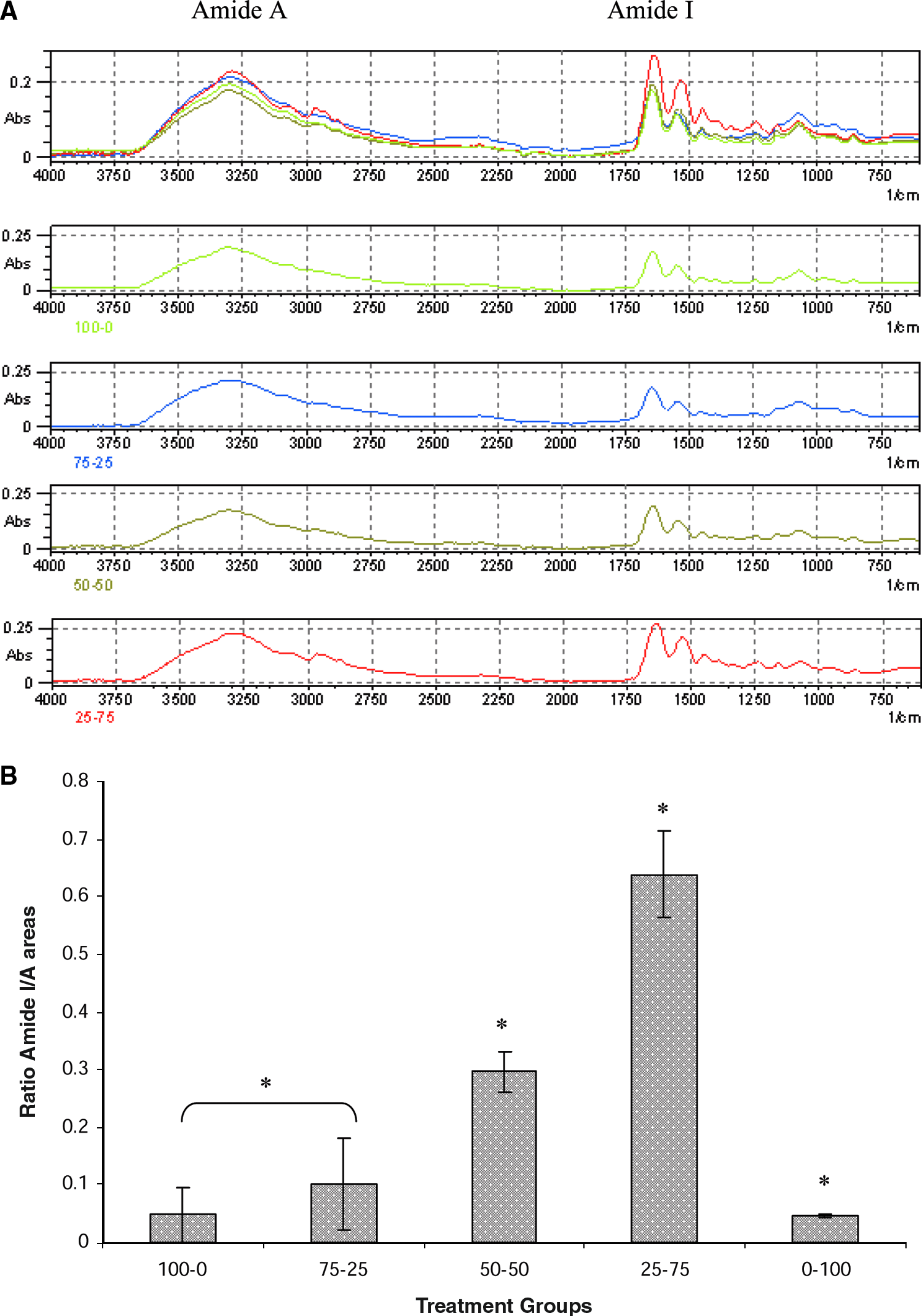
(
SDS-Page. The two gels (Fig. 3) provide visual evidence that ELP–collagen cross-linking occurs, and not just among collagen molecules. The first two columns are non-cross-linked and cross-linked collagen scaffolds with no obvious changes detected over time. Columns 3 and 4 correspond to 50-50 and 25-75, respectively, where several bands disappear over time as ELP gets incorporated into the collagen scaffold.

SDS-Page gel of four different treatment groups (1–4) cross-linked with 0.1 mg/mL of mTGase with the exception of column 1. Treatment groups are as follows: 1, collagen alone; 2, cross-linked collagen; 3, cross-linked 50–50% collagen–ELP; 4, cross-linked 75–25% collagen–ELP. Their incubation with the enzyme was interrupted at 20-min increments, and the effect on cross-linking is shown. At 0 min, time point different sizes bands are evident to disappear in subsequent time periods. Color images available online at www.liebertonline.com/ten.
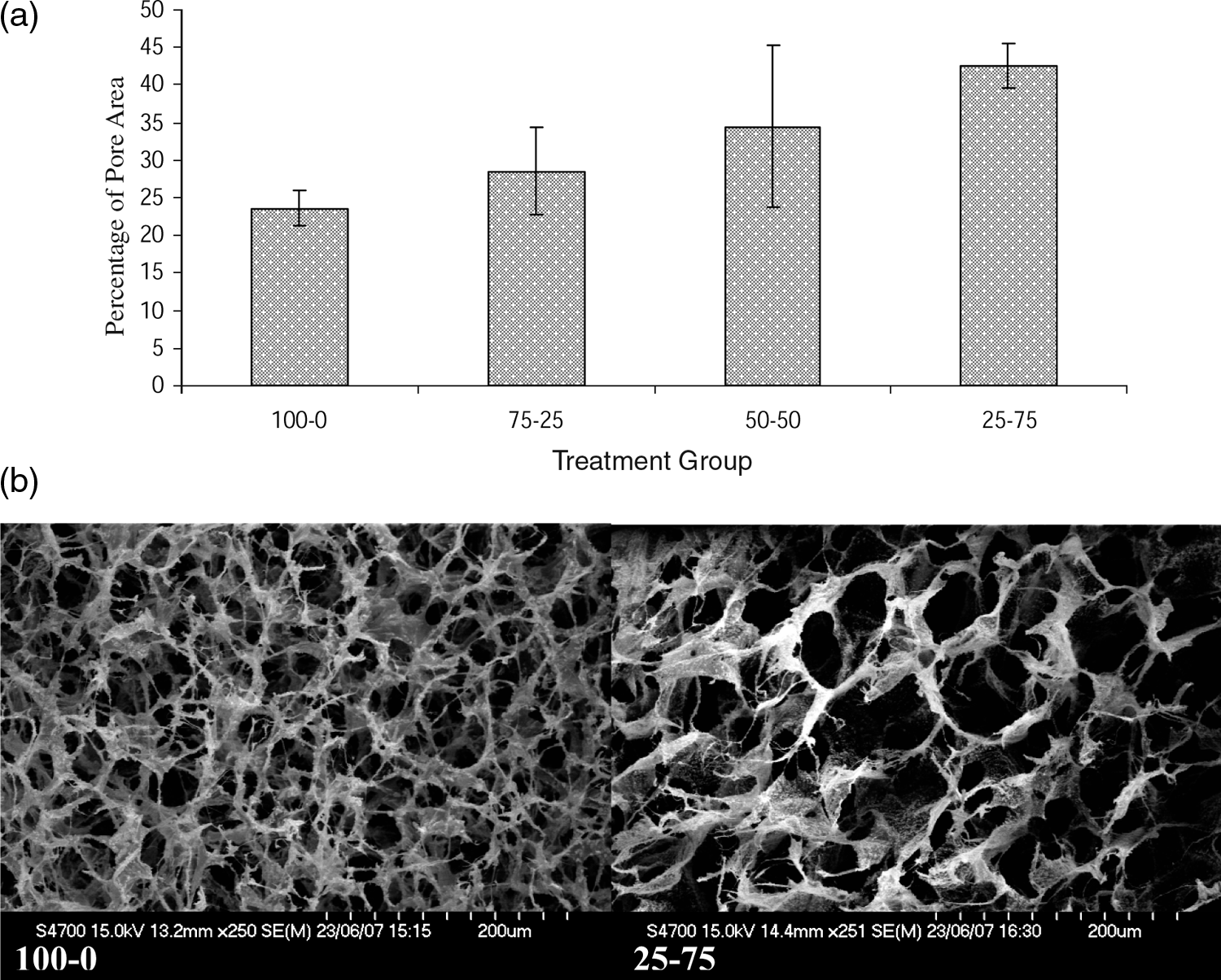
Porosity
Porosity was quantified using area fraction analysis. Pore area increases proportionally to the ELP concentration (Fig. 4a). However, these differences are not statistically significant, Figure 4b.
(
Thermodynamic behavior
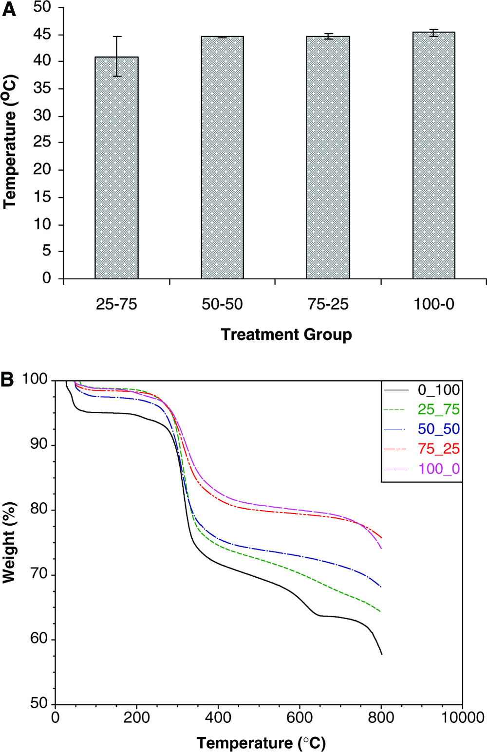
Changes in thermal properties analyzed with increasing quantities of ELP in SBF cannot be detected as seen in the DSC results shown in Figure 5A. However, the values obtained with SBF are smaller than the equivalent values obtained with PBS or distilled water (data not shown).
(
TGA calculates the onset temperature of the scaffolds' degradation, which increases as the amount of ELP increases (Table 2). On the other hand, the percentage of sample weight remaining after thermic treatment (Fig. 5B) is inversely proportional to the amount of ELP.
Temperature (°C).
Surface analysis
Topographical evaluation of the scaffolds using AFM showed that the fibril diameter of the collagen seems to be affected by the introduction of ELP (Fig. 6). The fibril diameter of the scaffolds with ELP is visibly smaller than the ones without. This infers that ELP interferes with collagen's fibrillogenesis.
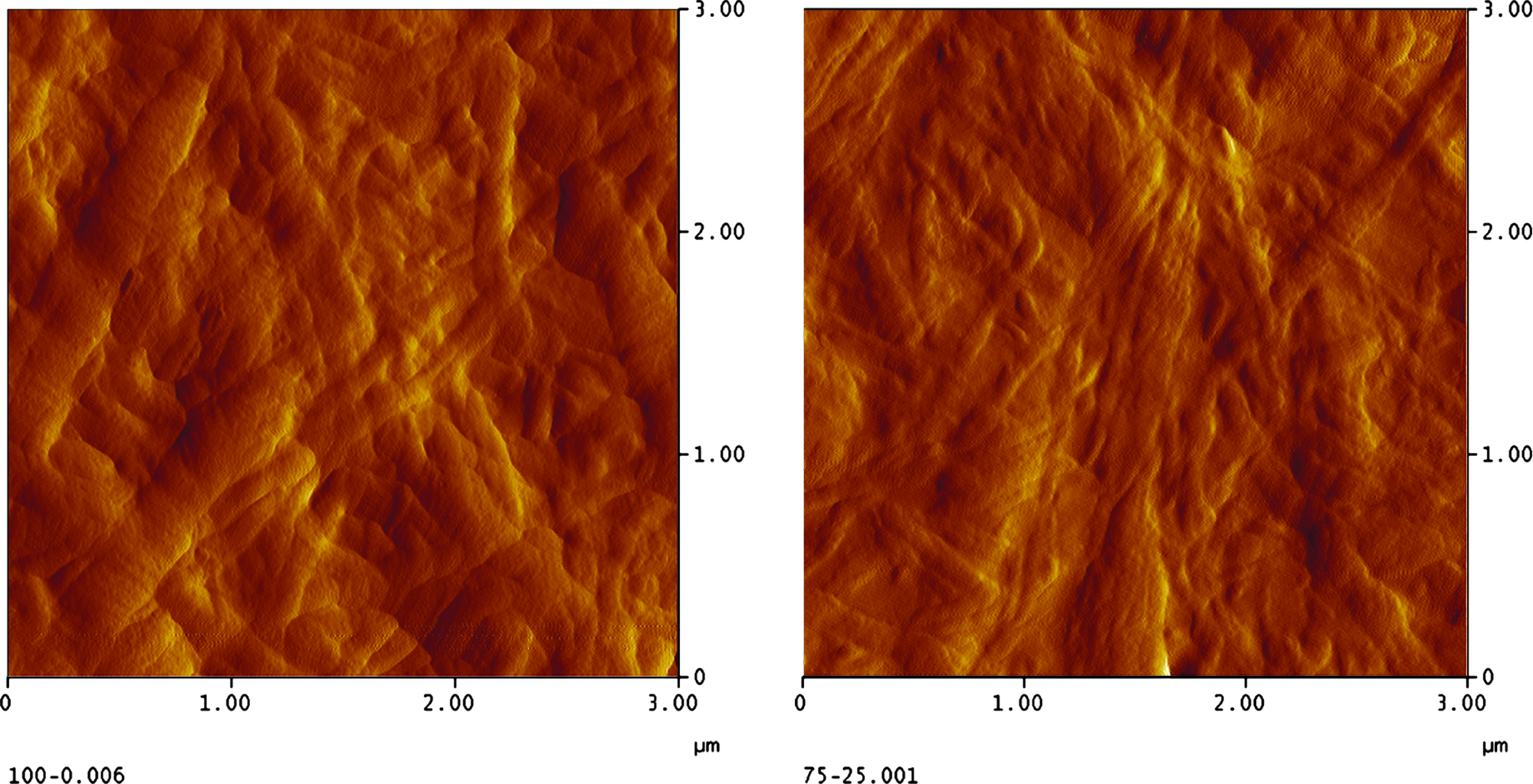
On the left: AFM image of collagen cross-linked with mTGase (100-0 indicates the percentage collagen–ELP; 006 is the sample number). On the right: AFM image of cross-linked collagen–ELP (75-25 indicates percentage collagen–ELP; 001 is the sample number). These AFM images show that the introduction of ELP into the scaffold decreases collagen fibril diameter. Color images available online at www.liebertonline.com/ten.
Mechanical characterization
G′ was found to increase proportionally to the concentration of ELP. The increase showed between 100-0 and 75-25 scaffolds does not appear to be significant, but stronger mechanical properties become statistically evident in 50-50 and 25-75 scaffolds, as shown in Figure 7.

The graph shows the rheological values of G′ plotted against treatment groups (indicated by collagen percentage–ELP percentage). The storage modulus increases with the proportion of ELP. Collagen alone and 75-25 do no show statistical differences in G′ values between themselves; however, they are when compared to 50-50 and 25-75 (n = 3; p ≤ 0.05).
Cell viability and behavior
AlamarBlue reduction assay
The metabolic activity of the different cellular types (fibroblasts, HUVECs, and SMC) seems affected by the inclusion of ELP, but only fibroblast's activity appear significantly reduced by the inclusion of ELP at both time periods, as shown in Figure 8.
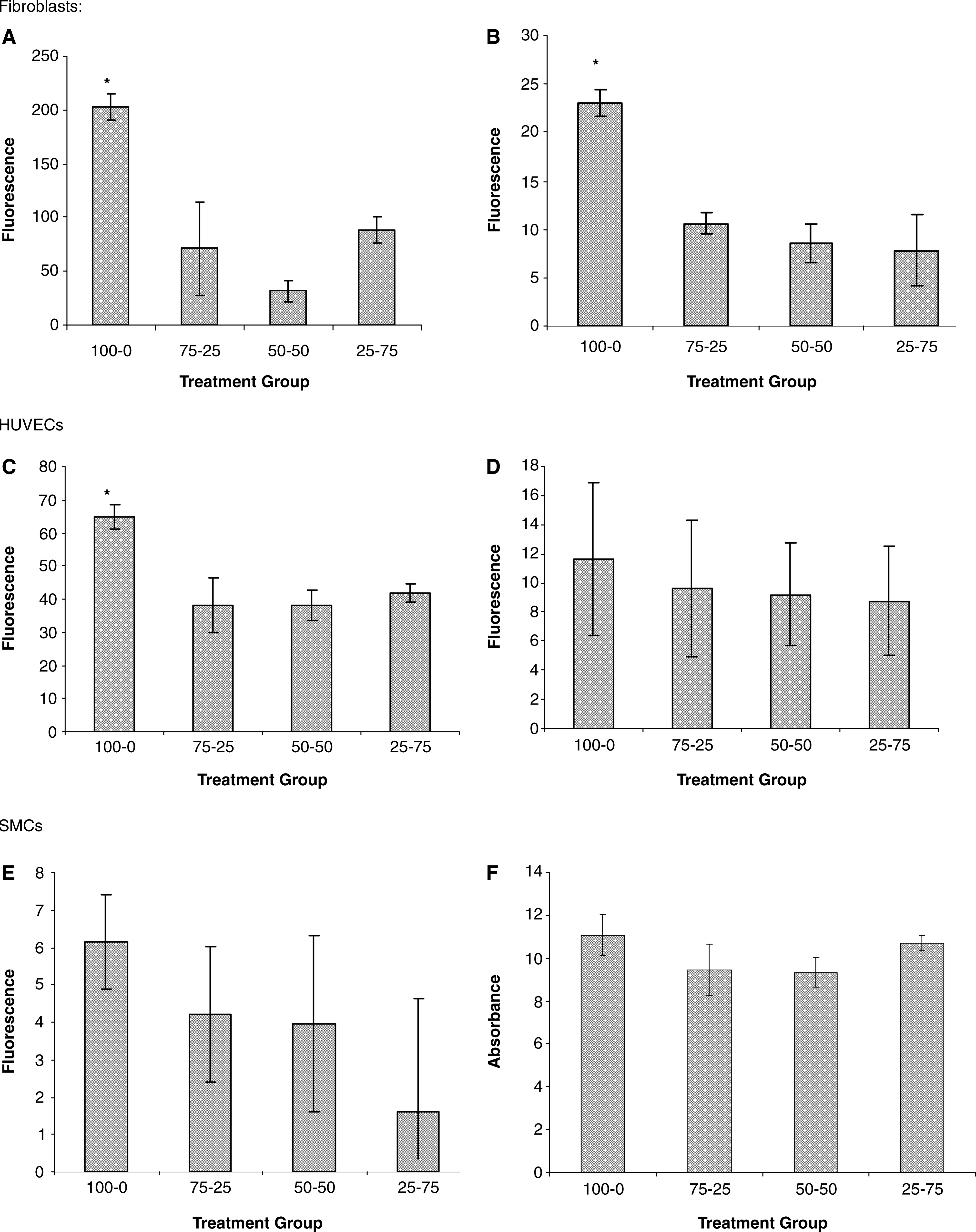
AlamarBlue™ reduction assay results for the different cell lines seeded on different scaffolds (indicated by collagen percentage–ELP percentage). (
Microscopy
The data obtained evaluating metabolic activity are reflected in phase contrast microscopy at early time periods. The fibroblastic distribution at 48 h on the different scaffolds was less uniform as the ELP proportion increased (Fig. 9). The cells were grouped, and the bipolar morphology became less evident in the presence of ELP, thus confirming the cell density data. These grouped structures/cores appear to be used as an anchorage system for radial proliferation. No turbidity or floating cells were observed at any daily scheduled monitoring times. HUVECs, on the other hand, had a more uniform distribution on the scaffolds, with no evidence of clumping or morphological changes as shown in confocal microscopy in Figure 10.

Phase contrast microscopy images of fibroblasts after 48 h of being seeded on different scaffolds (indicated by collagen percentage–ELP percentage). The antifouling properties of fibroblasts on ELP-based biomaterials are exhibited in phase contrast microscopy at × 200 magnification. Color images available online at www.liebertonline.com/ten.

Rhodamine Phalloidin-DAPI stained HUVECs seeded on 100-0 (
Bright field microscopy of SMC did not show any morphological differences between scaffolds; however, the overall density appears to be slightly reduced, even if not markedly significant (images not shown). Of the three cell lines, HUVECs seemed to be least affected by the presence of ELP, which was demonstrated in both bright field (images not shown) and confocal microscopy (Fig. 10). The latter showed clear actin stress fiber formation up to a 50:50 ratio of collagen to ELP. At higher concentrations of ELP the actin stress fibers appeared less well formed, but cells remained spread.
Discussion
Having studied and characterized transglutaminase cross-linked collagen scaffolds in previous work, 1 the introduction of another structural component that confers elastic and mechanical characteristics seemed a reasonable progression. The present investigation was undertaken to elucidate the effect of a tailored sequence of ELP on cell viability and morphology as well as on the physicochemical properties.
The incorporation of ELP into the scaffold that is designed to be covalently linked was proven using FTIR and SDS-Page. The FTIR spectra of the scaffolds show (Fig. 2a) the characteristic bands of protein structures. However, the main focus was on amide A and amide I. Their area ratios provide a clear indication of the cross-linking degree, going from free NH2, represented in Amide A, to covalent amide bonds, represented in Amide I. The amount of soluble collagen and enzyme added to the scaffold was fixed, so part of this increase of amide I/amide A ratios could be attributed to the increasing amounts of ELP and the possibility that the ELP cross-link itself. However, this contribution is minimal as seen in Figure 2b, 0-100 treatment group. It appears that self-cross-linking of the individual protein components account for small proportion of the total cross-linking.
Additionally, SDS-Page also demonstrated (Fig. 3) the creation of larger complexes unable to migrate into the resolving gel as incubation time progresses. The α and β bands characteristic of collagen molecules are visibly identified. Collagen is a relatively big protein that does not go far in this separating gel, and the effect of its cross-linking prevents its presence in the gel, thus probably eliminating it to the stacking gel. No protein marker was included in the SDS-Page, as the purpose of this assay was not the identification of proteins since the constituents are known. The lack of banding from 20 min onward suggests that ELP had cross-linked to collagen; however, the possibility that ELP binds to itself should also be considered. The AFM images (Fig. 6) aid to further support that ELP actually binds to collagen. ELP seems to interfere with the incorporation of more collagen molecules into the fibril. As a consequence, the fibrils are thinner in scaffolds with ELP.
This effect in collagen fibril thickness could be linked to the effect that ELP has on the scaffolds' porosity. Increased levels of enzymatic cross-linking are normally associated with increased collagen scaffold's porosity. 1 However, if ELP interferes with the compaction of the collagen fibers and if the concentration of substrate is incremented, a lack of statistical significance in the area fraction porosity might be justified.
Mechanical properties of the scaffolds cannot solely be attributed to the cross-linking effect. All mechanical measurements were done in gels after an overnight incubation period. The measurement of G′ as cross-linking was occurring was attempted. However, it was found that mechanical disturbances during gelation/fibrillogenesis and cross-linking in collagen scaffolds with no ELP were making the gels progressively weaker and the ELP containing scaffolds were getting increased G′ values and visibly dehydrated as the ELP content increased (data not shown). As a consequence, it was decided to enzymatically cross-link the scaffolds and measure the mechanical properties of the resulting gels after the standard overnight incubation period had elapsed. The measurement of the mechanical properties of the resulting gels produced more comparable data.
The increased mechanical properties are accompanied by thermal stability demonstrated by TGA of incremental quantities of ELP. However, if this thermal stability is due to an increase in ELP proportion or to an increase in the cross-linking degree cannot be concluded.
Polymer coacervation can be visually observed as an opaque cloudy solution appears when the temperature is raised above its ITT, an effect that completely reverses on lowering the temperature. 12 DSC is a method broadly used in the determination of ITT, enthalpy of the system. Under our working conditions, the scaffolds' ITT does not differ as the ELP concentration increases. The ions contained in the SBF might have decreased the transition temperature 12 to a point that the increase in substrate concentration did not have that much of an effect. An obvious advantage of this system is its potential for drug delivery at quasi body temperature. This system has been previously used in cancer therapy by dissolving an active principle in an aqueous solution and including it in the scaffold. 46 Upon implantation, the patient will receive an initial bolus release of the drug at body temperature or higher depending on the ITT, while coacervating. Over time, the patient will receive a sustained release of the drug via elastase degradation.12,46 In the system used for this work, the well-known stability of the elastin was overcome by the introduction of sequences for elastase activity to guarantee chronic release. 12 ELP, as well as PNIPAam due to its smart behavior, can also be used in recombinant protein purification by terminal tagging in recombinant systems. 47
After 4 days' incubation, the metabolic activity of fibroblasts, used as a measure of cell density, appeared to be more affected by the introduction of ELP than SMCs and endothelial cells. Both the SMCs and the endothelial cells plated onto the collagen–ELP mixtures showed no significant difference to the collagen control. Therefore, the presence of a normal morphology displayed by endothelial cells on the scaffold must be attributed to the REDV sequences present in the scaffolds. These findings also corroborate the chemoattracting effect that Long et al. had observed on bovine aortic endothelial cells in 1989 when using these sequences. 48 Further, a few years later, in 1997, Tajima et al. reported similar findings with chick vascular SMCs when using repeating sequences of VPGVG. 49
Conformational differences between different ELP-repeating patterns, polypentamers (VPGVG), or polyhexamers (VGVAPG) that affect fibroblasts in culture has been previously reported.50,51 Therefore, the detrimental effects on fibroblast's metabolic activity/cell density observed after 7 days' incubation might be justified. In addition, while elastin peptides or products of elastin degradation seem to enhance endothelial cell chemotaxis and proliferation, 6 ELP such as VPGVG monomer or polymer (pentapeptide repeating structure used in this work) have shown no effect on fibroblast proliferation corroborating also the in vivo studies where VPGVG type of polypentapeptides implants have demonstrated strong nonadhesive properties.52–54 Additionally, the collagen component of the scaffold and its subsequent effect on cell adhesion and proliferation cannot be forgotten.
Conclusions
The introduction of an elastic element into the collagen scaffold that can be cross-linked with mTGase while enhancing the scaffolds mechanical properties has proven to be successful. In addition, the tenability of differential colonization of the scaffold with a specific cell type makes this scaffold an attractive platform.
The lysine and glutamine residues of the ELP hydrophilic domains bind covalently to collagen, as confirmed by FTIR, AFM, and SDS-Page. Porosity and ITT do not seem to be affected by increasing concentrations of ELP. On the other hand, the storage modulus and onset of degradation temperatures increase with increasing amounts of ELP, while the residue obtained after thermal treatment decreases.
Fibroblasts did not display normal behavior and morphology or proliferate adequately in scaffolds with ELP. In view of these characteristics, if taken together with the normal endothelial cell behavior, anticipate the usefulness of these scaffolds in studying hypertrophic scar prevention, vascular tissue engineering, and cavity/tubular lining tissue-engineered constructs.
These scaffolds have proven in this in vitro characterization their potential in tissue engineering. Their final use will determine the appropriate concentration/percentage to be used. It could be speculated that 50-50 to 25-75 will be suitable for vascular and urogenital tissue engineering and 75-25 to 50-50 might be more suitable for skin wound healing where fibroblast growth as well as endothelial growth should need to be enhanced.
Footnotes
Acknowledgments
This experimental work has been funded by The Irish Higher Education Authority's Program for Research in Third-Level Institutions, Enterprise Ireland Research Innovation Fund, and EPSRC (grant reference GR/S21755/02).
Special thanks to Damien O'Halloran and Dr. Carlos Elvira for their help in the rheology and thermogravimetric work, respectively.
Disclosure Statement
No competing financial interests exist.